Pathology S1 U2
Study these flashcardsFree · spaced repetition · iPhone, Android, Mac & web
First 300 of 455 cards
1
initiating cause of a disease
etiology
2
refers to the sequence of molecular, biochemical, and cellulat evets that lead tp the development of disease
pathpgenesis
3
refer to the structural alterations in cells or tissues that are characteristic of a disease and diagnostic of an etiologic process
morphological changes
4
symptoms and signs) of disease, as well as its progression (clinical course and outcome).
clinical manifestations
5
eversible functional and structural responses to changes in physiologic states (e.g., pregnancy) and some pathologic stimuli, during which new but altered steady states are achieved, allowing the cell to survive and continue to function
adaptations
6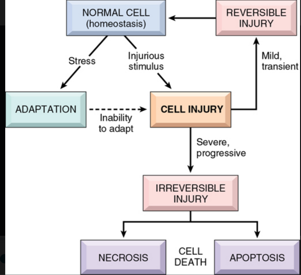
Image occlusion: name the hidden labels
7
Image occlusion: name the hidden labels
8
Image occlusion: name the hidden labels
9
Image occlusion: name the hidden labels
10
Image occlusion: name the hidden labels
11
Image occlusion: name the hidden labels
12
reduced blood flow (med term)
ischemia
13
calcium may be deposited at sites of cell death, causing this
pathological calcification
14
deficiency of oxygen, which causes cell injury by reducing aerobic oxidative respiration
hypoxia
15
causes of hypoxia (x3)
reduced blood flow (ischemia); inadequate oxygenation of the blood due to cardiorespiratory failure; and decreased oxygen-carrying capacity of the blood,
16
______________ deficiencies cause an appalling number of deaths, chiefly among lower-resource populations. Deficiencies of specific vitamins are found throughout the world
Proteoin-calporie
17
The integrated stress response (ISR) can be activated by 1-4 stress sensing kinases
Protein kinase R (PKR)
PRK like ER kinase (PERK)
GCN2
HRI
PRK like ER kinase (PERK)
GCN2
HRI
18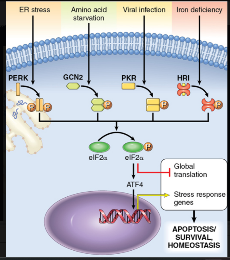
Image occlusion: name the hidden labels
19
Image occlusion: name the hidden labels
20
Image occlusion: name the hidden labels
21
Image occlusion: name the hidden labels
22
diases caused by misfolded proteins: Loss of CFTR leads to defects in chloride transport
cystic fobrosis
23
diseases caused by missfolded proteins: Loss of LDL receptor leads to hypercholesterolemia
familial hypercholesterolemia
24
diseases caysed by missfolded proteins: Lack of the lysosomal enzyme leads to storage of GM2 gangliosides in neurons
tay sachs disease
25
diseases caused by missfoldedproteins:Storage of nonfunctional protein in hepatocytes causes apoptosis; absence of enzymatic activity in lungs causes destruction of elastic tissue giving rise to emphysema
a1-antitrypsin deficiency
26
diseases caused by missfolded proteins: Abnormal folding of PrPsc causes neuronal cell death
creutzfeldt jacob disease
27
diseases caused by missfolded proteins: Abnormal folding of Aβ peptides causes aggregation within neurons and apoptosis
alzheimer disease
28
stephs of autophagy (x3)
• Nucleation and formation of an isolation membrane, also called a phagophore; the isolation membrane is believed to be derived from the ER, though other sources such as the plasma membrane and mitochondria may contribute
• Formation of a vesicle, called the autophagosome, from the isolation membrane, inside which intracellular organelles and cytosolic structures are sequestered
• Maturation of the autophagosome by fusion with lysosomes, to deliver digestive enzymes that degrade the contents of the autophagosome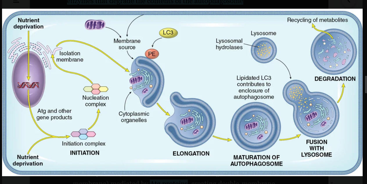
• Formation of a vesicle, called the autophagosome, from the isolation membrane, inside which intracellular organelles and cytosolic structures are sequestered
• Maturation of the autophagosome by fusion with lysosomes, to deliver digestive enzymes that degrade the contents of the autophagosome
29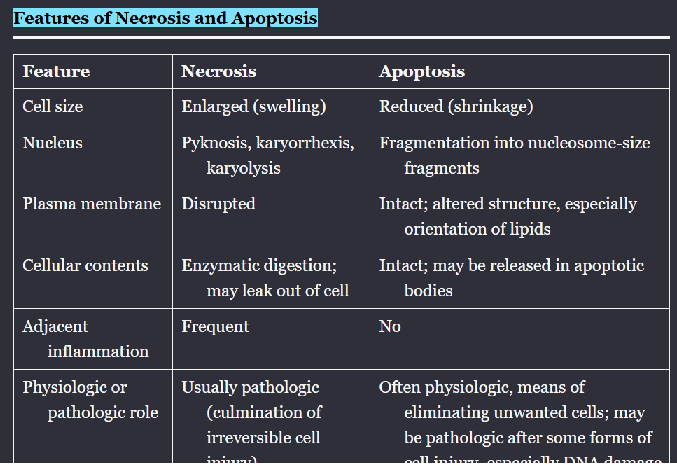
Image occlusion: name the hidden labels
30
Image occlusion: name the hidden labels
31
Image occlusion: name the hidden labels
32
Image occlusion: name the hidden labels
33
Image occlusion: name the hidden labels
34
Image occlusion: name the hidden labels
35
Image occlusion: name the hidden labels
36
Image occlusion: name the hidden labels
37
Image occlusion: name the hidden labels
38
Image occlusion: name the hidden labels
39
Image occlusion: name the hidden labels
40
Image occlusion: name the hidden labels
41
characterized by functional and structural alterations in early stages or mild forms of injury, which are correctable if the damaging stimulus is removed.
reversible cell injury
42
ultrastructural changes of reversile cell injury visible by electron micrpscopy (x5)
. Plasma membrane alterations, such as blebbing, blunting, and loss of microvilli
2. Mitochondrial changes, including swelling and the appearance of small amorphous densities
3. Accumulation of “myelin figures” in the cytosol composed of phospholipids derived from damaged cellular membranes
4. Dilation of the ER, with detachment of polysomes
5. Nuclear alterations, with disaggregation of granular and fibrillar elements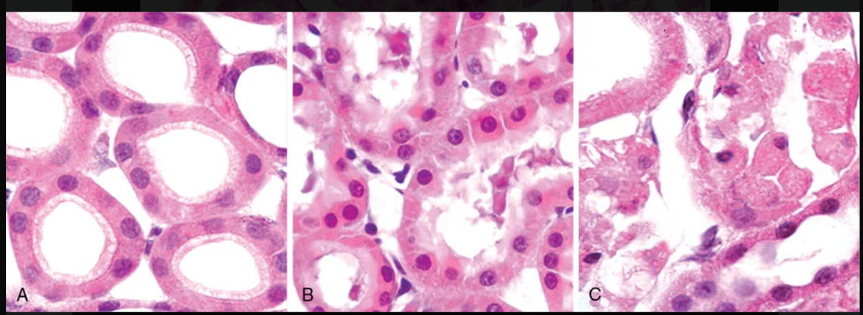
2. Mitochondrial changes, including swelling and the appearance of small amorphous densities
3. Accumulation of “myelin figures” in the cytosol composed of phospholipids derived from damaged cellular membranes
4. Dilation of the ER, with detachment of polysomes
5. Nuclear alterations, with disaggregation of granular and fibrillar elements
43
Severe mitochondrial damage with depletion of ATP and rupture of lysosomal and plasma membranes are typically associated with (apoptosis/necrosis)
necrosis
44
main causes of necrosis
loss of oxygen supply (ischemia), exposure to microbial toxins, burns and other forms of chemical and physical injury, and unusual situations in which active proteases leak out of cells and damage surrounding tissues (as in pancreatitis).
45
characterized by denaturation of cellular proteins, leakage of cellular contents through damaged membranes, local inflammation, and enzymatic digestion of the lethally injured cell
necrosis
46
substances released from injured, necrotic cells that include ATP, uric acid, and numeroud other molecykes that are usually confined within healthy cells and whose release is an indicator of severe cell injury
damage associated molecular patterns (DAMPs)
47
necrotic cells show (eosinophilla/neutrophilia) in H&E stains, attributable in part to the loss of cytoplasmic RNA and in part to accumulation of denatured cytoplasmic proteins
eosinophilia
48
nuclear changes: The basophilia of the chromatin may fade (______________), a change that presumably reflects loss of DNA because of enzymatic degradation by endonucleases.
karyolysis
49
nuclear changes: pattern (also seen in apoptotic cell death) is ______________, characterized by nuclear shrinkage and increased basophilia. Here the chromatin condenses into a dense, shrunken basophilic mass.
pyknosis
50
nuclear changes: third pattern, known as _________________, the pyknotic nucleus undergoes fragmentation. With the passage of time (1 or 2 days), the nucleus in the necrotic cell totally disappears.
karyorrhexis
51
a form of necrosis in which the architecture of dead tissue is preserved for a span of at least some days (Fig. 2.10). The affected tissue has a firm texture.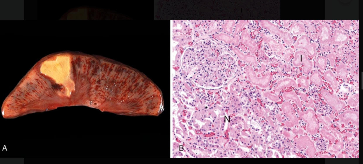
coagulative necrosis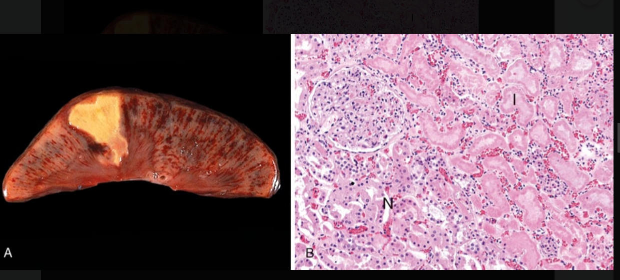
52
necrosis: characterized by digestion of the dead cells, resulting in transformation of the tissue into a viscous liquid. It is seen in focal bacterial or, occasionally, fungal infections, because microbes stimulate the accumulation of leukocytes and the liberation of enzymes from these cells.
liquefactive necrosis
53
necrosis: applied to a limb, generally Page 39Page 40the lower leg, that has lost its blood supply and has undergone necrosis (typically coagulative necrosis) involving multiple tissue planes
gangrenous necrosis
54
necrosis: encountered most often in foci of tuberculous infection (Chapter 8). The term caseous (cheeselike) is derived from the friable white appearance of the area of necrosis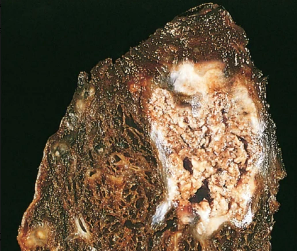
CASEOUS NECROSIS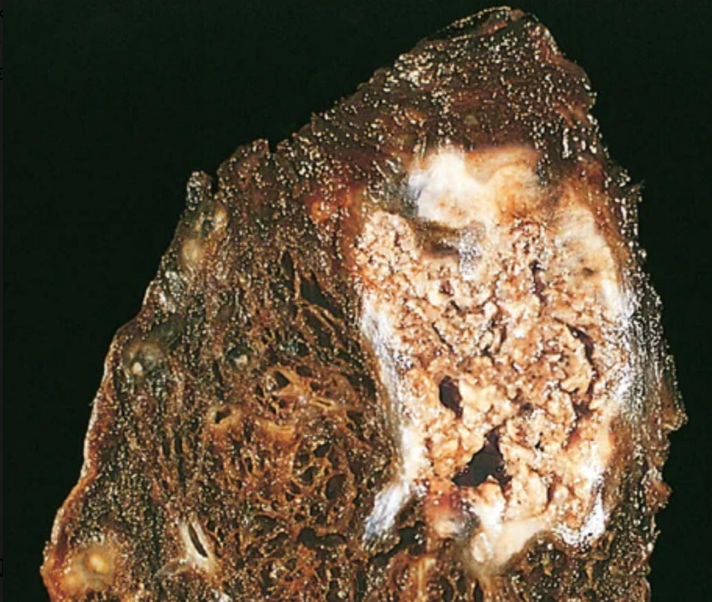
55
necrosis: refers to focal areas of fat destruction, typically resulting from release of activated pancreatic lipases into the substance of the pancreas and the peritoneal cavity. This occurs in the calamitous abdominal emergency known as acute pancreatitis
fat necrosis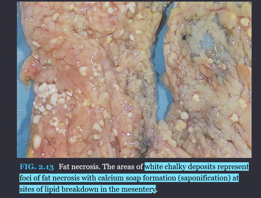
56
necrosis: a special form of vascular damage usually seen in immune reactions involving blood vessels (Chapter 11). It typically occurs when complexes of antigens and antibodies are deposited in the walls of arteries.
fibrinoid necrosis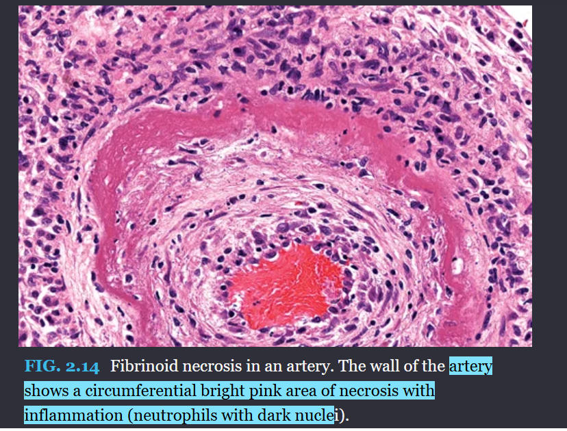
57
apoptosis: This is the most characteristic feature of apoptosis. The chromatin aggregates peripherally, under the nuclear membrane, into dense masses of various shapes and sizes (see Fig. 2.15B). The nucleus itself may break up into two or more fragments.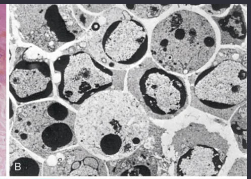
chromatin condensation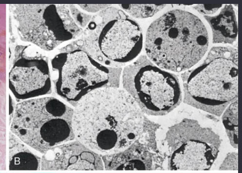
58
The apoptotic cell first shows extensive surface membrane blebbing, which is followed by fragmentation of the dead cells into membrane-bound apoptotic bodies composed of cytoplasm and tightly packed organelles, with or without nuclear fragments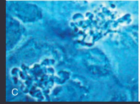
cytoplasmic blebs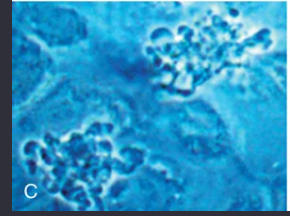
59
phases of apoptosis (x3)
initiation phase, during which some caspases become catalytically active and unleash a cascade of other caspases, and an execution phase, during which the terminal caspases trigger cellular fragmentation and demise.
60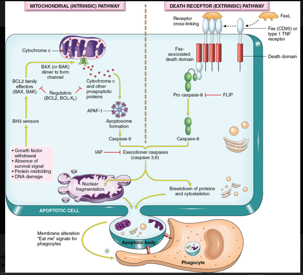
Image occlusion: name the hidden labels
61
Image occlusion: name the hidden labels
62
Image occlusion: name the hidden labels
63
Image occlusion: name the hidden labels
64
Image occlusion: name the hidden labels
65
apoptosis: The formation of apoptotic bodies breaks cells up into “bite-sized” fragments that are edible for ...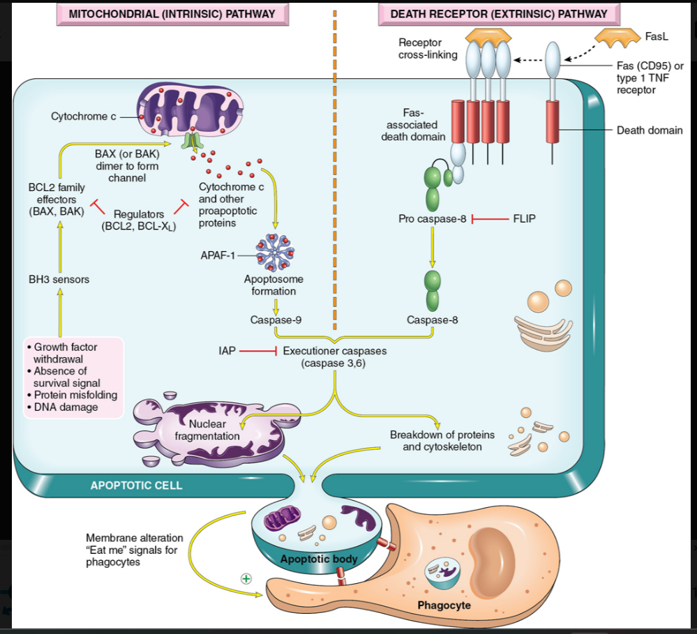
phagocytes
66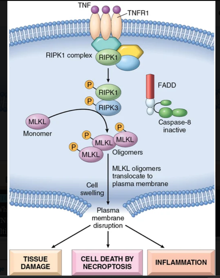
Image occlusion: name the hidden labels
67
Image occlusion: name the hidden labels
68
Image occlusion: name the hidden labels
69
Image occlusion: name the hidden labels
70
a lytic form of cell death accompanied by the release of the inflammatory mediators, including the fever-inducing cytokine IL-1 (pyro refers to fever)
pyroptosis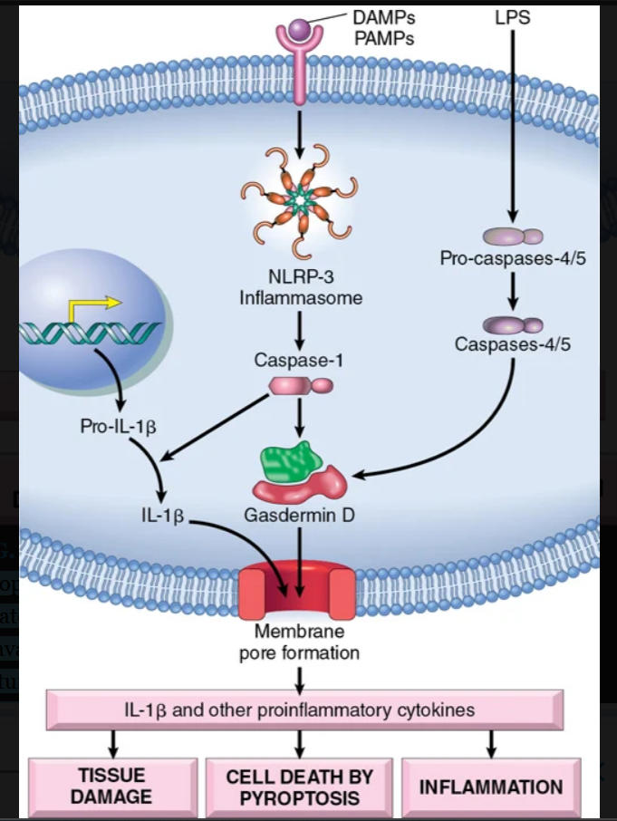
71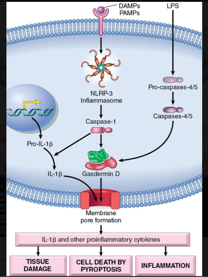
Image occlusion: name the hidden labels
72
Image occlusion: name the hidden labels
73
Image occlusion: name the hidden labels
74
Image occlusion: name the hidden labels
75
Image occlusion: name the hidden labels
76
Image occlusion: name the hidden labels
77
Image occlusion: name the hidden labels
78
Pyroptosis is activated by canonical and noncanonical pathways, both of which converge on the activation of pore-forming proteins called...
gasdermins
79
distinct form of cell death triggered when excessive intracellular levels of iron or ROS overwhelm the glutathione-dependent antioxidant defenses (discussed later) to cause unchecked membrane lipid peroxidation.
-can be prevented by reducing iron levels
-can be prevented by reducing iron levels
ferroptosis
80
Damage to nuclear DNA activates sensors that trigger _________________ dependent pathways
p53
81
fx of p53
rrests cells in the G1 phase of the cell cycle and activates DNA repair mechanisms. If these mechanisms fail to correct the DNA damage, p53 triggers apoptosis by the mitochondrial pathway.
82
how are free radicals made in cells?
1) The reduction-oxidation reactions that occur during normal metabolic processes.
2) UV, Xrays
3) enzyme metabolism of exogenous chemicals or drugs
4) transition metals that just leave them behind
5) nitric oxide (NO) that leaves electrons behind
2) UV, Xrays
3) enzyme metabolism of exogenous chemicals or drugs
4) transition metals that just leave them behind
5) nitric oxide (NO) that leaves electrons behind
83
pathways to remove free radicals from the bpdy
1) Antioxidants either block free radical formation or inactivate (e.g., scavenge) free radicals.
2) transition metals: reactivity of these metals is minimized by their binding to storage and transport proteins
3) Several enzymes
2) transition metals: reactivity of these metals is minimized by their binding to storage and transport proteins
3) Several enzymes
84
present in peroxisomes, decomposes H2O2 (2H2O2 → O2 + 2H2O).
2. Superoxidase dismutases (SODs) are found in many cell types and convert to H2O2 (2 + 2H → H2O2 + O2). This group of enzymes includes both manganese-SOD, which is localized in mitochondria, and copper-zinc-SOD, which is found in the cytoplasm.
2. Superoxidase dismutases (SODs) are found in many cell types and convert to H2O2 (2 + 2H → H2O2 + O2). This group of enzymes includes both manganese-SOD, which is localized in mitochondria, and copper-zinc-SOD, which is found in the cytoplasm.
catalase
85
protects against injury by catalyzing free radical breakdown (H2O2 + 2GSH → GSSG [oxidized glutathione] + 2H2O). The intracellular ratio of oxidized glutathione (GSSG) to reduced glutathione (GSH) reflects the oxidative state of the cell and is an important indicator of the cell’s ability to detoxify ROS.
Pathologic effects of free radicals
Pathologic effects of free radicals
glutathione peroxidase
86
what do ROS do to cell membranes (x3)
1) Lipid peroxidation in membranes. In the presence of O2, free radicals may cause peroxidation of lipids within plasma and organellar membranes. Oxidative damage is initiated when the double bonds in unsaturated fatty acids of membrane lipids are attacked by O2-derived free radicals
2) Oxidative modification of proteins. Free radicals promote oxidation of amino acid side chains, formation of covalent protein-protein cross-links (e.g., disulfide bonds), and oxidation of the protein backbone. Oxidative modifications may also damage the active sites of enzymes
3) Lesions in DNA. Free radicals can cause single- and double-strand breaks in DNA, cross-linking DNA strands, and forming adducts.
2) Oxidative modification of proteins. Free radicals promote oxidation of amino acid side chains, formation of covalent protein-protein cross-links (e.g., disulfide bonds), and oxidation of the protein backbone. Oxidative modifications may also damage the active sites of enzymes
3) Lesions in DNA. Free radicals can cause single- and double-strand breaks in DNA, cross-linking DNA strands, and forming adducts.
87
how can Ca2+ accumulation damage cells?
The accumulation of Ca2+ in mitochondria results in opening of the mitochondrial permeability transition pore and, as described earlier, failure of ATP generation.
• Increased cytosolic Ca2+ activates several enzymes with potentially deleterious effects on cells. These enzymes include phospholipases (which cause membrane damage), proteases (which break down both membrane and cytoskeletal proteins), endonucleases (which are responsible for DNA and chromatin fragmentation), and ATPases (thereby hastening ATP depletion).
• Increased cytosolic Ca2+ activates several enzymes with potentially deleterious effects on cells. These enzymes include phospholipases (which cause membrane damage), proteases (which break down both membrane and cytoskeletal proteins), endonucleases (which are responsible for DNA and chromatin fragmentation), and ATPases (thereby hastening ATP depletion).
88
promotes new blood vessel formation, stimulates cell survival pathways, and enhances glycolysis.
Hypoxia inducile factpr 1 (HIF-1)
89
what reaction? Reduced oxidative phosphorylation → ATP depletion → failure of Na pump → influx of sodium and water → organelle and cellular swelling (reversible).
mild ischemia
90
what reaction? Severe swelling of mitochondria, calcium influx into mitochondria and into the cell with rupture of lysosomes and plasma membrane. Death by necrosis and apoptosis due to release of cytochrome c from mitochondria.
severe ischemia
91
an increase in the size of cells that results in an increase in the size of the affected organ. The hypertrophied organ has no new cells, just larger cells. The increased size of the cells is due to the synthesis and assembly of additional intracellular structural components.
hypertrophy
92
what causes hypertrophy
a result of increased cellular protein production
93
an increase in the number of cells in an organ or tissue in response to a stimulus.
hyperplasia
94
hyperplasia due to the action of hormones or growth factors occurs when there is a need to increase functional capacity of hormone-sensitive organs, or when there is need for compensatory increase after damage or resection
physiologucal hyperplasia
95
hypetrplasia: caused by excessive or inappropriate actions of hormones or growth factors acting on target cells.
pathological hyperplasia
96
a reduction in the size of an organ or tissue due to a decrease in cell size and number.
atrophy
97
atrophy: When a fractured bone is immobilized in a plaster cast or when a patient is restricted to complete bed rest, skeletal muscle atrophy rapidly ensues.
decrease workload (disuse atrophy)
98
atrophy: The normal metabolism and function of skeletal muscle depend on its nerve supply. Damage to the nerves leads to atrophy of the muscle fibers supplied by those nerves
loss of innervatiom (deinervation atrophy)
99
atrophy results from ________ protein synthesis and _____ protein atriphy
decreased; increased
100
a reversible change in which one differentiated cell type (epithelial or mesenchymal) is replaced by another cell type
metaplasia
101
most common epithelial metaplasia
columnar to squamous (smokers)
102
esophageal squamous epithelium is replaced by intestinal-like columnar cells under the influence of refluxed gastric acid
barrett esophagus
103
describe abnormal accumulations of triglycerides within parenchymal cells.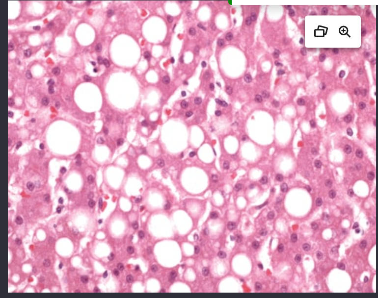
steatosis and fatty change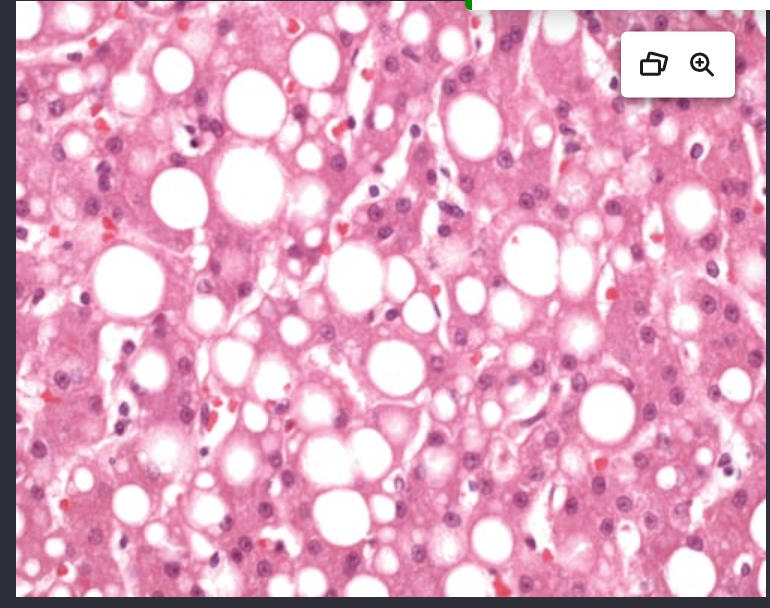
104
refers to the focal accumulations of cholesterol-laden macrophages in the lamina propria of the gallbladder
cholesterolosis
105
ntracellular accumulation of cholesterol within macrophages is also characteristic of acquired and hereditary hyperlipidemic states. Clusters of foamy cells are found in the subepithelial connective tissue of the skin and in tendons, producing tumorous masses known as xanthomas
xanthomas
106
not injurious to the cell or its functions. Its importance lies in its being a telltale sign of free radical injury and lipid peroxidation.
- insoluble pigment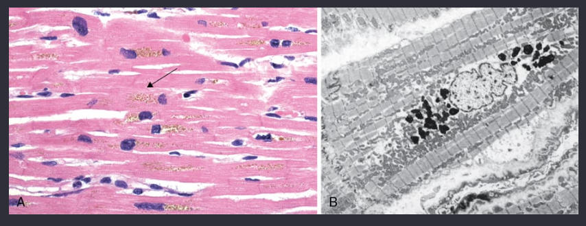
- insoluble pigment
lipofuscin granules
107
abnormal tissue deposition of calcium salts, together with smaller amounts of iron, magnesium, and other mineral salts.
pathological calcification
108
encountered in areas of necrosis, whether they are of coagulative, caseous, or liquefactive type, and in foci of enzymatic necrosis of fat. Calcification is almost always present in the atheromas of advanced atherosclerosis. It also commonly develops in aging or damaged heart valves, further hampering their function
dystrophic calcification
109
Deposition of calcium in normal tissues, caused by hypercalcemia (usually a consequence of parathyroid hormone excess)
metastatic calcification
110
accumulation of pleural fluid occurs in the following settings (x5)
• Increased hydrostatic pressure, as in congestive heart failure
• Increased vascular permeability, as in pneumonia
• Decreased osmotic pressure, as in nephrotic syndrome
• Increased intrapleural negative pressure, as in atelectasis
• Decreased lymphatic drainage, as in mediastinal carcinomatosis
• Increased vascular permeability, as in pneumonia
• Decreased osmotic pressure, as in nephrotic syndrome
• Increased intrapleural negative pressure, as in atelectasis
• Decreased lymphatic drainage, as in mediastinal carcinomatosis
111
pleural fluid causes: increased ____________ as in congestive heart failure
hydrostatic pressure
112
pleural fluid causes: increased ______- permiability as in pnemonia
vascular
113
pleureal fluid causes: decreased ________- pressure as in nephrotic syndrome
osmotic
114
pleural fluid causes: increased intrapleural __________ pressure, as in atelectasis
negative
115
pleural fluid causes: decreased ______ drainage, as in mediastinal carcinomatosis
lymphatic
116
are disorders associated with inflammation of the underlying lung, such as tuberculosis, pneumonia, lung infarction, lung abscess, and bronchiectasis.
pleuritis
117
Radiotherapy for tumors in the lung or mediastinum often causes a ....
serofibronous pleuritits
118
(empyema) usually results from bacterial or fungal seeding of the pleural space. Most commonly, this seeding occurs by contiguous spread of organisms from intrapulmonary infection
purulent pleural exudate (empyema)
119
when empyema results from an infection from elow the diaphragm, uts more likely to occur on what side?
right side
120
empyema is characterized by loculated, yellow-green, creamy pus composed of masses of __________ with other leukocytes
neutrophils
121
When ____________ pleuritis is encountered, a careful search should be made for the presence of tumor cells.
hemorrhagic
122
Noninflammatory collections of serous fluid within the pleural cavities are called
hydrothorax
123
most common cause of hydrothorax
heart failure
124
escapeof blood into the pleural cavity
hemothorax
125
an accumulation of milky fluid, usually of lymphatic origin, in the pleural cavity.
chylothorax
126
refers to air or gas in the pleural cavities and is most commonly associated with emphysema, asthma, and tuberculosis. It may be spontaneous, traumatic, or therapeutic
pneumothorax
127
pneumothorax is usually caused by some perforating injury to the chest wall, but sometimes the trauma pierces the lung and thus provides two avenues for the accumulation of air within the pleural spaces.
tramatic pneumothorax
128
This entity is encountered in relatively young people; seems to be due to rupture of small, peripheral, usually apical subpleural blebs; and usually subsides spontaneously as the air is resorbed. Recurrent attacks are common and can be quite disabling.
spontenous ideopathic pneumothrax
129
a soft tissue tumor with a propensity to occur in the pleura and, less commonly, in the lung, as well as other sites. It is highly associated with a cryptic inversion of chromosome 12 involving the genes NAB2 and STAT6.
solitary fibrous tumor
130
morphology of solitary fibrous tumor
Microscopically, the tumor shows whorls of reticulin and collagen fibers among which are interspersed spindle cells resembling fibroblasts. Rarely, this tumor may be malignant, marked by pleomorphism, mitotic activity, necrosis, and large size
-no relation to aasbestos exposre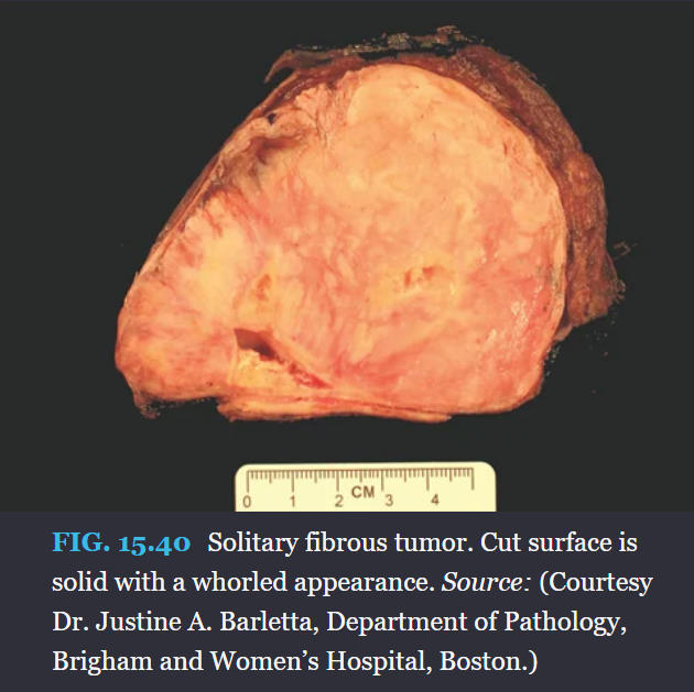
-no relation to aasbestos exposre
131
- increased incidence among people with heavy exposure to asbestos
-arises from either the visceral or the parietal pleura.
-Asbestos bodies (see Fig. 15.18A) are found in increased numbers and aspestos plaques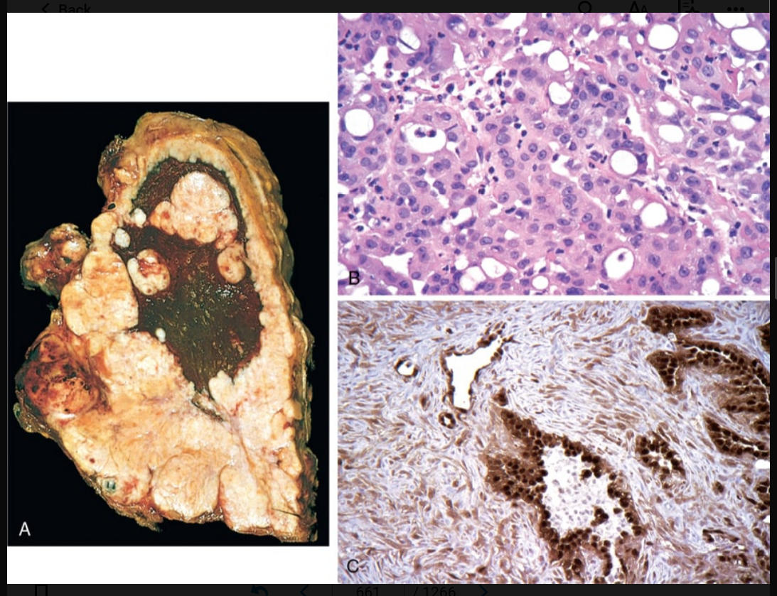
-arises from either the visceral or the parietal pleura.
-Asbestos bodies (see Fig. 15.18A) are found in increased numbers and aspestos plaques
malignant mesothelioma
132
types of malignant mesothelioma
epithelioid (60% to 80%), sarcomatoid (10% to 12%), or biphasic (10% to 15%)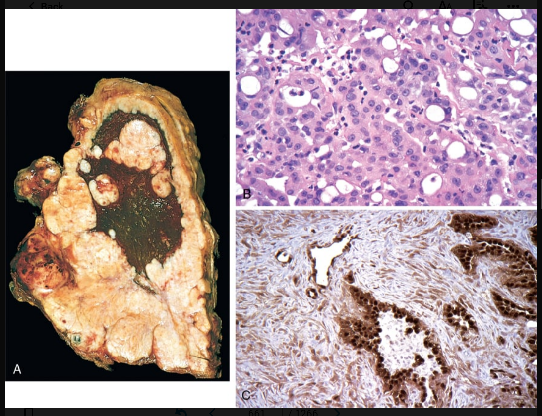
133
mesothelioma consists of cuboidal, columnar, or flattened cells forming tubular or papillary structures resembling adenocarcinoma
epithelioid type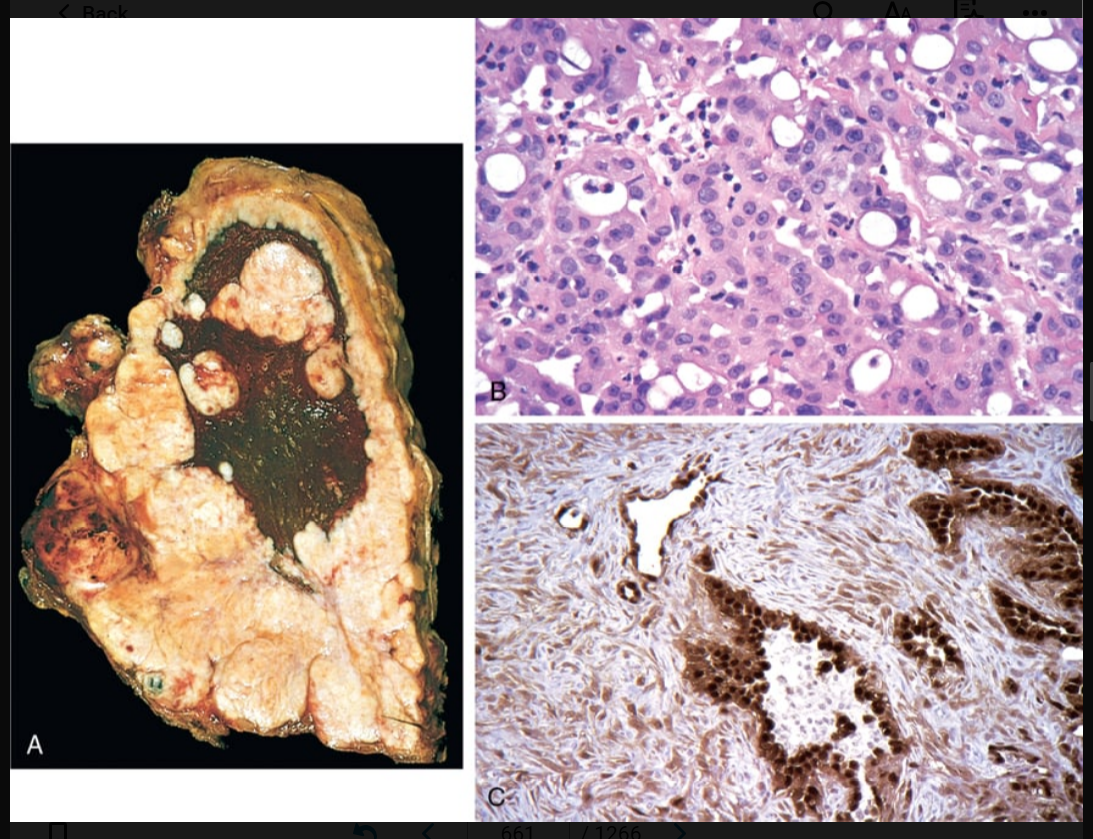
134
-mesothelioma has an appearance resembling fibrosarcoma and is usually extensively fibrotic and paucicellular.
-tend to have lower expression of many of the markers described above and some may be positive only for keratin
-tend to have lower expression of many of the markers described above and some may be positive only for keratin
sarcomatoid type
135
mesothelioma contains both epithelioid and sarcomatoid patterns in varying proportions and tends to behave similarly to sarcomatoid type
biphastic type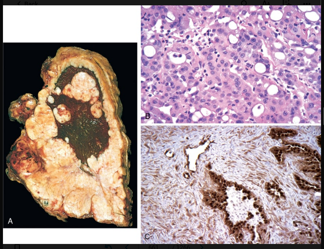
136
what are most mesotheliomas positive for?
keratin proteins, calretinin (Fig. 15.41C), Wilms tumor 1 (WT-1), cytokeratin 5/6, and podoplanin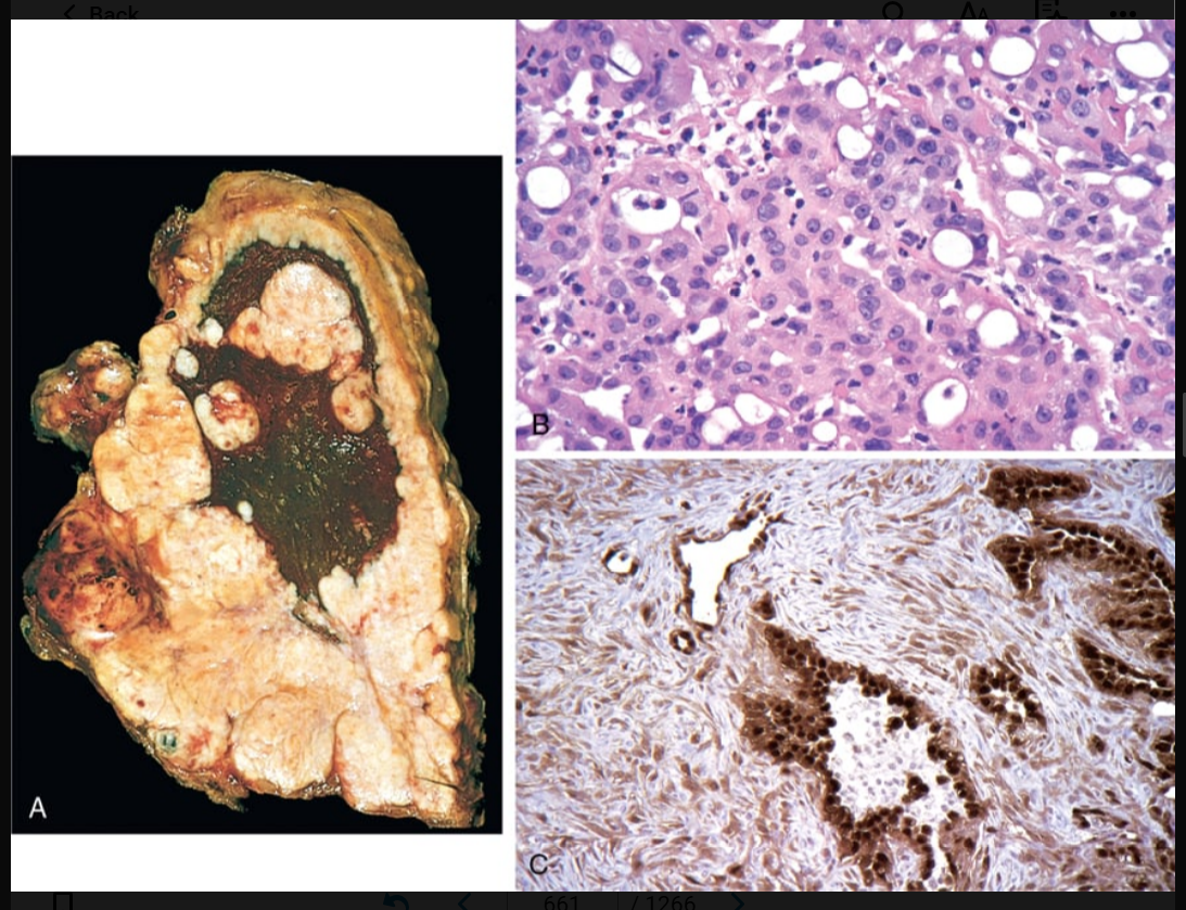
137
what are the presenting symptoms for mesothelioma (malignant) (x3)
chest pain, dyspnea, reccurent pleural effusions
138
important cause of morbidity and mortality, particularly in patients who are bedridden, but also in a wide range of conditions that are associated with hypercoagulability.
pulmonary embolism
139
where do pulmonary emboisms usually come fro?
thrombi in the deep veins of the leg
140
large-vessel pulmonary thromboses are rare and develop only in the presence of (x2)
pulmonary hypertension and heart failure
141
usually occurs in patients with a predisposing condition that increases the risk of thrombosis (Chapter 4) Patients often have cardiac disease or cancer or have been immobilized for several days or weeks prior to the appearance of a symptomatic embolism
pulmonary embolism
142
patients with these fractures are at particular high risk for pulmonary embolisms
hip
143
Indwelling ___________ can be a nidus for formation of right atrial thrombi, which can embolize to the lungs.
central venous lines
144
thee emboli are oten seen in patients who die after chest compressons performed firing resusvcitative efforts
small bone marrow emboli
145
what are the two deleterious pathophysiological consequences of emboli in the lungs?
-respiratory compromise due to the nonperfused, although ventilated, segment
-hemodynamic compromise due to increased resistance to pulmonary blood flow caused by the embolic obstruction.
-hemodynamic compromise due to increased resistance to pulmonary blood flow caused by the embolic obstruction.
146
Large emboli lodge in the main pulmonary artery or its major branches or at the bifurcation as a....
saddle embolism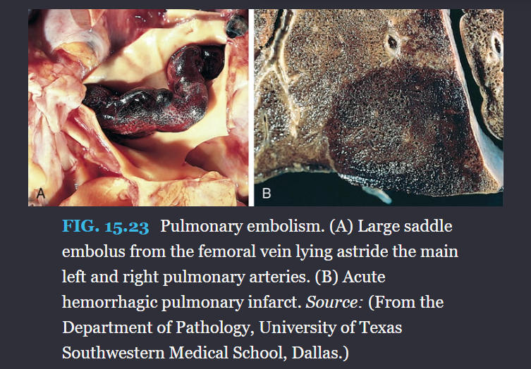
147
Overall, about 10% of emboli cause infarction. About 75% of infarcts affect these lobes, and in more than half, multiple lesions occur.
lower lobes
148
how do hemorrhagic areas show in the lungs
ischemic necrosis of the alveolar walls, bronchioles, and vessels.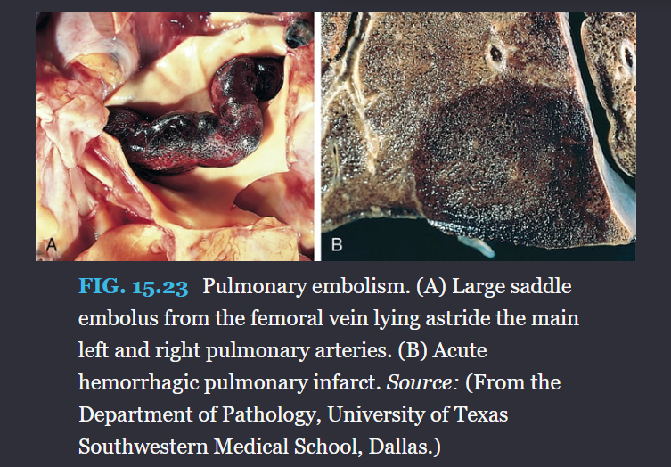
149
If the infarct is caused by an infected embolus, the neutrophilic inflammatory reaction can be intense. Such lesions are referred to as ________
septic infarcts
150
pulmonary embolism (large): During cardiopulmonary resuscitation in such instances, the patient frequently is said to have electromechanical dissociation (describe)
electrocardiogram has a rhythm, but no pulses are palpated because no blood is entering the pulmonary arterial circulation
151
symptoms of pulmonary embolism in descendoing order
dyspnea, pleuritic pain, and cough, accompanied in about half of cases by calf or thigh swelling or pain.
152
recurrent small emboli can gice rise to this
pulmonary hypertension
153
a normal _____- test level willl exclude a pulmonary embolism
D-dimer
154
this can be used to identify the obstructed pulmonary arteries in an embolism (imaging)
computed tomographic pulmonary angiogram
155
what is used to diagnose DVT?
duplex ultrasonograohy
156
In the presence of an underlying predisposing condition, patients with a pulmonary embolus have __________ chance of experiencing a second embolus.
30%
157
defined as a mean pulmonary artery pressure greater than or equal to 25 mm Hg at rest
pulmonary hypertension
158
5 groups of pulmonary hypertension
(1) pulmonary arterial hypertension, a diverse collection of disorders that all primarily impact small pulmonary muscular arteries;
(2) pulmonary hypertension due to left heart failure;
(3) pulmonary hypertension due to lung diseases and/or hypoxia;
(4) chronic thromboembolic pulmonary hypertension and other obstructions; and
(5) pulmonary hypertension with unclear and/or multifactorial mechanisms.
(2) pulmonary hypertension due to left heart failure;
(3) pulmonary hypertension due to lung diseases and/or hypoxia;
(4) chronic thromboembolic pulmonary hypertension and other obstructions; and
(5) pulmonary hypertension with unclear and/or multifactorial mechanisms.
159
this is associated with group 3 pulmonary hy[ertension and obliterates alveolar capilaries
chronic obstructive or interstitial lung diseases
160
this is associated with group 2 pulmonary hypertension and one ex is mitral stenosis
Antecedent congenital or acquired heart disease
161
this is associated with grouo 4 pulmonary hypertension and is caused by pulmonary emboli recurrence
Recurrent thromboemboli
162
this is associated with group 1 primary hypertemsion and involves the pulmonary vasculature and or the interstitum, leading to increased vascular resistace and pulmonary hypertension
autoimmune diseases
163
this is associated with group 1 primary hypertension and is associated with obsesity and hypizemia
obstructuve sleep apnea
164
what gene is associated with familial pulmomary arterial hypertension and pulmonart hypertension
BMPR2; specifically downregulation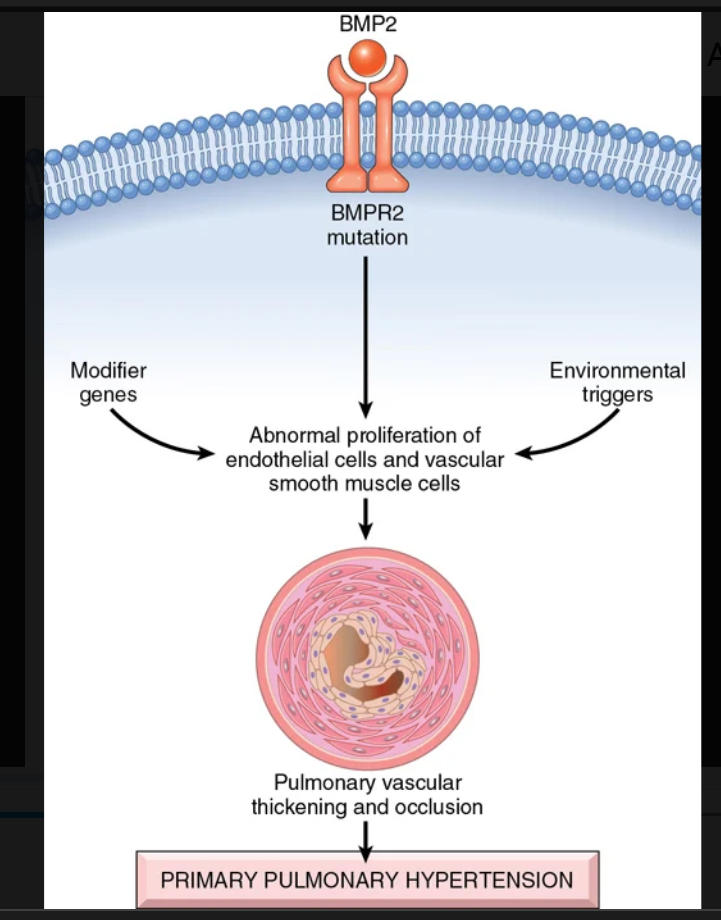
165
what are all forms of pulmonary hypertension associated with (morhology)
medial hypertrophy of the pulmonary muscular and elastic arteries and right ventricular hypertrophy
166
these are lesions oresent in some cases of pulmonary hypertension where a tuft of capillary formations is present, producing a network, or web, that spans the lumens of dilated thin-walled, small arteries and may extend outside the vessel
plexiform lesion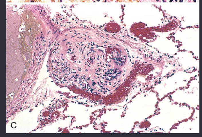
167
presenting features of all types of pulmonary hypertension
dyspnea and fatigue, but some patients have chest pain of the anginal type. Over time, severe respiratory distress, cyanosis, and right ventricular hypertrophy occur, and death from decompensated cor pulmonale, often with superimposed thromboembolism and pneumonia, ensues within 2 to 5 years in 80% of patients.
168
what is the definitive treatment for pulmonary hypertension
lung transplantation
169
what are the pulmonary hemorrhage syndromes?
goodpasture syndrome, idiopathic pulmonary hemosiderosis, vasculitis-associated hemorrhage
170
autoimmune disease in which kidney and lung injury is caused by circulating autoantibodies against the noncollagenous domain of the α3 chain of collagen IV.
goodpasture syndrome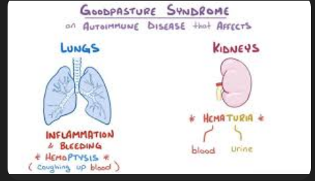
171
what is goodpasture syndrome called when only renal disease is caused by the antibodies
anti-glomerular basement membrane disease
172
what is the common clinical presentation of someone with goodpasture syndrome?
people in their teens or 20s, and in contrast to many other autoimmune diseases, there is a male preponderance. Most patients are active smokers.
173
what is the pathology og goodpasture syndrome?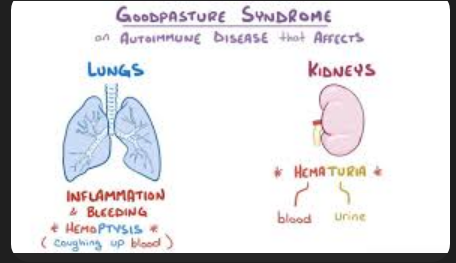
pathogenic antibodies initiate inflammatory destruction of the basement membrane in renal glomeruli and pulmonary alveoli, giving rise to rapidly progressive glomerulonephritis and a necrotizing hemorrhagic interstitial pneumonitis
174
symptomology of goodpasture syndrome
respiratory symptoms, principally hemoptysis, and radiographic evidence of focal pulmonary consolidations. Soon, manifestations of glomerulonephritis appear, leading to rapidly progressive renal failure. The most common cause of death is uremia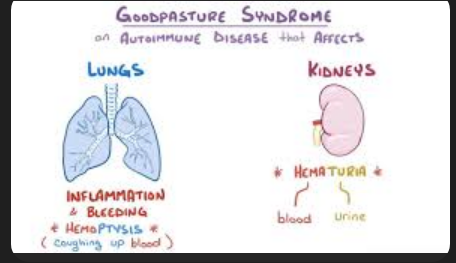
175
treatments for goodpasture syndrome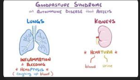
-intensive plasmapheresis to remove the anti basement membrane antibodies wiut simultaneous immunosupressive therapy
176
rare disorder characterized by intermittent, diffuse alveolar hemorrhage. Most cases occur in young children, although the disease has been reported in adults as well
idiopathic pulmonary hemosuderosis
177
symptomology idiopathic pulmonary hemosiderosis
insidious onset of productive cough, hemoptysis, and anemia associated with diffuse pulmonary infiltrates.
178
treatment and potential cause of idiopathic pulmonary hemosiderosis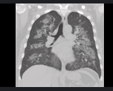
favorable response to long-term immunosuppression with prednisone and/or azathioprine indicates that an immunologic mechanism may be involved
179
autoimmune disease most often involves the upper respiratory tract and/or the lungs, with hemoptysis being the common presenting symptom.
granulomatosis with polyangiitis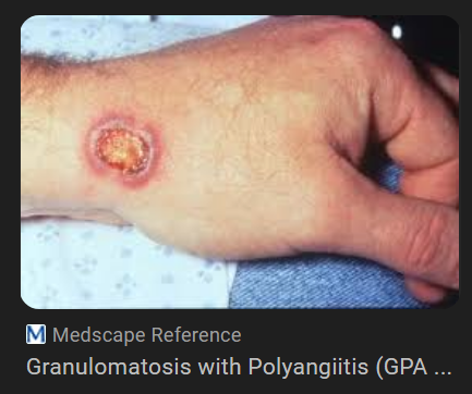
180
most common presenting symptom of granulomatosis with polyangiitis
hemoptysis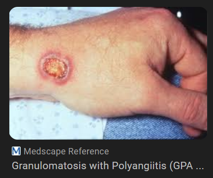
181
most imp histological geatures of granulomatosis with polyangiitis
capillaritis and scattered, poorly formed granulomas
182
is the process of blood clotting that prevents excessive bleeding after blood vessel damage. Inadequate hemostasis may result in hemorrhage, which can compromise regional tissue perfusion and, if massive and rapid, may lead to hypotension, shock, and death.
hemostasis
183
accumulation of fluid in tissues
edema
184
accumulation of fluid in body cavities
effusion
185
Elevated____________ or diminished ________ disrupts this balance and results in increased movement of fluid out of vessels.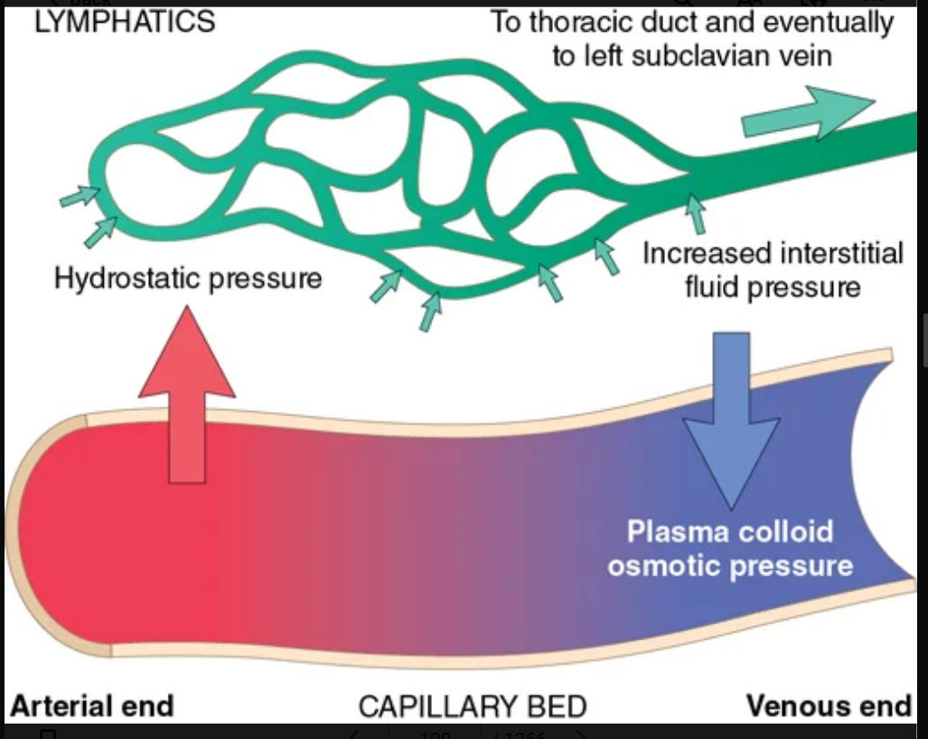
hydrostatic pressure; colloid osmotic pressure
186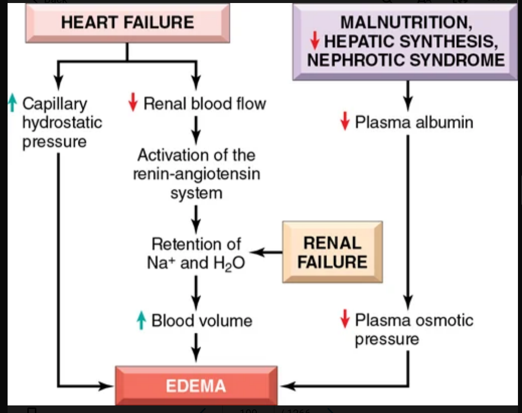
Image occlusion: name the hidden labels
187
Image occlusion: name the hidden labels
188
Image occlusion: name the hidden labels
189
conditions leading to inadequate Page 101synthesis or increased loss of _________ from the circulation are common causes of reduced plasma oncotic pressure.
albumin
190
when does reduced albumin sythesis occur?
severe liver disease and protein malnutrition
191
Increased salt retention—with obligate retention of associated water—causes both increased hydrostatic pressure (due to _______________) and diminished vascular colloid osmotic pressure (due to _________).
intravascular fluid volume expansion; dilution
192
clearing and separation of the extracellular matrix (ECM) and subtle cell swelling.
edema
193
can be diffuse or more conspicuous in regions with high hydrostatic pressures. Its distribution is often influenced by gravity (e.g., it appears in the legs when standing and the sacrum when recumbent)
subcutanepus edema
194
edema: Its distribution is often influenced by gravity (e.g., it appears in the legs when standing and the sacrum when recumbent), a feature termed
dependent edema
195
edema: Finger pressure over markedly edematous subcutaneous tissue displaces the interstitial fluid and leaves a depression, a sign called
pitting edema
196
edema: edema resulting from renal dysfunction often appears initially in parts of the body containing loose connective tissue, such as the eyelids; _______ is thus a characteristic finding in severe renal disease
periorbital edema
197
edema: brain exhibits narrowed sulci and distended gyri, which are compressed by the unyielding skull
brain edema
198
effusion involving the pleural cavoty
hydrothorax
199
effusion in the pericardial cavity
hydropericardium
200
peritoneal cavity effusion
hydroperitoneum or ascites
201
effusion: typically protein poor, translucent, and straw colored
transudative effusion
202
when is pulmonary edema most often scene
left venricular failure
203
what can subcutaneous edema signal
cardiac or renal disease
204
an active process in which arteriolar dilation (e.g., at sites of inflammation or in skeletal muscle during exercise) leads to increased blood flow.
hyperemia
205
a passive process resulting from reduced venous outflow of blood from a tissue.
congestion
206
clinical presentation of congested tissues
an abnormal blue-red color (cyanosis) that stems from the accumulation of deoxygenated hemoglobin in the affected area.
207
congestion: marked by engorged alveolar capillaries, alveolar septal edema, and focal intraalveolar hemorrhage
acute pulmonary congestion
208
congestion: often caused by congestive heart failure, the septa are thickened and fibrotic, and the alveoli often contain numerous macrophages laden with hemosiderin (heart failure cells) derived from phagocytosed red cells.
chronic pulmonaru congestion
209
heart failure cells
hemosiderin
210
congestion: the central vein and sinusoids are distended. Because the centrilobular area is at the distal end of the hepatic blood supply, centrilobular hepatocytes may undergo ischemic necrosis, and the periportal hepatocytes—better oxygenated because of proximity to hepatic arterioles—may only develop fatty change.
acute hepatic congestion
211
congestion: the centrilobular regions are grossly red-brown and slightly depressed (because of cell death) and are accentuated against the surrounding zones of uncongested tan liver (nutmeg liver)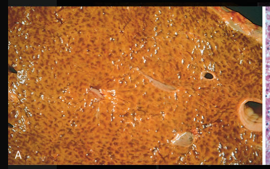
chronicpassive hepatic congestion
212
occurs immediately and markedly reduces blood flow to the injured area (see Fig. 4.4A). It is mediated by reflex neurogenic mechanisms and may be augmented by the local secretion of factors such as endothelin, a potent endothelium-derived vasoconstrictor.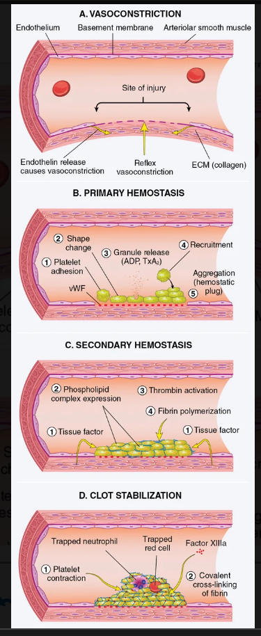
vasoconstriction
213
the formation of the platelet plug. Disruption of the endothelium exposes subendothelial von Willebrand factor (vWF) and collagen, which promote platelet adherence and activation.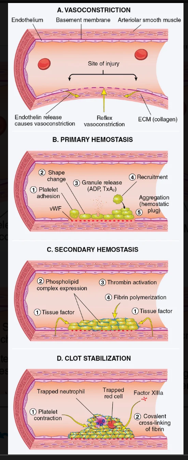
promary hemostasis
214
deposition of fibrin. Vascular injury exposes tissue factor (TF) at the site of injury. Tissue factor is a membrane-bound procoagulant glycoprotein that is normally expressed by subendothelial cells in the vessel wall, such as smooth muscle cells and fibroblasts. Tissue factor binds and activates factor VII (see later), setting in motion a cascade of reactions that culminates in thrombin generation.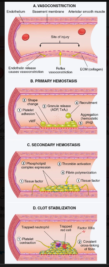
secondary hemostasis
215
Polymerized fibrin and platelet aggregates undergo contraction to form a solid, permanent plug that prevents further hemorrhage. 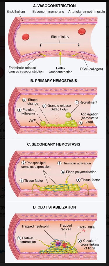
clot stabilization and resorption
216
Image occlusion: name the hidden labels
217
Image occlusion: name the hidden labels
218
Image occlusion: name the hidden labels
219
Image occlusion: name the hidden labels
220
two types of cytoplasmic granules released from platelets
a-granules and dense granules
221
have the adhesion molecule P-selectin on their membranes (Chapter 3) and contain proteins involved in coagulation, such as fibrinogen; coagulation factors V, XI, and vWF; as well as protein factors that may be involved in wound healing, such as fibronectin, platelet factor 4 (PF4, a heparin-binding chemokine), platelet-derived growth factor (PDGF), and transforming growth factor-β (TGF-β).
a-granules
222
contain adenosine diphosphate (ADP), adenosine triphosphate (ATP), ionized calcium, serotonin, and epinephrine
dense granules
223
what is platelet adhesion mediated by?
interactions between GpIb and vWF in the subendothelial matrix
224
what triggers platelet activation?
coagulation factor
thrombin: special G protein–coupled receptor referred to as protease-activated receptor-1 (PAR-1), which is switched on by a proteolytic cleavage carried out by thrombin
ADP: acts by binding two G protein–coupled receptors (P2Y1 and P2Y12). (recruitment)
thrombin: special G protein–coupled receptor referred to as protease-activated receptor-1 (PAR-1), which is switched on by a proteolytic cleavage carried out by thrombin
ADP: acts by binding two G protein–coupled receptors (P2Y1 and P2Y12). (recruitment)
225
an inherited deficiency of GpIIb and GpIIIa results in a bleding disorder called this
Glanxmann thrombasthenia
226
assesses the function of the proteins in the extrinsic pathway (factors VII, X, V, II [prothrombin], and fibrinogen). In brief, tissue factor, phospholipids, and calcium are added to plasma, and the time for a fibrin clot to form is recorded.
prothrombin time
227
assesses the function of the proteins in the intrinsic pathway (factors XII, XI, IX, VIII, X, V, II, and fibrinogen). In this assay, clotting of plasma is initiated by the addition of negatively charged particles (e.g., ground glass) that activate factor XII (Hageman factor) together with phospholipids and calcium, and the time to fibrin clot formation is recorded.
partial thromboplastin time
228
Based on the effects of various factor deficiencies in humans, it is believed that, in vivo, _______________ complex is the most important activator of factor IX and that ______________complex is the most important activator of factor X.
factor VIIa/tissue factor; factor IXa/factor VIIIa
229
factors limiting coagulation: blood flowing past the site of injury washes out activated coagulation factors, which are rapidly removed by the liver
dilution
230
factors limiting coagulation: mainly provided by platelets that have been activated by contact with subendothelial matrix at sites of vascular injury.
negatively charged phospholipds
231
bind thrombin and protein C, respectively, in a complex on the endothelial cell surface. When bound in this complex, thrombin loses its ability to activate coagulation factors and platelets, and instead cleaves and activates protein C, a vitamin K–dependent protease that requires a cofactor, protein S. Activated protein C/protein S complex is a potent inhibitor of coagulation cofactors Va and VIIIa
thrombomodulin and endothelial prtein c receptor
232
Activated protein C/protein S complex is a potent inhibitor of coagulation cofactors (x2)
Va and VIIIa
233
how do defects of primary hemostasis show clinically
often present with small bleeds in skin or mucosal membranes, which typically take the form of petechiae, minute 1- to 2-mm hemorrhages (Fig. 4.11A), or purpura, which are slightly larger (≥3 mm) than petechiae.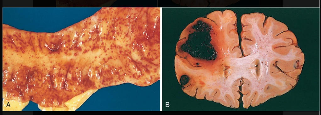
234
how do defects of secondary hemostasis present clinically?
present with bleeds into soft tissues (e.g., muscle) or joints. Bleeding into joints (hemarthrosis) following minor trauma is particularly characteristic of hemophilia
235
how do generalized defects involving small vessels present clinically
“palpable purpura” and ecchymoses. Ecchymoses (sometimes simply called bruises) are hemorrhages of 1 to 2 cm in size. In both purpura and ecchymoses, the volume of extravasated blood may be large enough to create a palpable mass of blood known as a hematoma
236
how much % of blood does an adult need to lose to cause problems
20%
237
intracranial hemorrhage may increase intracranial pressure to a level that compromises the blood supply or causes
herniation of the brainstem
238
chronic or recurrent external blood loss (e.g., peptic ulcer or menstrual bleeding) causes __-
iron loss or iron deficient anemia
239
primary abnormalities that lead to thrombosis (virchows triad)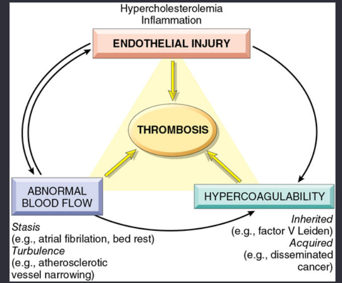
(1) endothelial injury,
(2) stasis or turbulent blood flow, and
(3) hypercoagulability of the blood
(2) stasis or turbulent blood flow, and
(3) hypercoagulability of the blood
240
Aortic and arterial dilations called ____________` result in local stasis and are therefore fertile sites for thrombosis
aneurisms
241
results in areas of noncontractile myocardium and sometimes in cardiac aneurysm; both are associated with stasis and flow abnormalities that promote the formation of cardiac mural thrombi
acute myocardial infarctions
242
increases resistance to flow and causes small vessel stasis
hyperviscovity
243
what type of mutation is the factor V leuden mutation
single nucleotide mutation (dominant autosomal)
244
A single nucleotide change (G20210A) in the 3′-untranslated region of the prothrombin gene is another common mutation (1% to 2% of the population) associated with hypercoagulability.
prothrombin gene mutation
245
Elevated levels of homocysteine may be inherited or acquired. Marked elevations of homocysteine may be caused by an inherited deficiency of cystathione β-synthetase
homocysteinemia
246
what causes hypercoagulbility in oral contraceptive use?
increased hepatic synthesis of coagulation factors and reduced anticoagulant synthesis
247
a serious, potentially life-threatening disorder that occurs following the administration of unfractionated heparin.
heparin-induced thrombocytopenia (HIT) syndrome
248
an extremely rare complication that can occur in those who received certain adenoviral-vector-based vaccines for coronavirus disease 2019 (COVID-19).
-characterized by PF4 antibodies
-characterized by PF4 antibodies
Vaccine-induced thrombotic thrombocytopenia (VITT)
249
an autoimmune disorder characterized by:
• Presence of one or more antiphospholipid (aPL) autoantibodies.
• Venous or arterial thromboses, or pregnancy complications such as recurrent miscarriages, unexplained fetal death, and premature birth.
• Presence of one or more antiphospholipid (aPL) autoantibodies.
• Venous or arterial thromboses, or pregnancy complications such as recurrent miscarriages, unexplained fetal death, and premature birth.
antiphospholipid syndrome (APS)
250
antiphospholipid syndrome: patients exhibit only the manifestations of a hypercoagulable state and lack evidence of other Page 112well-defined autoimmune disorders.
primary antiphospholipiud syndrome
251
clinical manifestations of Antiphospholipid syndrome
recurrent thromboses, repeated miscarriages, cardiac valve vegetations, and thrombocytopenia.
252
Thrombi often have grossly and microscopically apparent laminations called __________, which are pale platelet and fibrin deposits alternating with darker red cell–rich layers.
lines of Zahn
253
Thrombi occurring in heart chambers or in the aortic lumen are designated
mural thrombi
254
what causes/promotes mural thrombi?
-Abnormal myocardial contraction (arrhythmias, dilated cardiomyopathy, or myocardial infarction)
-endomyocardial injury (myocarditis or catheter trauma)
-endomyocardial injury (myocarditis or catheter trauma)
255
venous thrombi or red thrombi are called this becayuse they vcontain more
RBCs
256
clots: elatinous and have a dark-red dependent portion where red cells have settled by gravity and a yellow “chicken fat” upper portion and are usually not attached to the underlying vessel wall.
post mortem clots
257
thrombi in the heart valves; may be unfected or sterile
vegetations
258
heightened thrombotic risks during prehnancy
decreased venous return from leg veins and systemic hypercoagulability associated with the hormonal changes of late pregnancy and the postpartum period.
259
a major cause of arterial thromboses because it is associated with loss of endothelial integrity and with abnormal blood flow
atherosclerosis
260
widespread thrombosis within the microcirculation that may be of sudden or insidious onset. It is not a specific disease but rather a complication of many conditions associated with systemic activation of thrombin.
-runaway thrombosis “uses up” platelets and coagulation factors (hence the synonym consumptive coagulopathy) and often activates fibrinolytic mechanisms. Thus, symptoms initially related to thrombosis can evolve into a bleeding catastrophe, such as hemorrhagic stroke or hypovolemic shock
-runaway thrombosis “uses up” platelets and coagulation factors (hence the synonym consumptive coagulopathy) and often activates fibrinolytic mechanisms. Thus, symptoms initially related to thrombosis can evolve into a bleeding catastrophe, such as hemorrhagic stroke or hypovolemic shock
Disseminated intravascular coagulation (DIC)
261
Sudden death, acute right-heart failure (cor pulmonale), or cardiovascular collapse occurs when emboli obstruct ______ or more of the pulmonary circulation.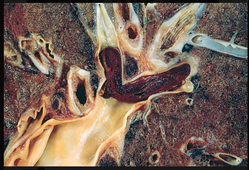
60%
262
refers to the presence of microscopic fat globules—sometimes with associated hematopoietic bone marrow—in the vasculature, typically after fractures of long bones or, rarely, in the setting of soft tissue trauma and burns.
fat embolism
263
when are fat embolisms common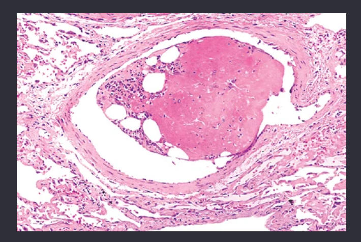
fractures of long bones; severe skeletal injurt
264
characterized by pulmonary insufficiency, neurologic symptoms, anemia, and thrombocytopenia, and is fatal in 5% to 15% of cases. Typically, 1 to 3 days after injury, there is a sudden onset of tachypnea, dyspnea, tachycardia, irritability, and restlessness, which ultimately can progress to delirium or coma.
fat embolism syndrome
265
A particular form of gas embolism, called _____________-, occurs when individuals experience sudden decreases in atmospheric pressure. Scuba and deep-sea divers, underwater construction workers, and persons in unpressurized aircraft who undergo rapid ascent are all at risk.
decompression sickness
266
The rapid formation of gas bubbles within skeletal muscles and supporting tissues in and about joints is responsible for the painful condition called
the bends
267
In the lungs, gas bubbles in the vasculature cause edema, hemorrhage, and focal atelectasis or emphysema, leading to a form of respiratory distress called
the chokes
268
-(named for the pressurized vessels used in bridge construction; workers in these vessels suffered both acute and chronic forms of decompression sickness).
-persistence of gas emboli in the skeletal system leads to multiple foci of ischemic necrosis; the more common sites are the femoral heads, tibia, and humeri.
-persistence of gas emboli in the skeletal system leads to multiple foci of ischemic necrosis; the more common sites are the femoral heads, tibia, and humeri.
caisson disease
269
how is acute decompression sickness treated?
being placed in a chamber under sufficiently high pressure to force the gas bubbles back into solution.
270
symptomology amniotic fluid embolism
sudden severe dyspnea, cyanosis, and shock, followed by neurologic impairment ranging from headache to seizures and coma, and by DIC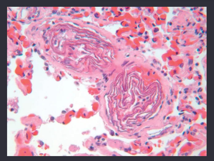
271
an area of ischemic necrosis caused by occlusion of either the arterial supply or the venous drainage.
infarct
272
(Fig. 4.18A) occur (1) with venous occlusions (e.g., testicular torsion, Chapter 19), (2) in loose, spongy tissues (e.g., lung) where blood can collect in the infarcted zone, (3) in tissues with dual circulations (e.g., lung and small intestine) that allow blood to flow from an unobstructed parallel supply into a necrotic zone, (4) in tissues previously congested by sluggish venous outflow, and (5) when flow is reestablished to a site of previous arterial occlusion and necrosis (e.g., following angioplasty of an arterial obstruction)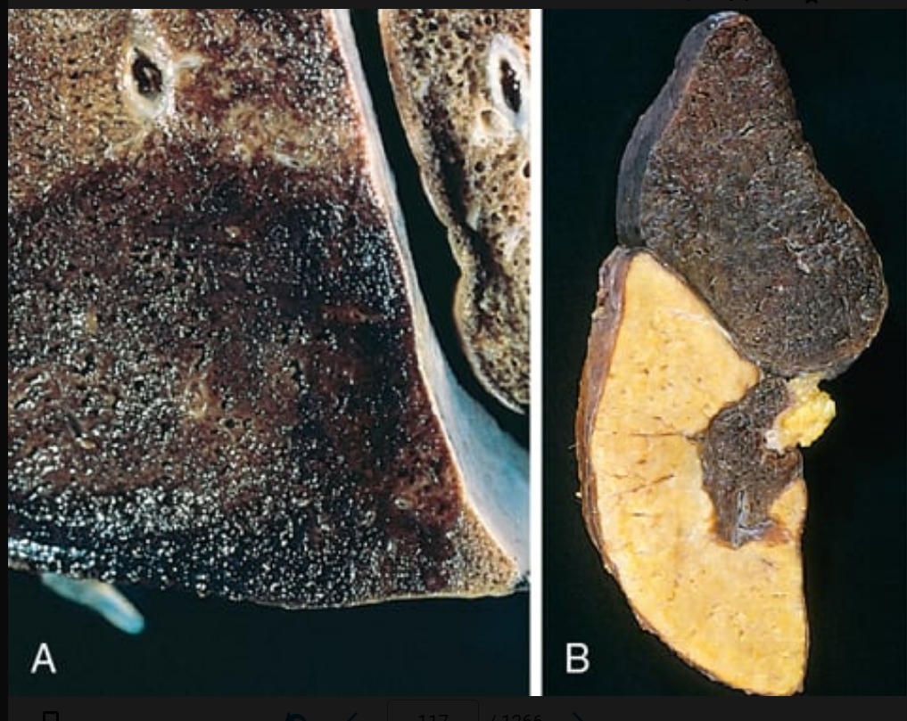
red infarcts
273
(see Fig. 4.18B) occur with arterial occlusions in solid organs with end-arterial circulation (e.g., heart, spleen, and kidney), and where tissue density limits the seepage of blood from adjoining capillary beds into the necrotic area.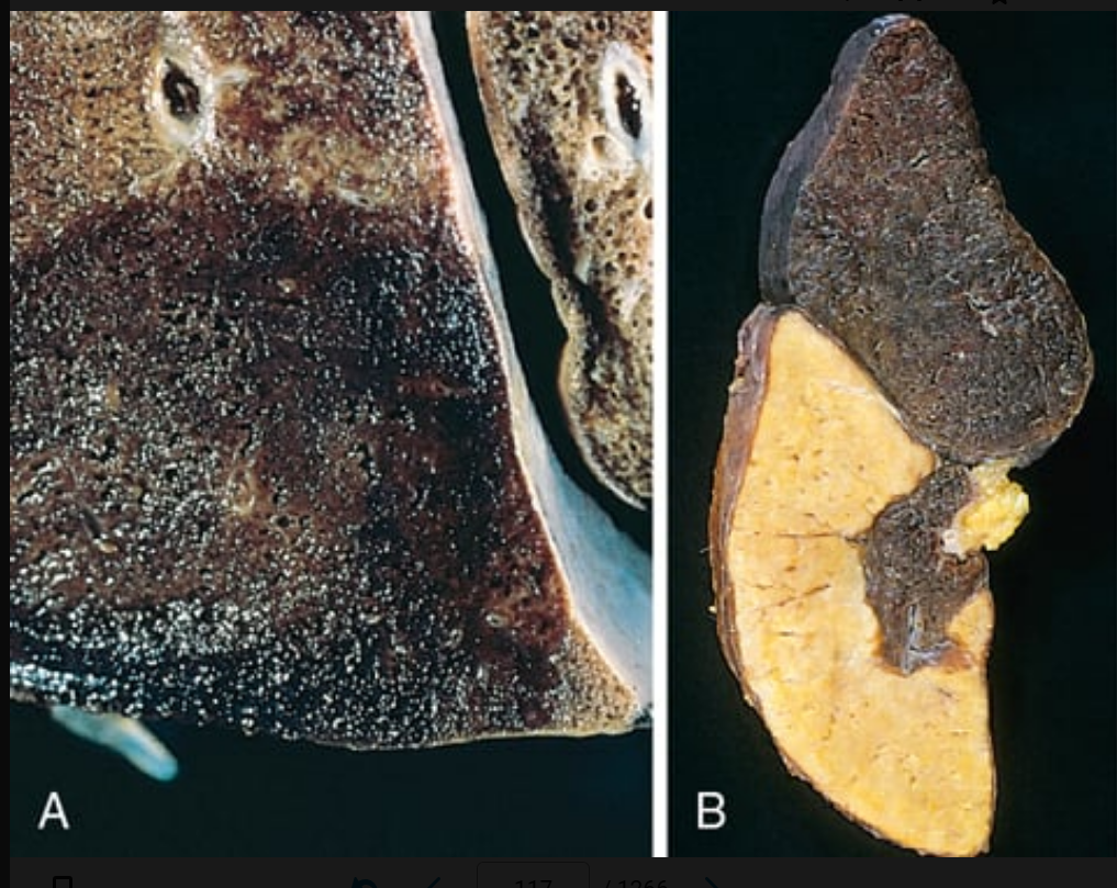
white infarcts
274
dominant necrosis seen in infarcts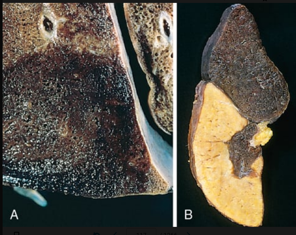
coagulative necrosis
275
infarctation in the brain results in this necrosis
liquefactive necrosis
276
occur when infected cardiac valve vegetations embolize or when microbes seed necrotic tissue. In these cases, the infarct is converted to an abscess, with a correspondingly greater inflammatory response
septic infarctions
277
a state of circulatory failure that impairs tissue perfusion and leads to cellular hypoxia. At the outset, the cellular injury is reversible; however, prolonged shock eventually leads to irreversible tissue injury and can be fatal
shock
278
results from low cardiac output due to myocardial pump failure
cardiogenic shock
279
shock: results from low cardiac output due to low blood volume, such as can occur with massive hemorrhage or fluid loss from severe burns.
hypovolemic shock
280
life-threatening organ dysfunction caused by a dysregulated host response to infection
sepsis
281
a subset of sepsis in which particularly profound circulatory, cellular, and metabolic abnormalities are associated with a greater risk of mortality than with sepsis alone.
septic shock
282
a sepsis-like condition associated with systemic inflammation that may be triggered by a variety of nonmicrobial insults, such as burns, trauma, and/or pancreatitis.
systemic inflammatory response syndrome (SIRS)
283
what bacteria most usually trgers septoc shock
gram positive followed by gram negative and fungal
284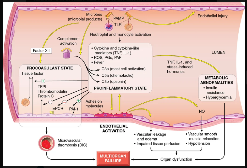
Image occlusion: name the hidden labels
285
Image occlusion: name the hidden labels
286
Image occlusion: name the hidden labels
287
Image occlusion: name the hidden labels
288
polyclonal T-lymphocyte activators that induce the release of high levels of cytokines that result in a variety of clinical manifestations, ranging from a diffuse rash to vasodilation, hypotension, shock, and death.
superantigens
289
syages of shock (x3)
• An initial nonprogressive stage during which reflex compensatory mechanisms are activated and vital organ perfusion is maintained
• A progressive stage characterized by tissue hypoperfusion and onset of worsening circulatory and metabolic derangement, including acidosis
• An irreversible stage in which cellular and tissue injury is so severe that even if the hemodynamic defects are corrected, survival is not possible
• A progressive stage characterized by tissue hypoperfusion and onset of worsening circulatory and metabolic derangement, including acidosis
• An irreversible stage in which cellular and tissue injury is so severe that even if the hemodynamic defects are corrected, survival is not possible
290
generic term for arterial wall thickening and loss of elasticity.
arteriosclerosis
291
arteriosclerosis: characterized by calcifications of the medial walls of muscular arteries, typically starting along the internal elastic membrane. Adults older than age 50 years are most commonly affected, particularly those with renal insufficiency and secondary hypercalcemia.
mockenberg medial scleroisius
292
occurs in muscular arteries larger than arterioles. It is driven by inflammation (as in a healed arteritis or transplant-associated arteriopathy; see Chapter 12) or by mechanical injury (e.g., associated with stents or balloon angioplasty; see later) and can be considered a healing response.
fibromuscular intimal hyperplasia
293
obstrutive thrombus
294
when do females exceed males on risk for atherosclerosis related diseases
after menopause
295
a class of drugs that lower circulating cholesterol levels by inhibiting hydroxymethylglutaryl coenzyme A (HMG CoA) reductase, the rate-limiting enzyme in hepatic cholesterol biosynthesis
statins
296
rate limiting enzyme in hepatic cholesterol biosynthesis
coenzyme A (HMG CoA)
297
this is present during all stages of atherogenesis and is intimately linked with atherosclerotic plaque formation and rupture
inflammation
298
caused by a rare inborn error of metabolism, results in elevated circulating homocysteine (>100 µmol/L) and is associated with premature vascular disease.
homocystinuria
299
chronic inflammatory and healing response of the arterial wall to endothelial injury.
atherosclerosis
300
atheroschlerosis progression
• Endothelial injury and dysfunction, causing (among other things) increased vascular permeability, leukocyte adhesion, and thrombosis.
• Accumulation of lipoproteins (mainly LDL and its oxidized forms) in the vessel wall.
• Monocyte adhesion to the endothelium, followed by migration into the intima and transformation into macrophages and foam cells.
• Platelet adhesion.
• Factor release from activated platelets, macrophages, and vascular wall cells, inducing SMC recruitment, either from the media or from circulating precursors.
• SMC proliferation, ECM production, and recruitment of T cells.
• Lipid accumulation both extracellularly and within cells (macrophages and SMCs).
• Calcification of ECM and necrotic debris late in the pathogenesis.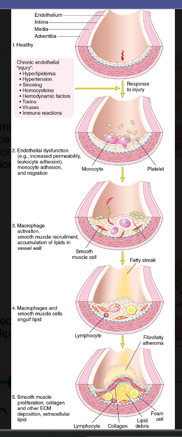
• Accumulation of lipoproteins (mainly LDL and its oxidized forms) in the vessel wall.
• Monocyte adhesion to the endothelium, followed by migration into the intima and transformation into macrophages and foam cells.
• Platelet adhesion.
• Factor release from activated platelets, macrophages, and vascular wall cells, inducing SMC recruitment, either from the media or from circulating precursors.
• SMC proliferation, ECM production, and recruitment of T cells.
• Lipid accumulation both extracellularly and within cells (macrophages and SMCs).
• Calcification of ECM and necrotic debris late in the pathogenesis.
155 more cards in OutLearn. Open the full deck