Embryology exam 1
Study these flashcardsFree · spaced repetition · iPhone, Android, Mac & web
All cards
1
what are the periods of development?
early development (fert-2wks)
embryonic period (3-8wks)
fetal period (9-end)
embryonic period (3-8wks)
fetal period (9-end)
2
what happens during the psriod of early development? (x5)
fertilization and implantation
proliferation
formation of trilaminar embryo
placenta is formed
determination of body axes
proliferation
formation of trilaminar embryo
placenta is formed
determination of body axes
3
what happens during the embryonic period?
organs and body parts form
4
what is succeptable to birth defects in the fetal period?
brain, eyes, ears, teeth, pallette, external genitalia
5
parental and materal strands
diploid
6
one copy (either parental or maternal or a mix)
haploid
7
what is the final product of spermatogenesis?
4 spermatids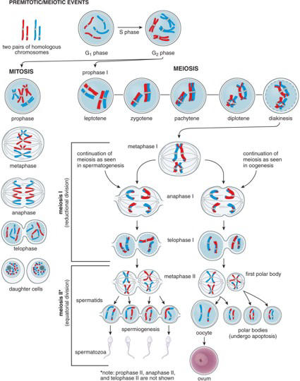
8
what is the final product of oogenesis
ovum and 3 polar bodies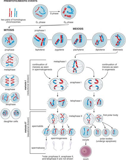
9
stem cells that can form sperm or eggs, unfertilized at this point
primordial germ cells
10
where do the primordial germ cells travel to differentiate?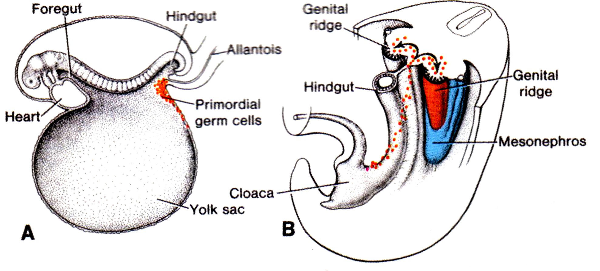
epiblast to yolk sac to genital ridge
11
what forms in/around the genital ridge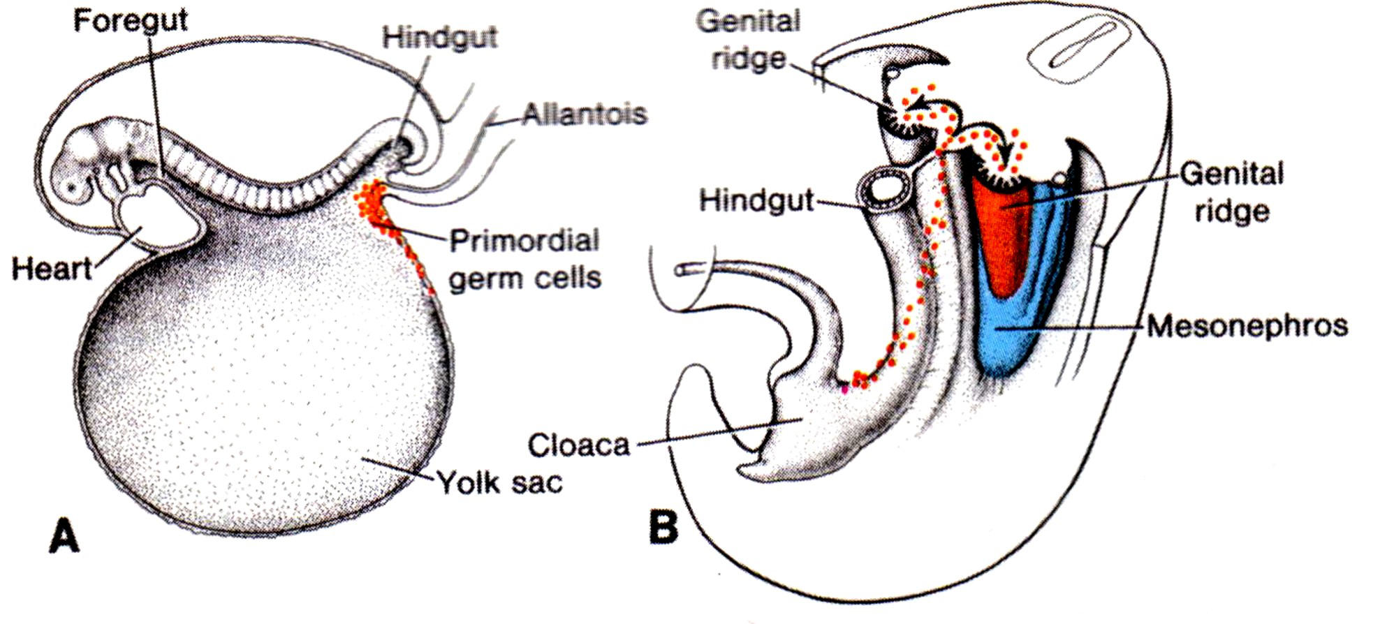
gonads and kidneys
12
what do the primordial germ cells becme when they enter the genital ridge
primitive sex cords
13
what surrounds the primative sex cords if they are destined to become testes
sertoli cells
14
what surrounds the primative sex cords if they are destined to become oocytes
follicular cells
15
when do the primordial germ cells actovate in males
puberty
16
what is the pathway to primordial germ cell activation in males
LH activates leydig cells that make testosterone that activates primordial germ cells
17
what is the blue?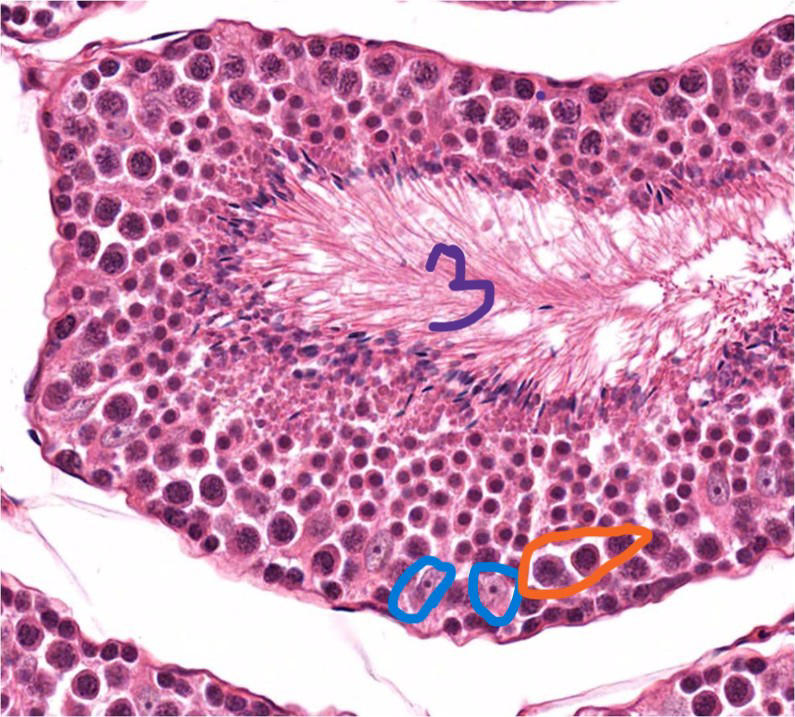
sertoli cells
18
what is the orange?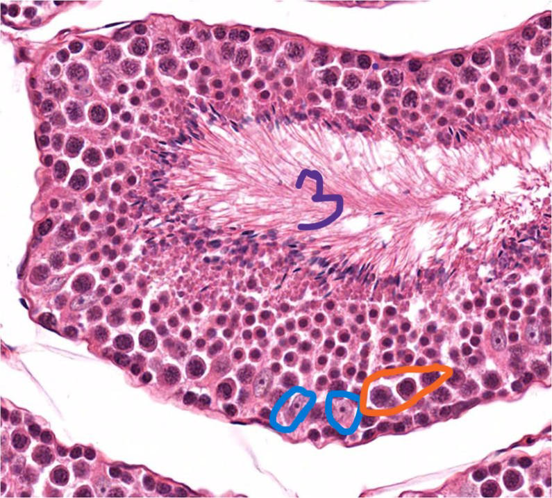
sperm making cells
19
what is the 3 at the center?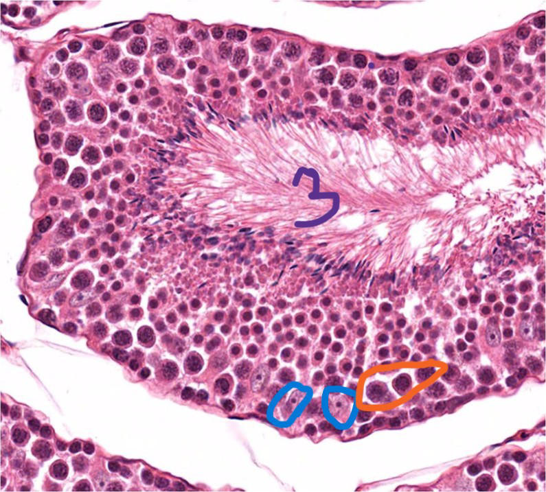
lumen
20
sperm making pathway
spermatogonia via mitosis to primary spermatocyte to 2nd spermatocyte to spermatid
primary to spermatid is meiosis
primary to spermatid is meiosis
21
conversion of spermatids into mature sperm
spermiogenesis
22
what does the process of spermiogenesis include
cytoplasm shed, condense nucleus, neck and tail formation, acrosome formed
23
where does capacitation occur
vagina and uterus
24
what does capacitation do?
prunes glyconalyx proteins on the sperm
25
By week _________________ of development, a population of oogonia enter the first meiotic prophase and become dormant until ________________________
12; puberty
26
oocyte and follicle cells together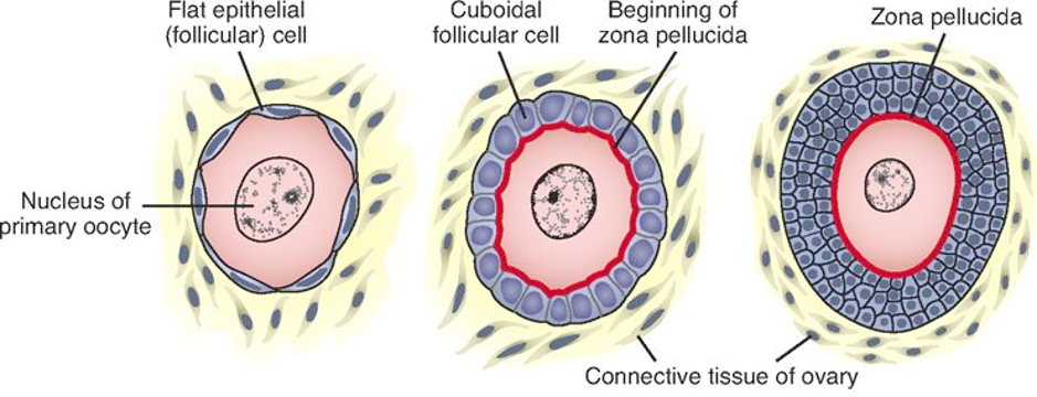
ovarian follicle
27
what secreted by the folicular cells prevents any further progression of the cell cycle
OM1
28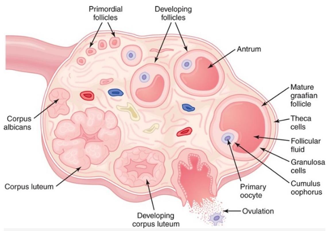
Image occlusion: name the hidden labels
29
Image occlusion: name the hidden labels
30
Image occlusion: name the hidden labels
31
what specific primary cell type?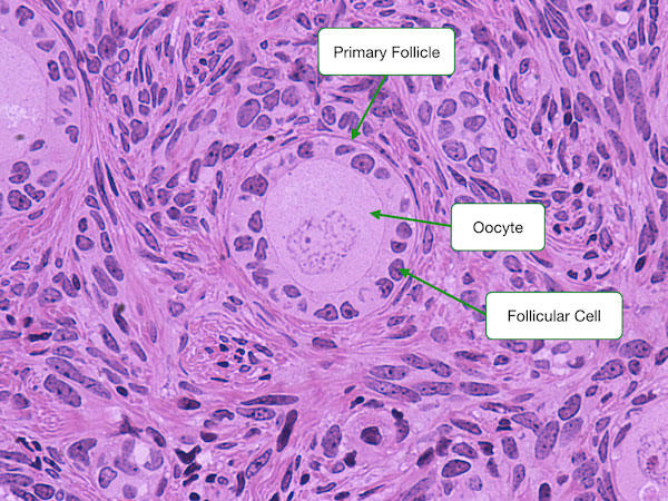
primordial
32
primary? secondary? definitive?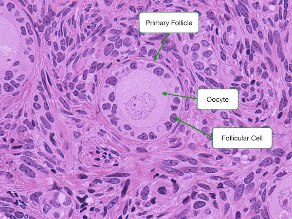
primary
33
specific primary folicle type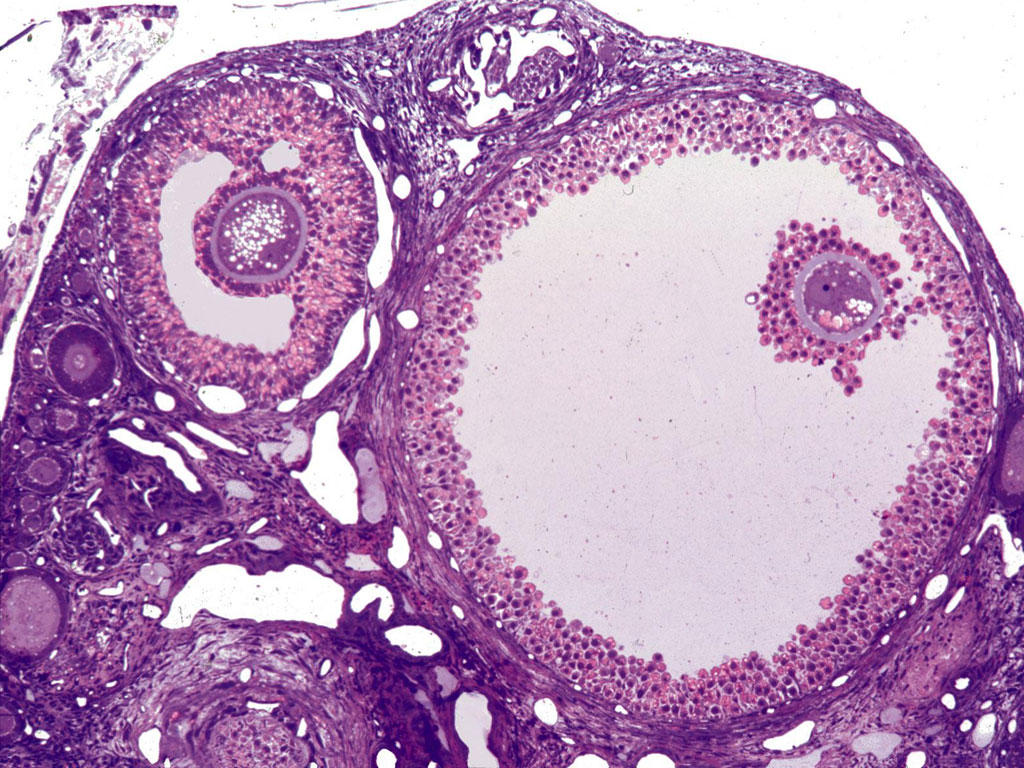
graafian
34
The hypothalamus releases this to control levels of pituitary hormone and ovulation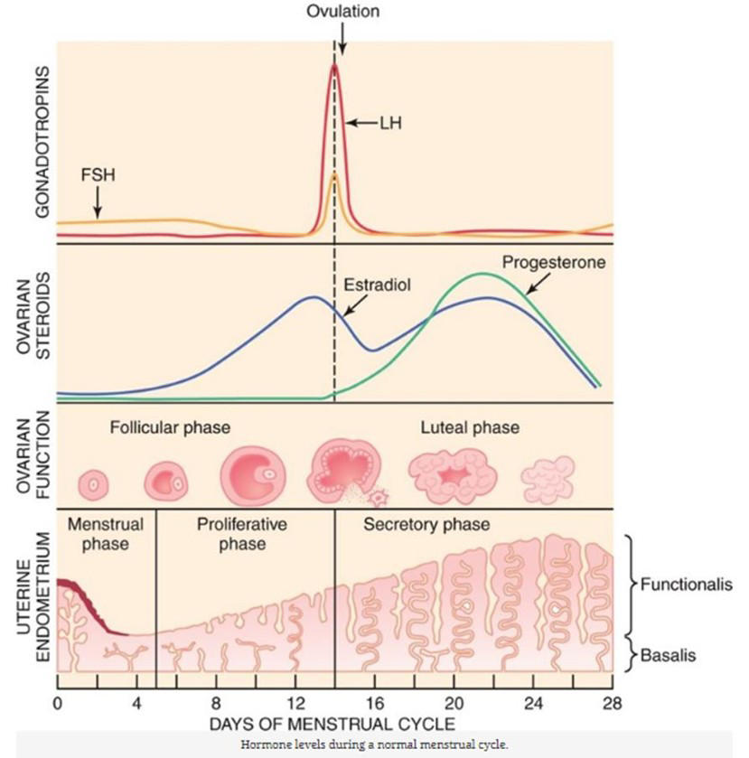
GnRH
35
when do LH and FSH spike causing an estradiol spike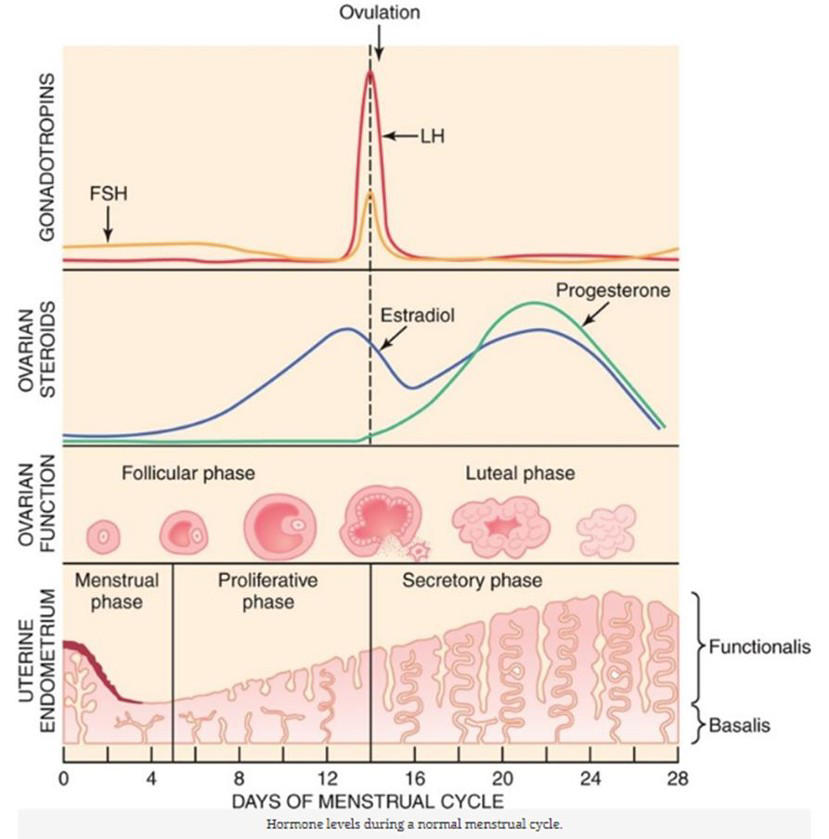
ovulation
36
what type of follicule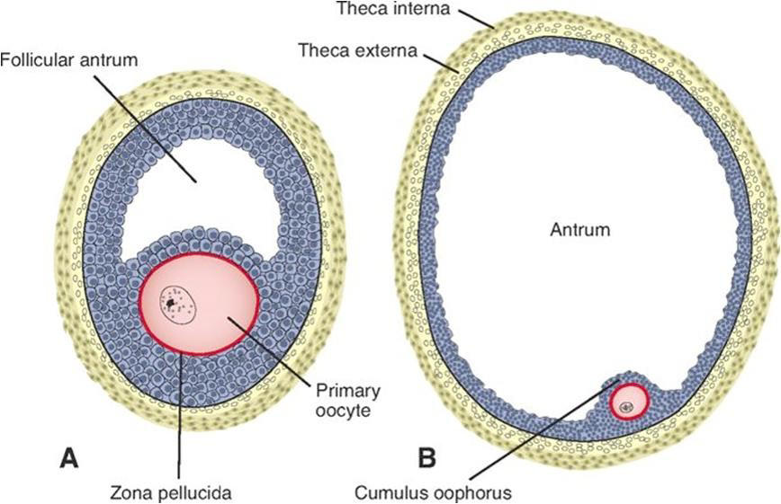
graafian
37
what oocyte detaches into the atrium during ovulation
2ndary oocyte
38
what is the summary of female egg production
oogonia via mitosis to primary oocyte to 2ndary oocyte to definitive oocyte (meiosis is 1st to definitive)
2nd oocyte made at ovulation
definitive oocyte made at fertilization
2nd oocyte made at ovulation
definitive oocyte made at fertilization
39
what happens if there is implantation (corpus luteu)
secretes chorionic gonadotropic hormone (hCG); this keeps the corpus luteum (corpus luteum of pregnancy) producing progesterone (required for maintaining pregnancy) for about five months, after which the placenta produces progesterone
40
Trisomy 21?
Down syndrome
41
X0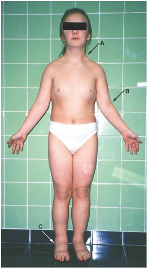
turner syndrome
42
XXY?
kleinfelters syndrome
43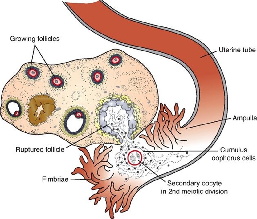
Image occlusion: name the hidden labels
44
what are the phases of fert
sperm penitrates corona radiata
sperm penitrates zona pellucida
fusion of sperm and oocyte membranes which completes the 2nd meiotic division of the oocyte
sperm penitrates zona pellucida
fusion of sperm and oocyte membranes which completes the 2nd meiotic division of the oocyte
45
what is the zona rxn
oocyte change zona pelucida rxn so more sperm cant enter
46
what is the cortical reaction
oocyte change PM to prevent further sperm fertilization
47
what are the zygote cells called in the first week of division
blastomeres
48
what happens during compaction
increase surface area, making gap junctions, shifting nuclei towards the center
49
blastocyst
what stage of division does the cell invade the wall of the uterus
50
when does implantation occur
5.5-6 days
51
what is an ectopic pregnancy
pregnancy occurs in the wrong spot
52
implantation in the cervix
placenta previa
53
detachedleeding of the uterus
placenta abruption
54
placenta penitrates too deply into the uterine wall
placenta accreta
55
placenta penitrates too deeply into the uterine muscle
polacenta increta
56
placenta penitrates too deeply through the uterne wall into the organs
placenta percreta
57
what develops from the embryoblast?
epiblast and hypoblast
58
what does the embryoblast make
embryo
59
what does the trophoblast make
placenta
60
what tissues form from the trophoblast (cell types)
syncytiotrophoblast and cytotrophoblast
61
what two spaces develop in the 2nd week
amnion and yolk sac
62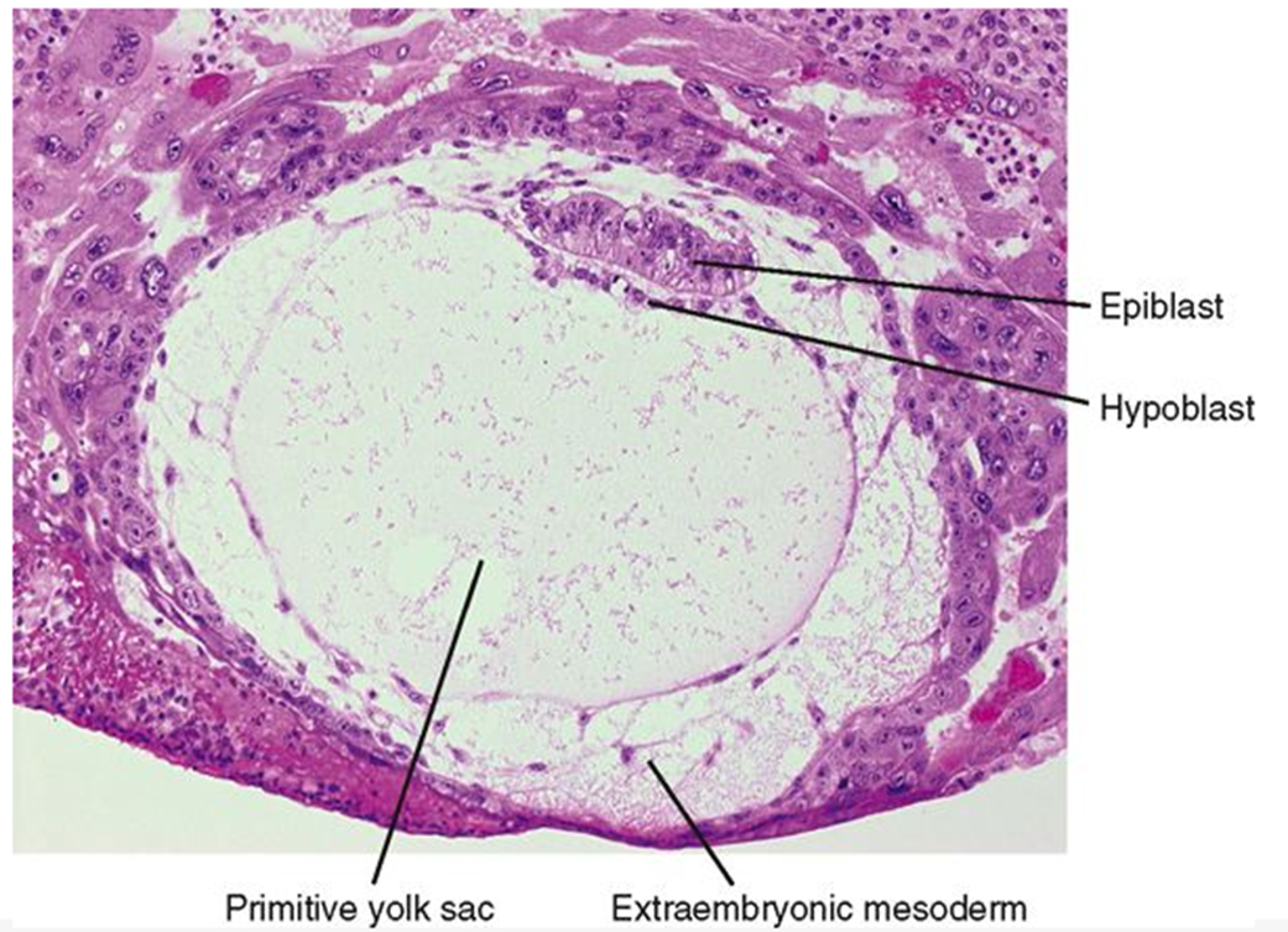
Image occlusion: name the hidden labels
63
Image occlusion: name the hidden labels
64
Image occlusion: name the hidden labels
65
what is the asterix (cavity)?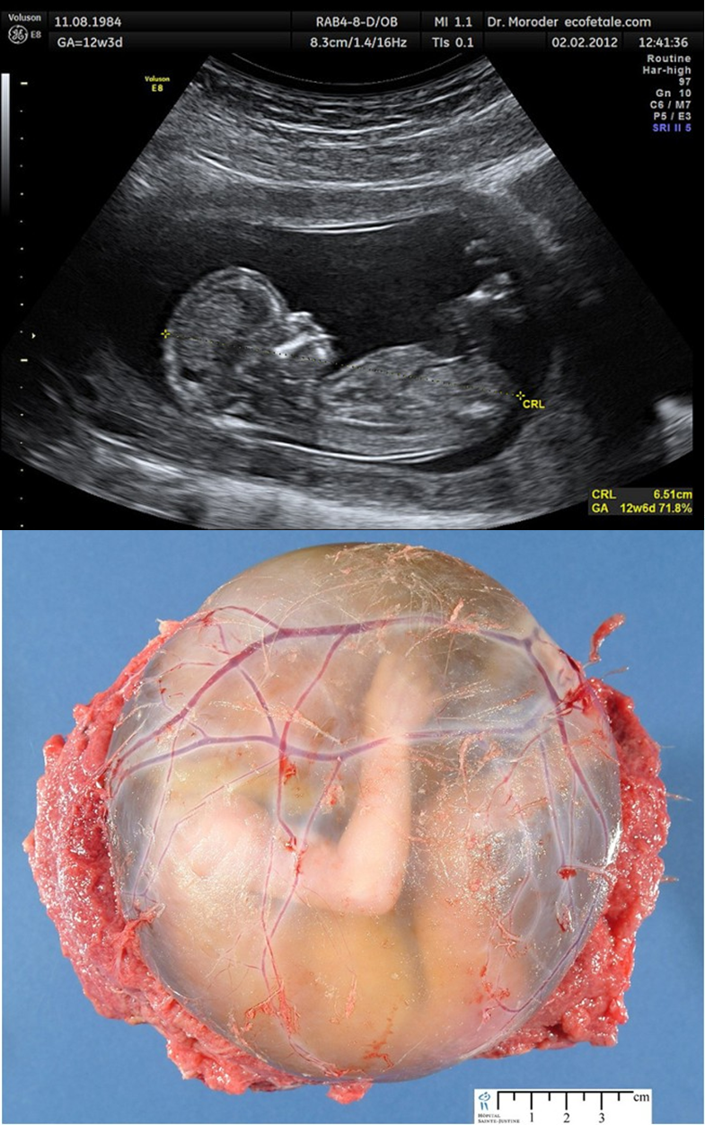
amniotic cavity
66
where does the amniotic fluid come from?
fetus and placenta
67
too much amniotic fluid
polyhydraminos
68
too little amniotic fluid
oligohydraminous
69
these form from the hypoblast and line the primary yolk sac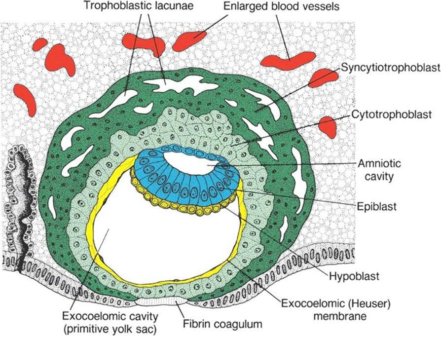
Heuser membrane (line whole yolk sac), extraembryonic endoderm
70
what are the layers of the extraembryonic mesoderm?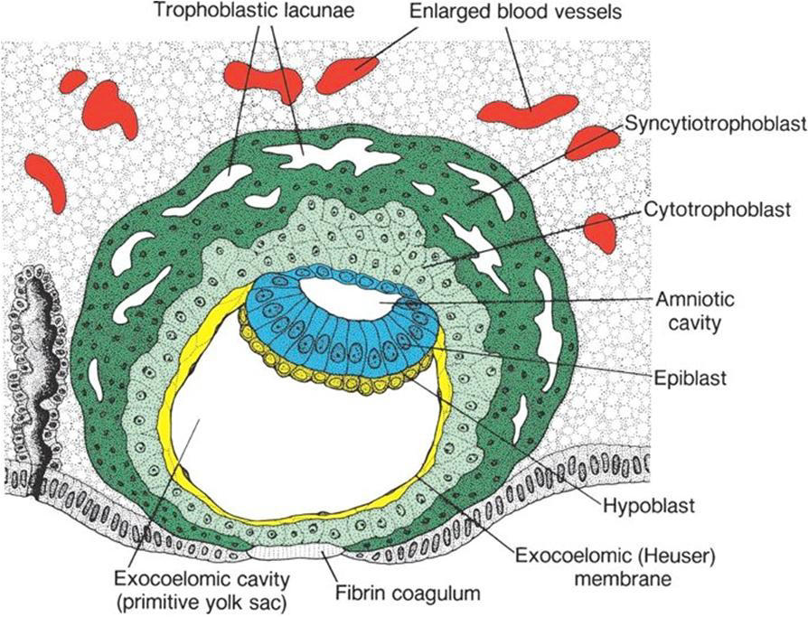
somatic, splanchnic
71
this secretes HCG
syncytiotrophoblast
72
what could increased HCG levels indicate?
Pregnancy
Ectopic pregnancy (typically in the low range)
Hydatidiform mole (typically very high)
Choriocarcinoma
Germ cell tumor (testes, ovaries)
Hepatoma
Lymphoma
Ectopic pregnancy (typically in the low range)
Hydatidiform mole (typically very high)
Choriocarcinoma
Germ cell tumor (testes, ovaries)
Hepatoma
Lymphoma
73
what could decreased HCG levels on repeated testing mean
**indicative of decreased viability of the placenta**
High risk of spontaneous abortion
Dead fetus
High risk of spontaneous abortion
Dead fetus
74
these in the syncytiotrophoblast, fill with blood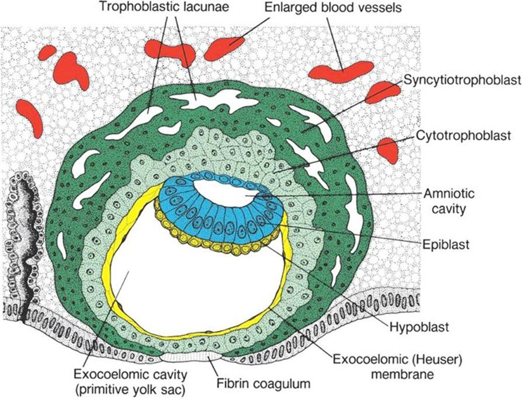
trophoblastic lacunae
75
what layers must materials initially cross tp resach mother to fetus
syncithiotrophoblast to cytotrophoblast tpo connective tissue to fetal blood vessels
76
what tissues is the chorion composed of?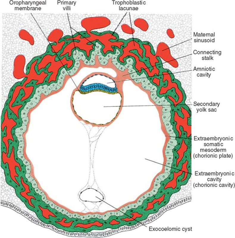
syncitiotrophoblast, cytotrophoblast, mesoderm
77
the X genome develops ___________________ and the Y genome develops ____________
embryoblast tissue; placental tissue
78
what are the propose mechanisms for molar pregnancy (x2)
1) empty egg fert with only sperm
2) 2 sperm fertilize an egg
2) 2 sperm fertilize an egg
79
what are some key signs for molar pregnancy
Bleeding in the first half of pregnancy
Lower abdominal pain
Pre-eclampsia before 24 weeks’ gestation
Hyperemesis gravidarum
Uterus “large for dates” (only 50% of cases)
Absent fetal heart sounds, no fetal parts present
Expulsion of “grape-like” vesicles (hydropic villi)
Lower abdominal pain
Pre-eclampsia before 24 weeks’ gestation
Hyperemesis gravidarum
Uterus “large for dates” (only 50% of cases)
Absent fetal heart sounds, no fetal parts present
Expulsion of “grape-like” vesicles (hydropic villi)
80
intellectual disability, congenital heart defects, low-set ears, and flexion of fingers and hands (Fig. 2.10). In addition, patients frequently show micrognathia, renal anomalies, syndactyly, and malformations of the skeletal system.
Trisomy 18
81
intellectual disability, holoprosencephaly, congenital heart defects, deafness, cleft lip and palate, and eye defects, such as microphthalmia, anophthalmia, and coloboma; 90% die days after birth
trisomy 13
82
only monosomy compatible with life. Even then, 98% of all fetuses with the syndrome are spontaneously aborted. The few that survive are unmistakably female in appearance (Fig. 2.12) and are characterized by the absence of ovaries (gonadal dysgenesis) and short stature. Other common associated abnormalities are webbed neck, lymphedema of the extremities, skeletal deformities, and a broad chest with widely spaced nipples.
Turner syndrome
83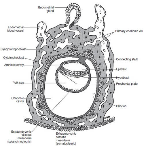
Image occlusion: name the hidden labels
84
Image occlusion: name the hidden labels
85
Image occlusion: name the hidden labels
86
Image occlusion: name the hidden labels
87
Image occlusion: name the hidden labels
88
Image occlusion: name the hidden labels
89
what three layers form the third week
amnion, yolk sac, chotrion
90
when does gastrulation occur?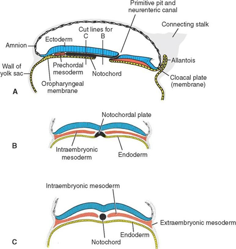
day 16
91
what is the stody of gastrulation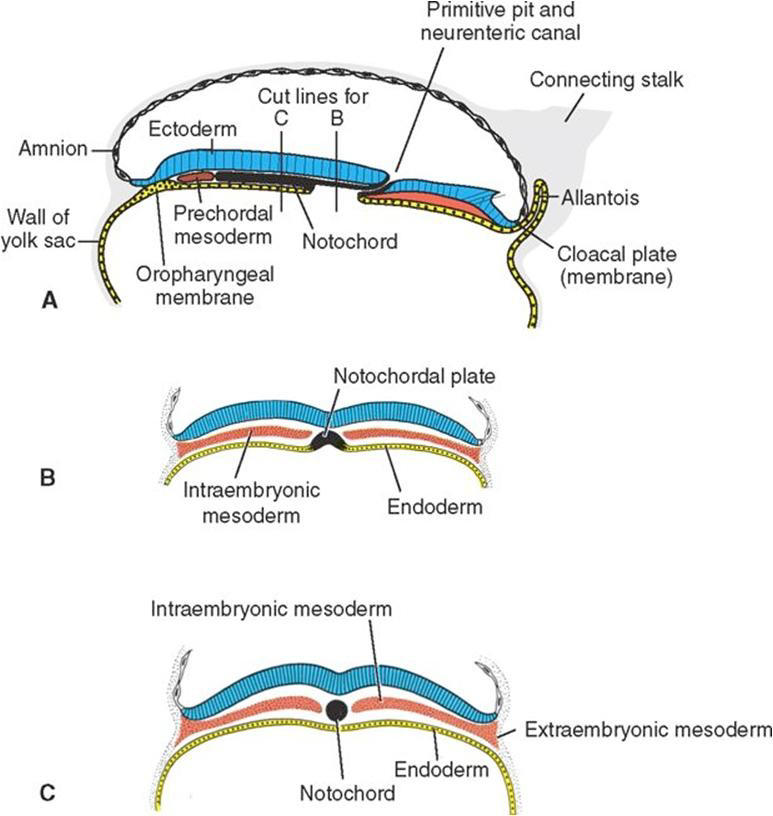
cells of epiblast divide and migrate under the primitive strek, hypoblast reoplaced with endoderm, mesoderm formed inbetween, epiblast replaced with ectoderm
92
how does the notochord form?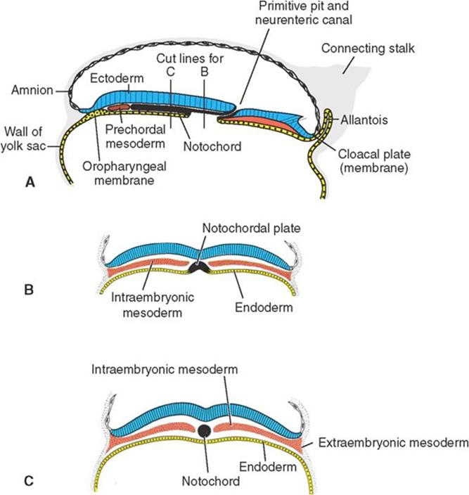
invaginates from primitive pit and extends, fuses witj endoderm and becomes notochordal plate, detaches and forms notochord
93
what are the functions of the notochord (x4)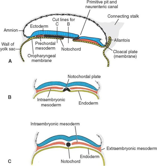
1. directs CNS development
2. development of the vertebral column
3. anatomic midline
4. forms the nucleus pulposis
2. development of the vertebral column
3. anatomic midline
4. forms the nucleus pulposis
94
what are the two types of embryonic folding?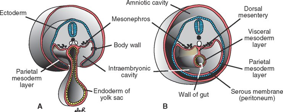
longitudinal fold: head to tail
transverse fold: ectoderm fuses wth each other
transverse fold: ectoderm fuses wth each other
95
heart forms outside the heart due to improper folding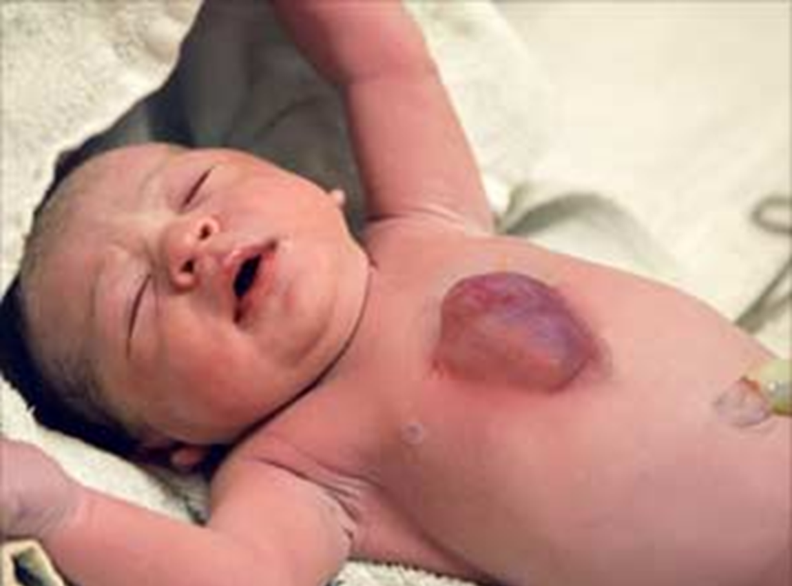
ectopic cordis
96
heart is on the wrong side of the body due to a midline defect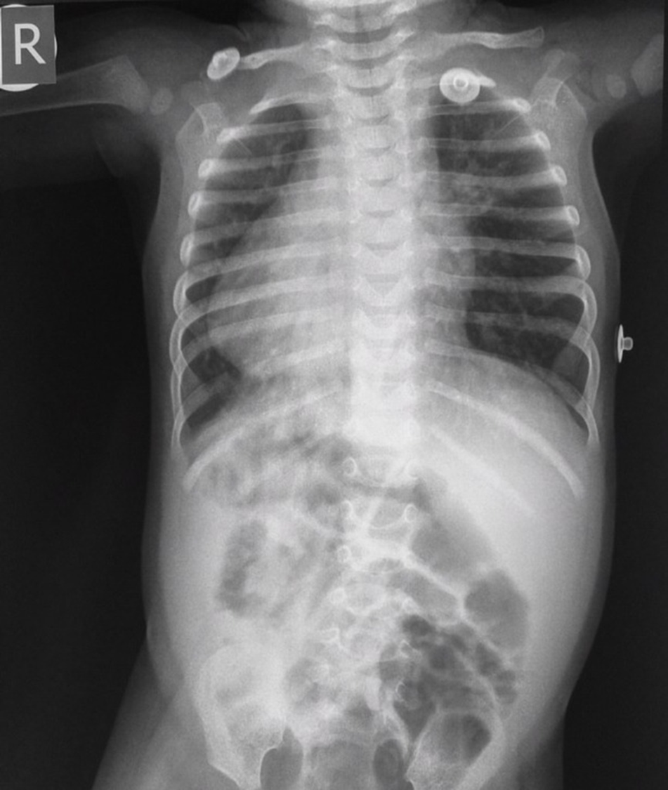
dextrocardia
97
everything forms on the ride side of the body (organs) due to midline defectsody axis defects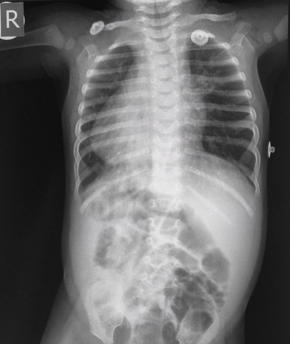
situs inversus
98
epithalial mesoderm benign tumor
angiomas
99
epithelial mesoderm malignant tumor
sarcomas
100
epithalial endoderm/ectoderm benign tumors
papillomas/adenomas
101
epithalial endo/ecto malig tumors
carcinomas
102
connective tissue benign tumotr
fibroma, lipoma, chondroma, osteoma
103
connective toissue malig timor
sarcoma
104
what do neural crest cells form
ganglia cells
105
what happens weeks 9-12 dev
wk9: liver hematopoetic organ
post wk9: external genitals male/female
post wk9: external genitals male/female
106
wks 13-16 dev?
ossification evident
eyes movement and eyes face forward
eyes movement and eyes face forward
107
wks 17-20 dev
primary oocytes formed
testes descend
testes descend
108
wks 21-25 dev
surfacant secreted in lungs
109
wks 26-29 lungs
lungs breath
CNS can control breath and body temp
spleen makes blood but bone marrow can now
CNS can control breath and body temp
spleen makes blood but bone marrow can now
110
wks 35-38 fetal dev
final prep of cardio and resp tissus
111
what uterine layers must you penitrate in amniocentesis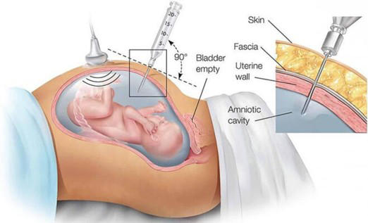
perimetrium, myometrium, endometrium, chorion, amnion
112
what is the pathway of somite development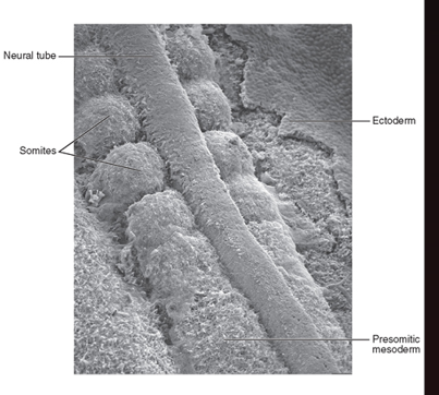
paraxial mesoderm to somitomeres to somites
113
what do somites differentiate into? (x2)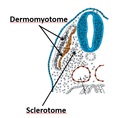
sclerotomes and dermomyotomes
114
process where sclerotome portion of each somite's caudal half grows and fuses with the cranial half of the adjacent sclerotome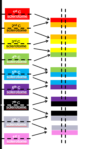
resegmentation
115
the cranial portion of the first cervical sclerotome helps to form the ______________
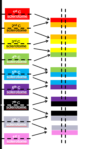
occipital bone
116
the intervertebral discs forms from these (x2)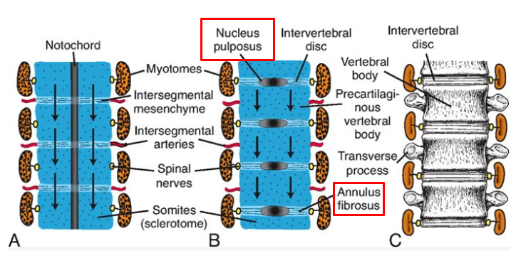
notochord and sclerotomes
117
the dermomyotome splits and becomes (x2)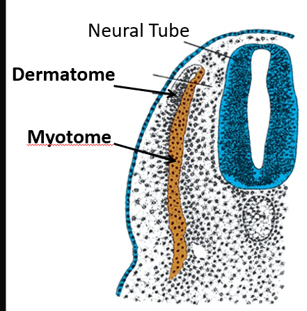
dermatome and myotome
118
muscles of the head are formed from
somitomeres
119
muscles of the limbs are formed by
somites
120
form Muscles of the back, shoulder girdle, and intercostal muscles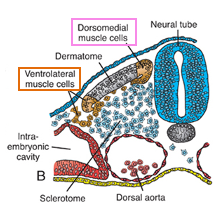
primaxial muscle cells
121
form body wall, infrahyoid, and limb muscles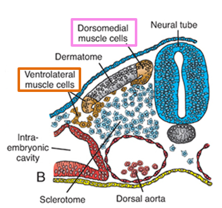
abaxial muscle cells
122
what does the ventral ramus inivate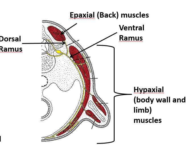
abdominal wall muscles and lumbs
123
what is the most likely diagnosis?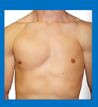
poland anomaly
124
if the cranial portion of the neuropore doesnt close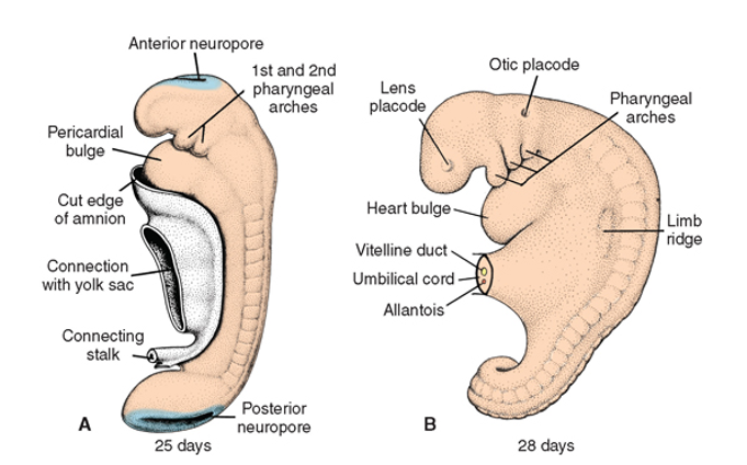
anancephally
125
if the caudal portion of the neuropore doesnt clsoe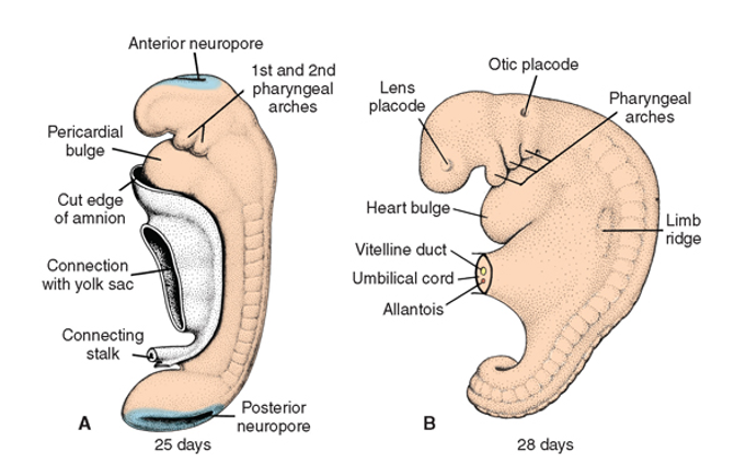
spina bifida
126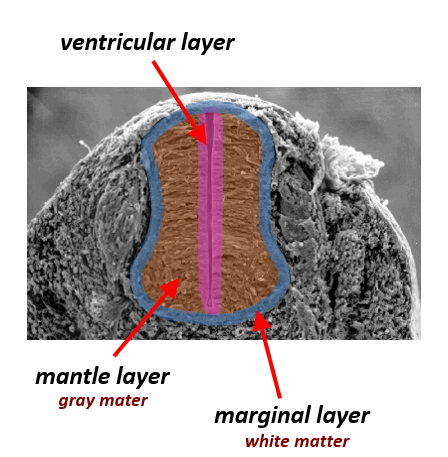
Image occlusion: name the hidden labels
127
Image occlusion: name the hidden labels
128
Image occlusion: name the hidden labels
129
these cells differentiate into mesenchymal cells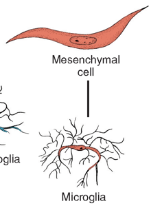
microglia
130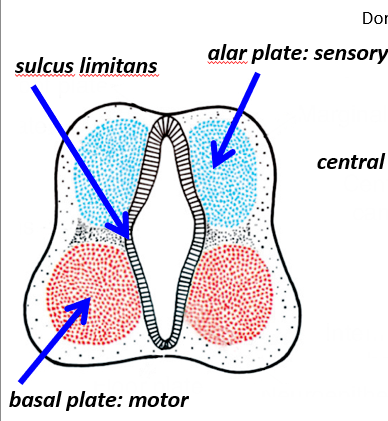
Image occlusion: name the hidden labels
131
Image occlusion: name the hidden labels
132
Image occlusion: name the hidden labels
133
what is the developmental sequence of nerves
1) GSE [control to muscles]
2) GVE [the outgoing part of the autonomic nervous system; only at certain levels!]
3) Sensory [returning information from skin, muscles, etc]
2) GVE [the outgoing part of the autonomic nervous system; only at certain levels!]
3) Sensory [returning information from skin, muscles, etc]
134
what do neural crest cells give rise to specifically (5)
DRG, sympathetic chain, suprarenal g., prevertebral g., enteric ganglia
135
neural tube doesnt close and neural tissue is sealed out of spinal column and not covered
spina bifida with meningocele
136
neural tube doesnt close and neural tissue is sealed out of spinal column and is covered
spina bifida with myeloschisis
137
occur during organogenesis and result in abnormal strcture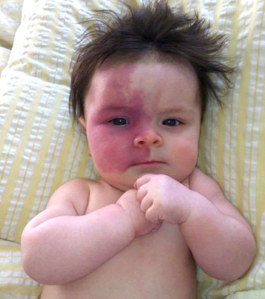
malformations
138
results from external influences that cause alterations of completely formed structures or organs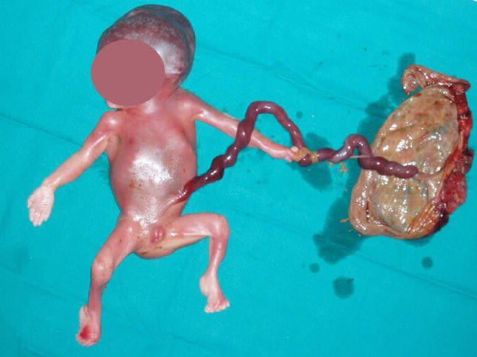
disruptions
139
abnornal structure resulting from mechanocal forces (congenital abnormality)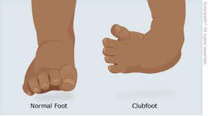
deformations
140
abnormal organization of cells into tissue (congenital abnormality)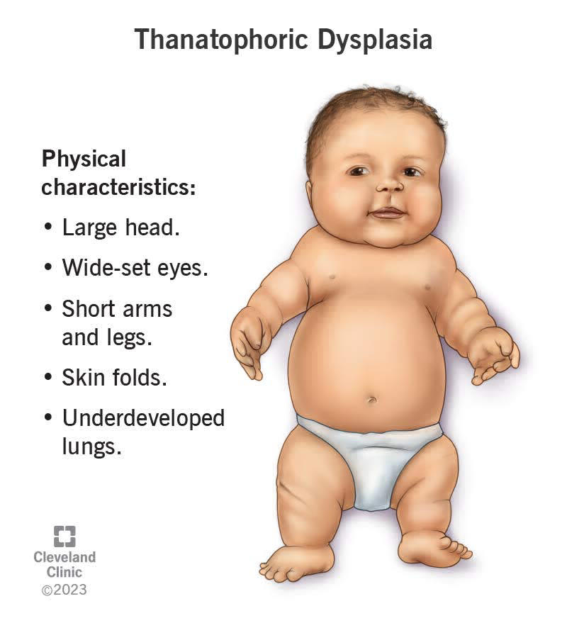
dysplasia
141
absence of organ
agenesis
142
cells to form an organ never formed (migrating cells)
aplasia
143
anomalies occuring tofether without determined cause
association
144
absence of opening or lumen
atresia
145
overgrowth of an organ
hyperplasia
146
underdevelopment of an organ
hypoplasia
147
functions to stimulate the development of ovarian follicles and production of estrogen
FSH
148
serves as a trigger for ovulation and stimulates the production of progesterone
LH
149
secreted by the trophoblast and appears in materna;l blood 24-48hrs after fertilization, can be used for pregnancy tests during the first 10 days
Early pregnancy factor
150
masses of endodernally derived tissue commonly associateds with the spinal cprd; derived from neurenteric canal where ectoderm and endoderm are closely associated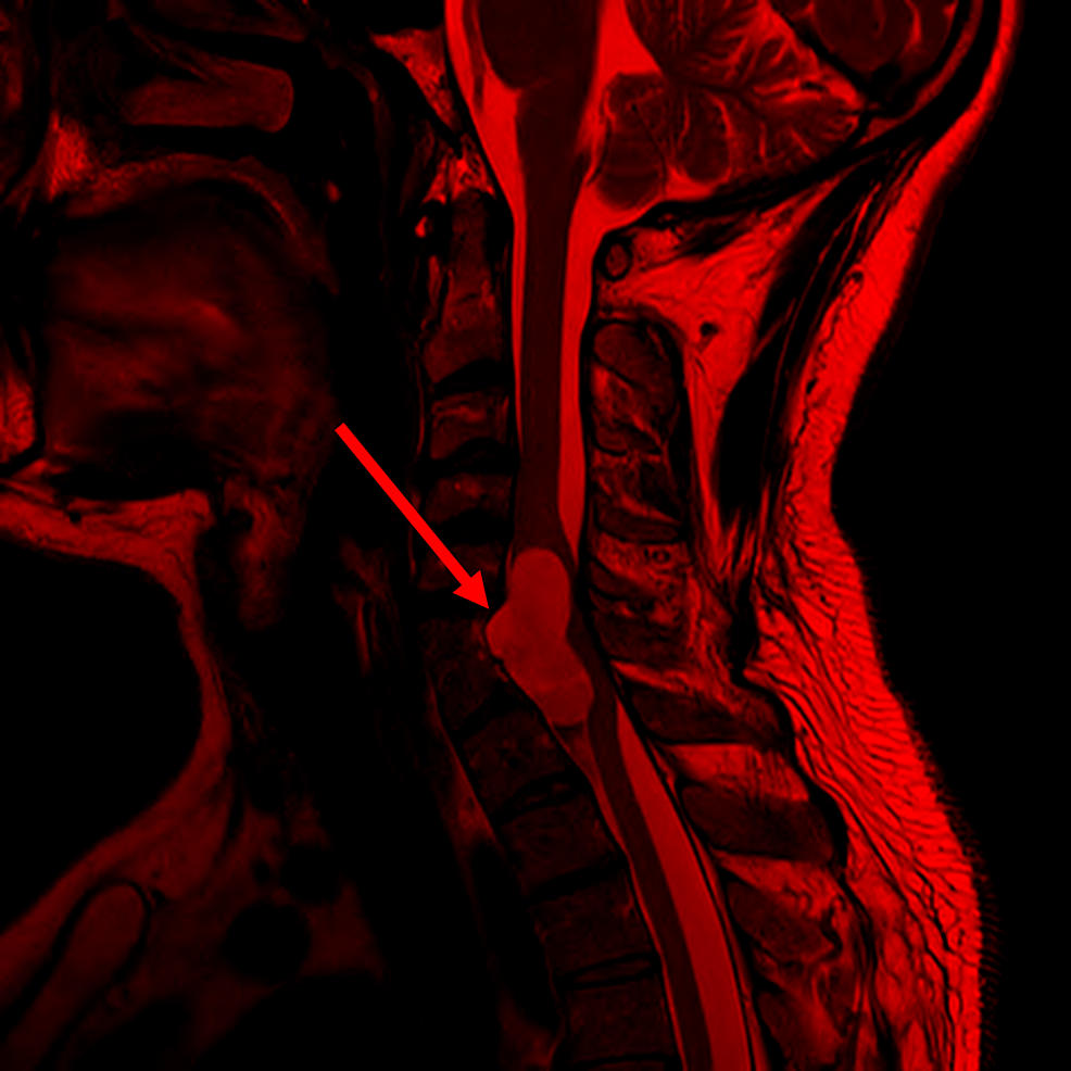
neurenteric cysts
151
results from the innapropriate death of midline cells, causes a deficiencyt of midline head structures
holoprosencephally
152
a neurological condition where the spinal cord is abnormally attached to surrounding tissues, limiting its movement and causing neurological problems
tethered cord syndrome
153
vascular derivatives of the mesoderm
hemangioblasts: give rise to blood vessels
hematopoietic stem cells: give rise to blood cell precursors
hematopoietic stem cells: give rise to blood cell precursors