Radiology S1 U2
Study these flashcardsFree · spaced repetition · iPhone, Android, Mac & web
All cards
1
how many ribs do you want to be visible in a chest CT/xray
8-10
2
how do you calculate cardiothoracic ratio? 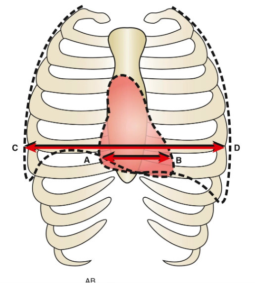
measure the transverse diameter of the hear and divide by the transvetrse thoracc movement
3
In severe left atrial enlargement, the right lateral border of the left atrium is superimposed over the right atrial shadow, producing a
double density sign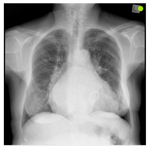
4
A lower left than right hilum indicates ...
left lower lobe collapse
5
the concave space immediately below the aortic arch and above the left pulmonary artery
aortopulmonary window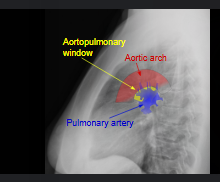
6
how is the vascular pedicle determined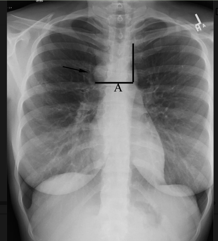
drawing a horizontal line (A) from the junction of the azygos vein (arrow) and SVC over to a perpendicular line drawn from the left subclavian artery inferiorly along the transverse portion of the thoracic aorta (aortic arch).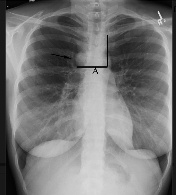
7
due to the superimposition of the aerated upper lobes, whereas the right middle lobe and the lingular segment of the left lobe are obscured by the cardiac silhouette.
retrosternal lucency
8
Placing a patient on their side (decubitus position) and obtaining a film across the chest in the AP direction is described as a
decubitus view
9
the anatomical plane, which divides the body into right and left parts.
sagittal plane
10
divides the body into ventral and dorsal (front and back) parts
coronal plane
11
allows imaging of the chest within one breath hold (5–10 seconds).
multidetector CT (MDCT)
12
what does this show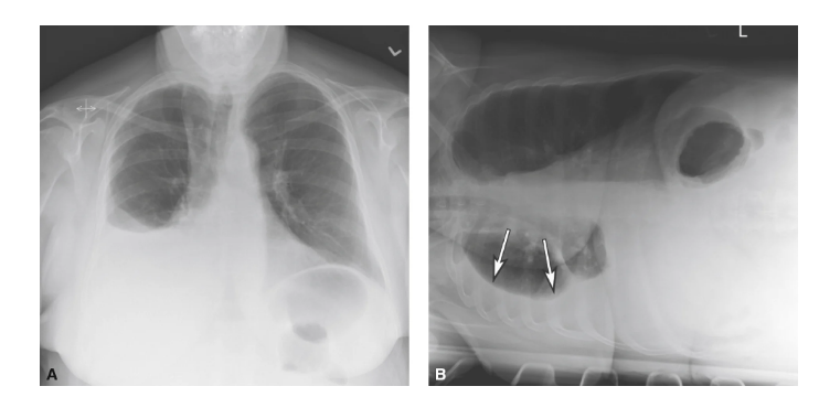
right pleural effusion
13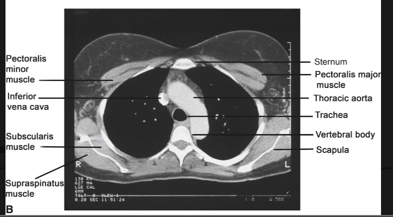
Image occlusion: name the hidden labels
14
Image occlusion: name the hidden labels
15
Image occlusion: name the hidden labels
16
Image occlusion: name the hidden labels
17
Image occlusion: name the hidden labels
18
Image occlusion: name the hidden labels
19
Image occlusion: name the hidden labels
20
Image occlusion: name the hidden labels
21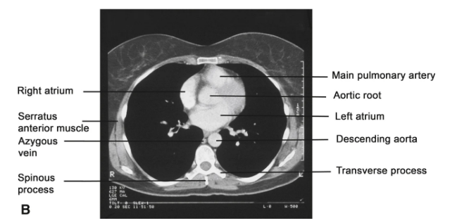
Image occlusion: name the hidden labels
22
Image occlusion: name the hidden labels
23
Image occlusion: name the hidden labels
24
Image occlusion: name the hidden labels
25
Image occlusion: name the hidden labels
26
Image occlusion: name the hidden labels
27
cardiac imaging (CT or MR) is best performed using ECG gating, which allows data acquisition typically during _______ when the heart is not moving.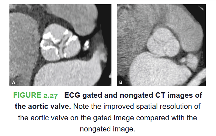
diastole
28
used to evaluate the aorta and myocardium where it is helpful in the diagnosis of cardiac masses and cardiomyopathy.
cardiac MRI
29
most common thoracic aortic anomaly is a ________(Fig. 2.29). On the PA view, the right-sided arch appears more cephalad (higher) than a normal left-sided arch. 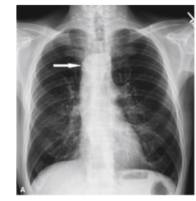
right sided aortic arh
30
a stenosis at the junction of the aortic arch and the descending thoracic aorta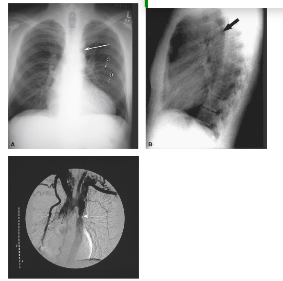
coarticulation of the aorta
31
For IV access, the optimal location of central venous lines is between
mid SVC and mid right atrium
32
The correct position of the endotracheal tube (ETT)
optimal location of the tip is in the mid trachea, approximately 5 cm above the carina if the patient’s neck is in the neutral position
33
what does this show?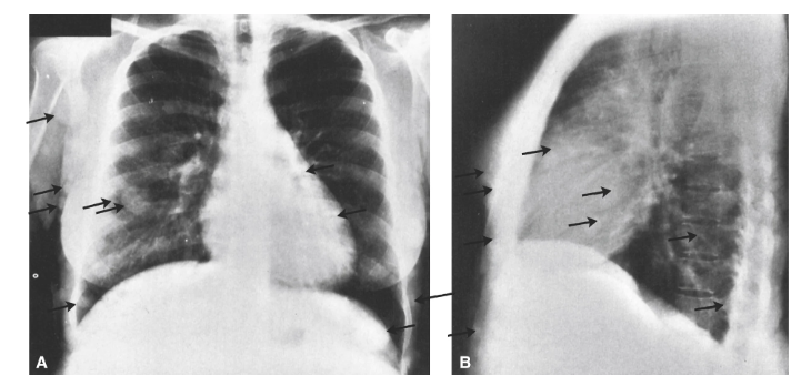
Neurofibromatosis. Chest PA (A) and lateral (B) radiographs. Multiple well-defined subcutaneous soft tissue nodules (arrows) project over the thorax which should not be mistaken for pulmonary nodules.
34
what does this show?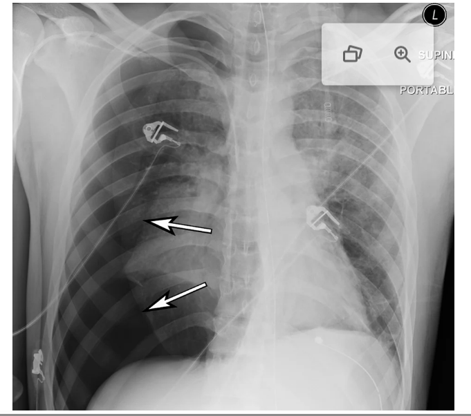
Right tension pneumothorax. Portable AP radiograph shows a large right pneumothorax with near complete collapse of the right lung (arrows). Note the depression of the right diaphragm and the deep right lateral costophrenic sulcus or angle (deep sulcus sign). Also, note the slight deviation of the cardiac silhouette to the left side.
35
a collection of air within the mediastinum, the significance of which is variable but can be ominous in patients with a history of trauma or vomiting, suggesting rupture of either the esophagus or trachea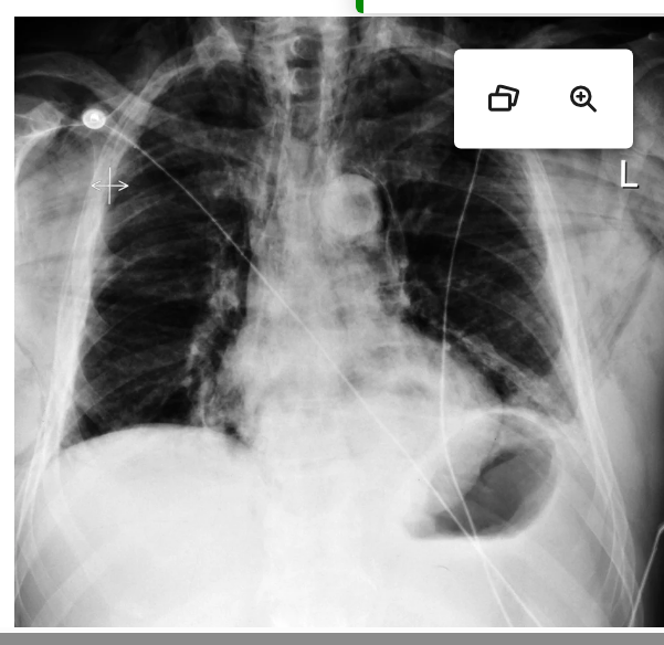
pneumoediastinum
36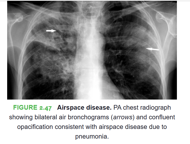
Image occlusion: name the hidden labels
37
what does this show? 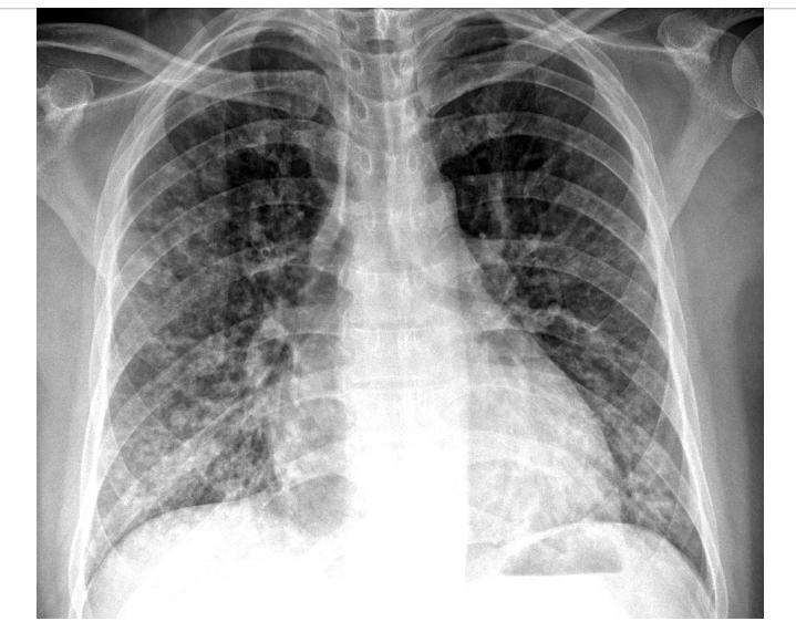
Interstitial (nodular) lung disease. PA chest radiograph showing extensive increased nodular markings consistent with interstitial lung disease.
38
a CT pattern of a hazy opacity that does not obscure the underlying bronchial structures or pulmonary vessels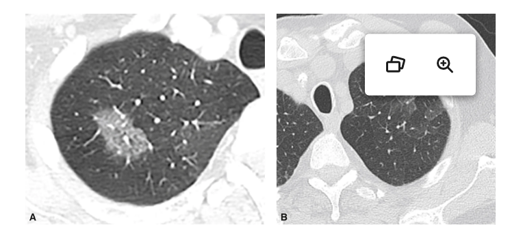
groundglass opacity
39
A structure’s outline is only seen when an adjacent structure in the same anatomic plane is of a different radiographic tissue density giving ris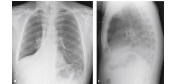
silhouette sign
40
represents a loss of aeration and varies in extent from being subsegmental appearing as a linear density (subsegmental or discoid atelectasis)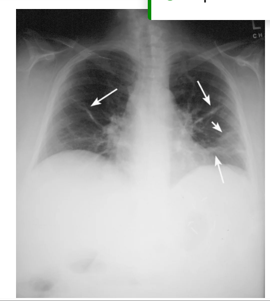
atelectasis
41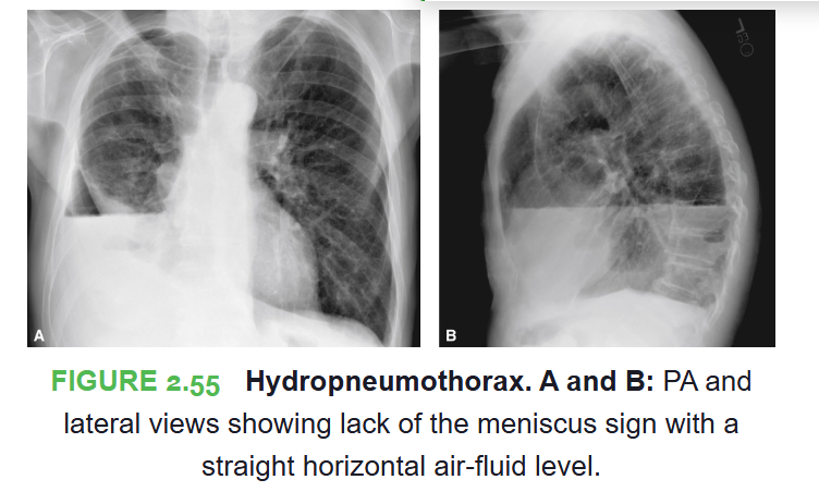
Image occlusion: name the hidden labels
42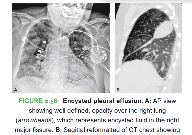
Image occlusion: name the hidden labels
43
calcified pleural plaques are often benign, especially when bilateral, indicating asbestos exposure
-more extensive or unilateral may be due to healed empyema or tuberculosois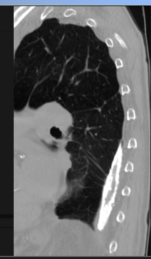
-more extensive or unilateral may be due to healed empyema or tuberculosois
pleural calcifications
44
what does tjis show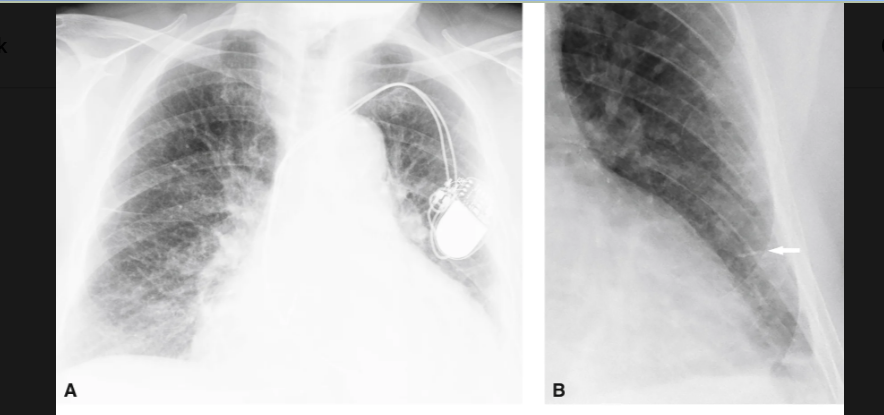
Cardiogenic pulmonary edema. A: Upper lobe venous diversion. B: A magnified view of the left lung shows interstitial edema (arrow) due to transudation of fluid into the avascular connective tissues
45
(formally known as “Kerley lines”), which are identified on chest films as small perpendicular lines at the lung periphery, most commonly in the lower zones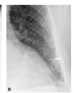
interstistial edema
46
what does this show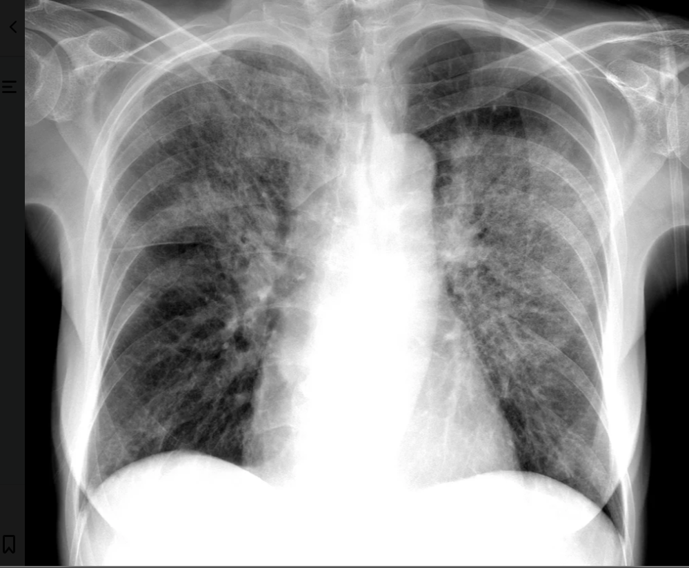
FIGURE 2.60 Noncardiogenic pulmonary edema. Similar appearance with increase in perihilar airspace disease to cardiogenic pulmonary edema, but with a normal heart size.
47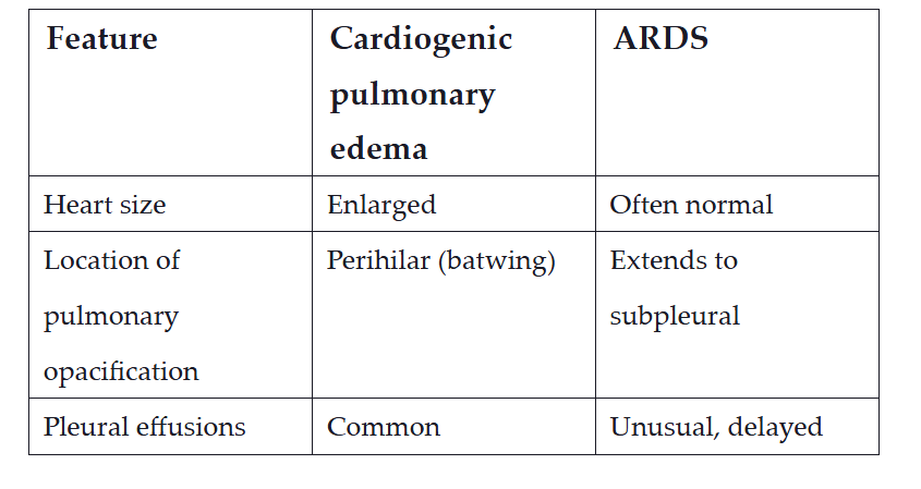
Image occlusion: name the hidden labels
48
Image occlusion: name the hidden labels
49
Image occlusion: name the hidden labels
50
a rare but serious syndrome characterized by sudden acute respiratory distress following transfusion and is the leading cause of blood product transfusion–related mortality in the United States. Chest film shows noncardiogenic pulmonary edema with bilateral patchy infiltrates, which may rapidly progress to complete “white out” indistinguishable from ARDS.
transfusion related acute lung injury (TRALI(
51
Image occlusion: name the hidden labels