Anatomy S1 U1
Study these flashcardsFree · spaced repetition · iPhone, Android, Mac & web
All cards
1
ear onsisting of the part attached to the lateral aspect of the head and the canal leading inward.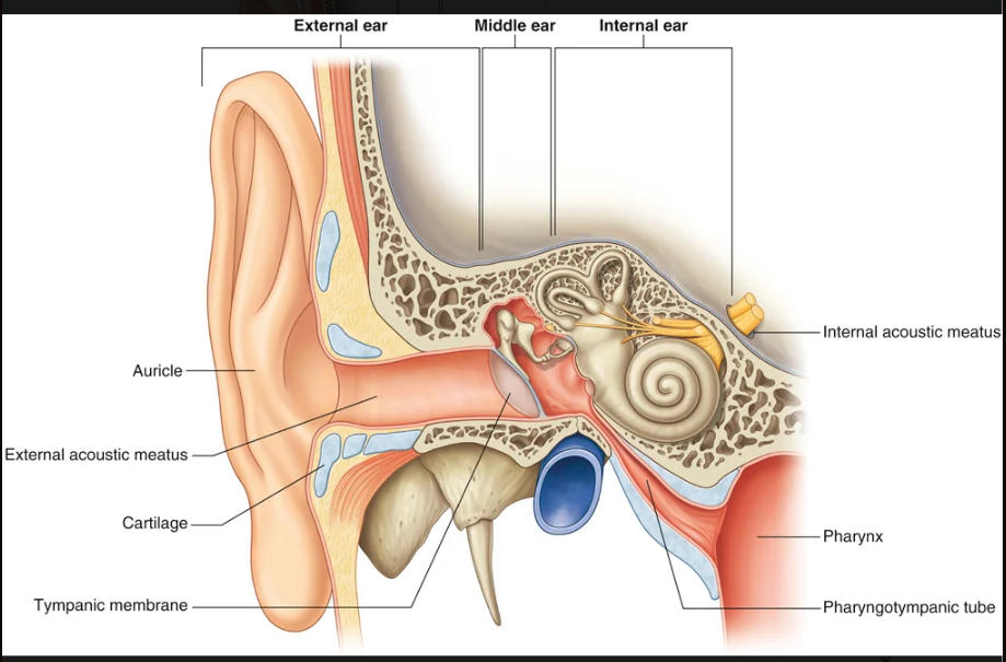
external ear
2
ear: a cavity in the petrous part of the temporal bone bounded laterally, and separated from the external canal, by a membrane and connected internally to the pharynx by a narrow tube.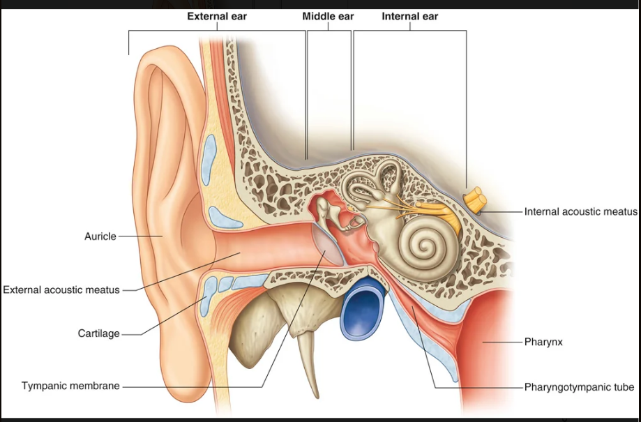
middle ear
3
ear consisting of a series of cavities within the petrous part of the temporal bone between the middle ear laterally and the internal acoustic meatus medially.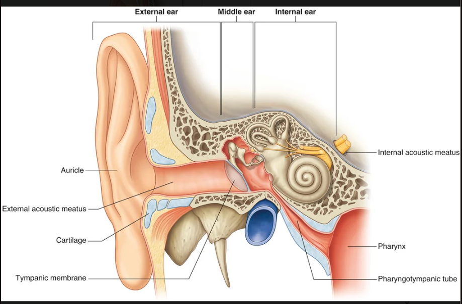
internal ear
4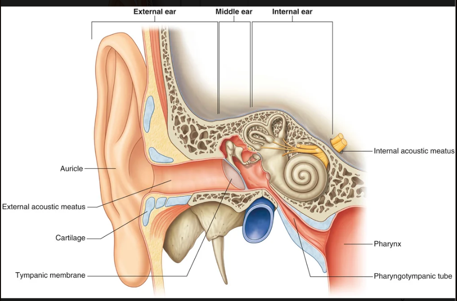
Image occlusion: name the hidden labels
5
Image occlusion: name the hidden labels
6
Image occlusion: name the hidden labels
7
Image occlusion: name the hidden labels
8
Image occlusion: name the hidden labels
9
Image occlusion: name the hidden labels
10
ear: on the side of the head and assists in capturing sound. It consists of cartilage covered with skin and arranged in a pattern of various elevations and depressions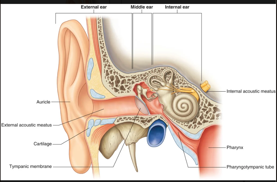
auricle
11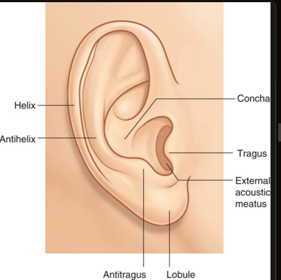
Image occlusion: name the hidden labels
12
Image occlusion: name the hidden labels
13
Image occlusion: name the hidden labels
14
Image occlusion: name the hidden labels
15
Image occlusion: name the hidden labels
16
Image occlusion: name the hidden labels
17
Image occlusion: name the hidden labels
18
muscles associated with the auricle of the ear (x2)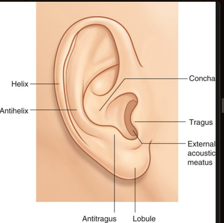
▪ The intrinsic muscles pass between the cartilaginous parts of the auricle and may change the shape of the auricle.
▪ The extrinsic muscles, the anterior, superior, and posterior auricular muscles, pass from the scalp or skull to the auricle and may also play a role in positioning of the auricle
▪ The extrinsic muscles, the anterior, superior, and posterior auricular muscles, pass from the scalp or skull to the auricle and may also play a role in positioning of the auricle
19
what innervates the intrinsic and extrinsic muscles of the ear (associated with the auricle)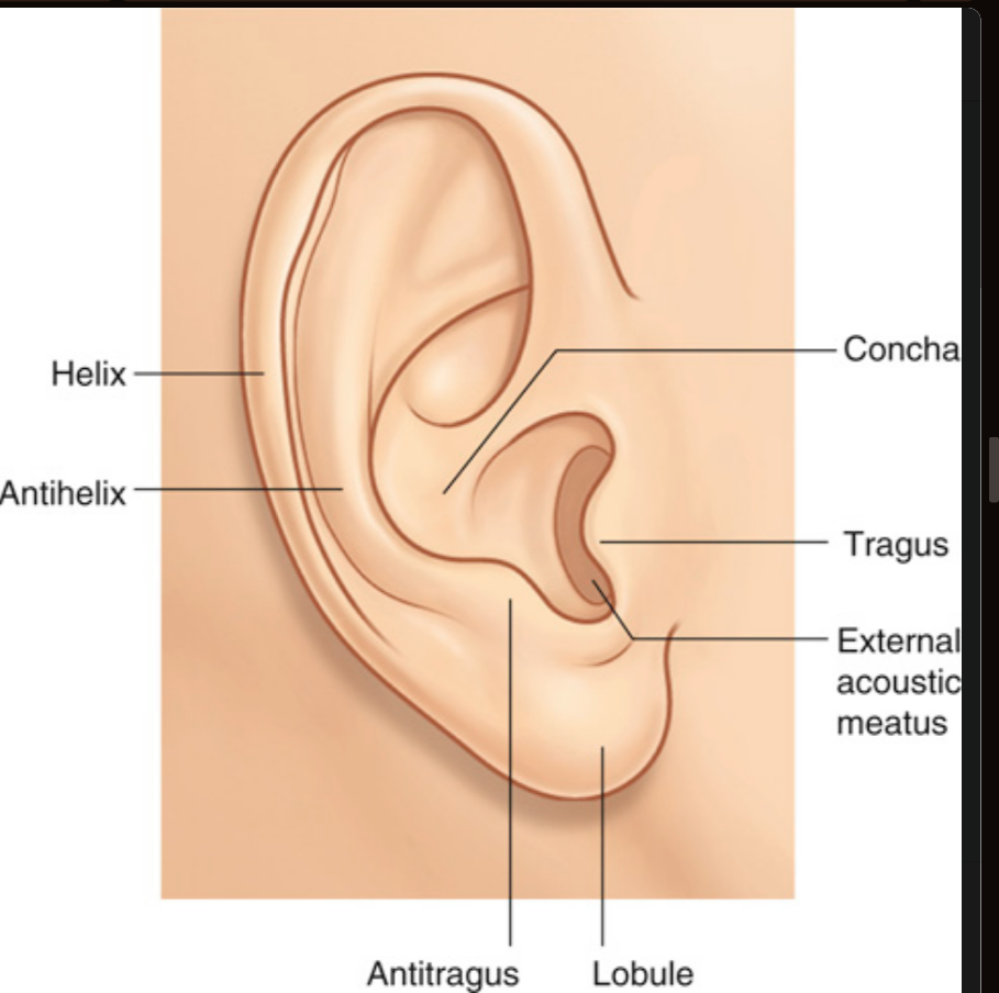
facial n (VII)
20
what supplies the sensory info to the outermost superficial surfaces of the auricle
he great auricular nerve (anterior and posterior inferior portions) and the lesser occipital nerve (posterosuperior portion) from the cervical plexus and the auriculotemporal branch of the mandibular nerve [V3] 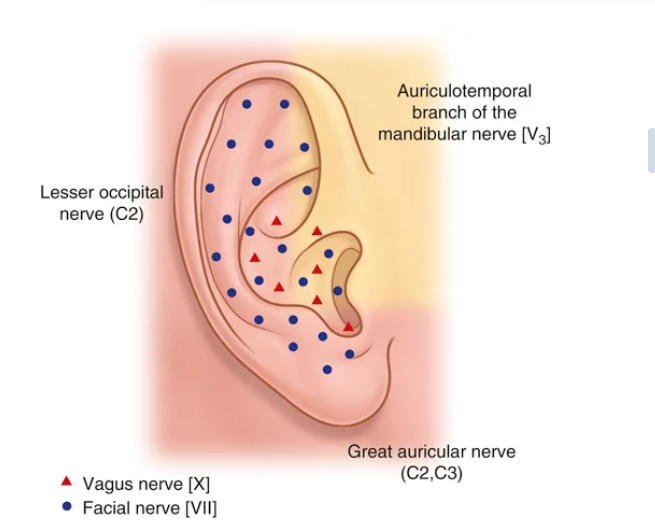
21
what supplies the sensory innervation to the deeper parts of the auricle?
vagus nerve [X] (the auricular branch) and the facial nerve [VII] (which sends a branch to the auricular branch of the vagus nerve [X]).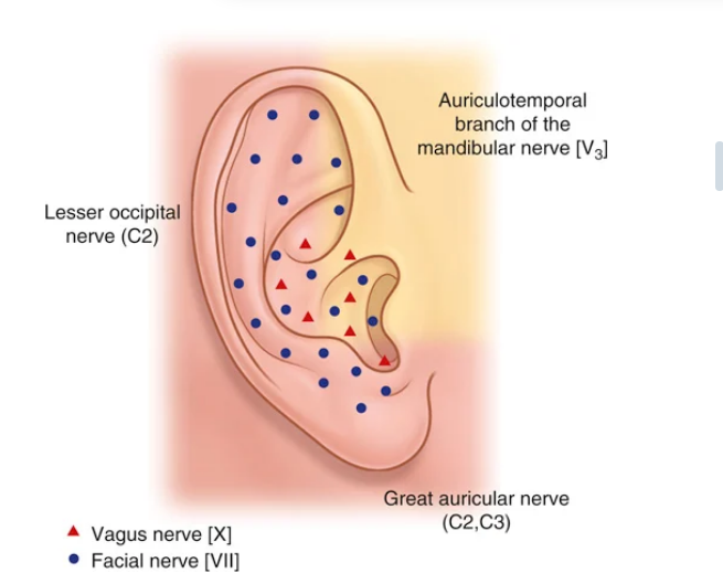
22
what lymph nodes drain the auricle
anteriorly into parotid nodes and posteriorly into mastoid nodes, and possibly into the upper deep cervical nodes.
23
anterior openings to the nasal cavity
nares
24
posterior openings to the nasal cavoty
choanae
25
an area between the posterior aspect (ramus) of the mandible and a flat region of bone (lateral plate of the pterygoid process) just posterior to the upper jaw (maxilla). This fossa, bounded by bone and soft tissues, is a conduit for one of the major cranial nerves—the mandibular nerve (the mandibular division of the trigeminal nerve [V3]),
infratemporal fossa
26
on each side is just posterior to the upper jaw. This small fossa communicates with the cranial cavity, the infratemporal fossa, the orbit, the nasal cavity, and the oral cavity. A major structure passing through the pterygopalatine fossa is the maxillary nerve (the maxillary division of the trigeminal nerve [V2]).
pterygopalatine fossa
27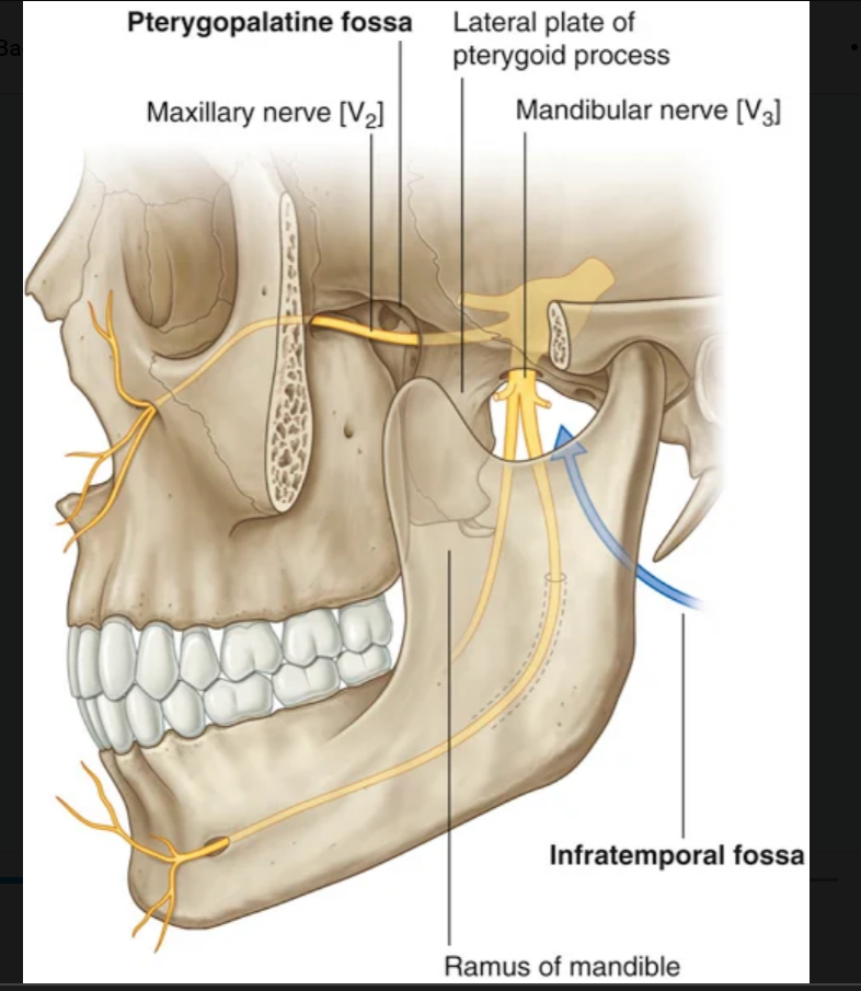
Image occlusion: name the hidden labels
28
Image occlusion: name the hidden labels
29
Image occlusion: name the hidden labels
30
Image occlusion: name the hidden labels
31
what are the 4 major compartments of the neck and what is contained in them?
▪ The vertebral compartment contains the cervical vertebrae and associated postural muscles.
▪ The visceral compartment contains important glands (thyroid, parathyroid, and thymus), and parts of the respiratory and digestive tracts that pass between the head and thorax.
▪ The two vascular compartments, one on each side, contain the major blood vessels and the vagus nerve.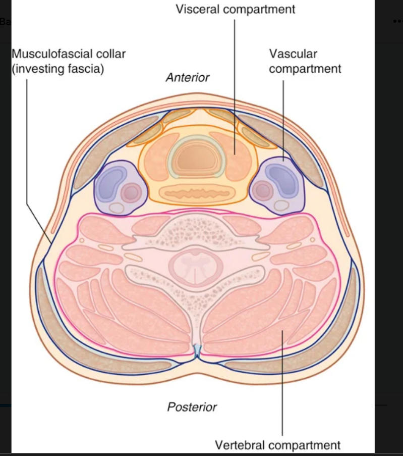
▪ The visceral compartment contains important glands (thyroid, parathyroid, and thymus), and parts of the respiratory and digestive tracts that pass between the head and thorax.
▪ The two vascular compartments, one on each side, contain the major blood vessels and the vagus nerve.
32
upper opening of the larynx; tilted posteriorly and continuous with the pharynx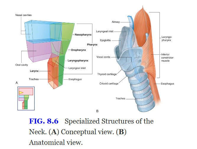
laryngeal inlet
33
what are the functions of fontanelles (x2)
▪ the head to deform during its passage through the birth canal, and
▪ postnatal growth.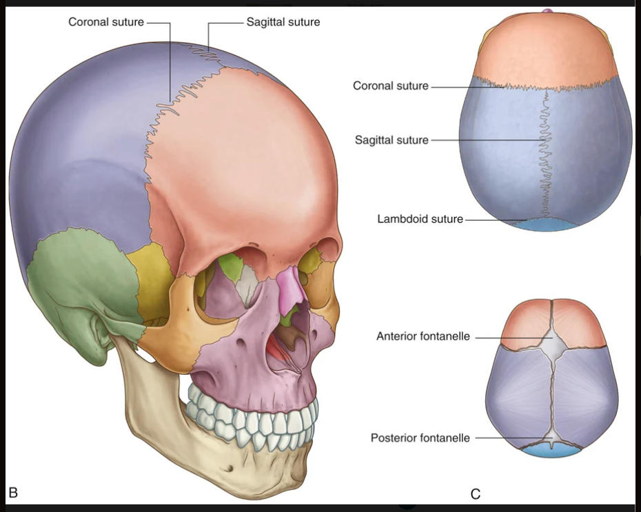
▪ postnatal growth.
34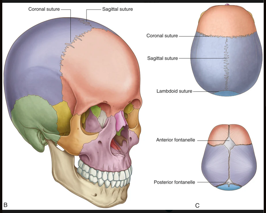
Image occlusion: name the hidden labels
35
Image occlusion: name the hidden labels
36
Image occlusion: name the hidden labels
37
Image occlusion: name the hidden labels
38
Image occlusion: name the hidden labels