Lower Limb
Study these flashcardsFree · spaced repetition · iPhone, Android, Mac & web
All cards
1
what are the articulations of the LE designed for?
weight-bearing and gait
2
what is at risk of injury is there is damage to the fibular neck
common fibular n
3
what is at risk of injury if there is damage to the tarsal tunnel
distal tibial n.
4
what attaches to the ischial tuberosity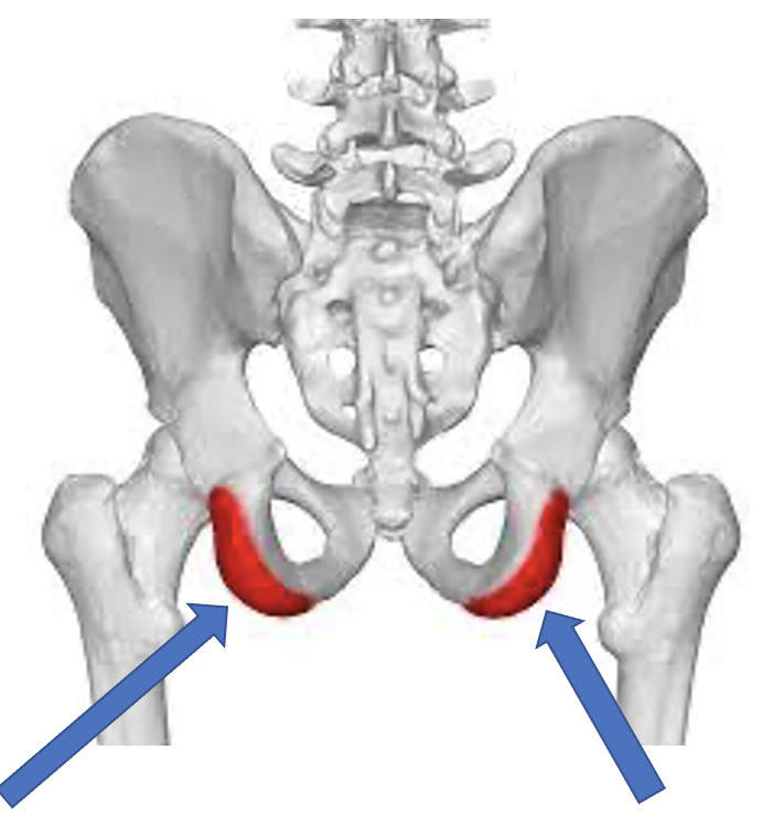
hamstrings, adductor magnus
5
what attaches to the ASIS
inguinal ligament, obliquus, sartorius
6
what attaches to the AIIS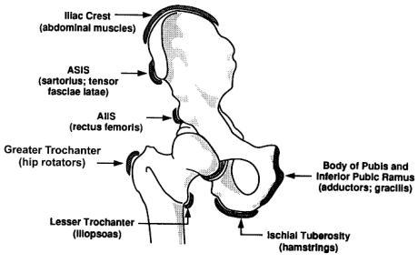
rectus femoris
7
what attaches to the greater trochanter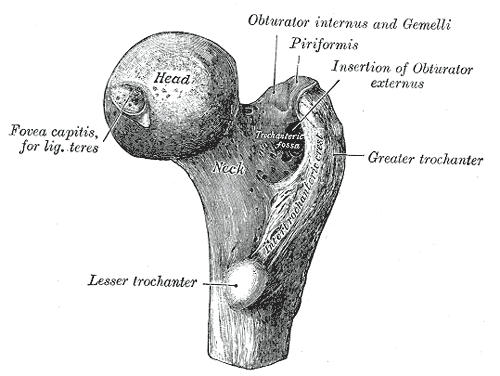
gluteus medius, orburator, piriformis, orturator internus and externus, gemmeli
8
what attaches to the adductor tubercle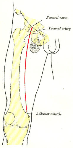
adductor magnus
9
what ttaches to the tibial tuberosity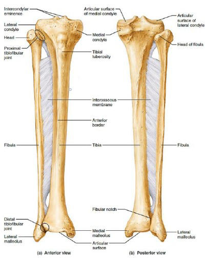
patellar ligament
10
what attaches to the base of the 1st metatarsal
fibularis longus
11
what attaches to the base of the 5th metatarsal
fibularis brevis
12
lower limb fascia layers (x3)
skin, superficial fascia, investing fascia
13
what lives in the superficial fascia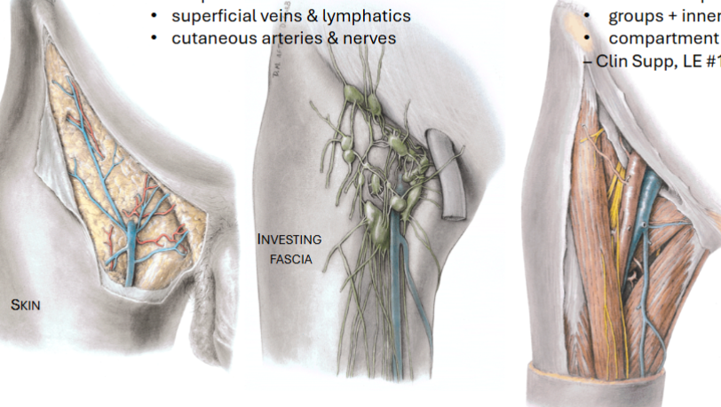
supoerficial veins, lymphatics, cutaneous nerves and arteries
14
what is the fumction of the investing fascia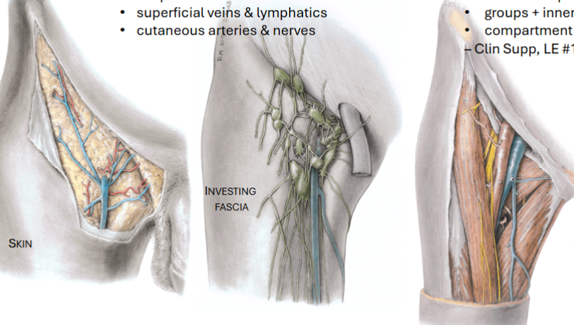
group muscles and innervations
15
a painful condition caused by increased pressure within a muscle compartment, potentially restricting blood flow and causing nerve and muscle damage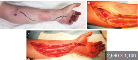
compartment syndrome
16
where does blood supply from the lower limb come from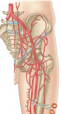
internal and external iliac arteries
17
places on the leg where pulse can be assessed 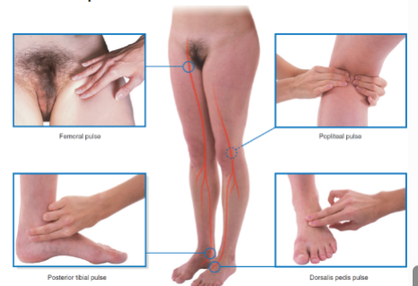
femoral, popiteal, posterior tibial, dorsal pads
18
normal pulse
2/4
19
what causes varicose veins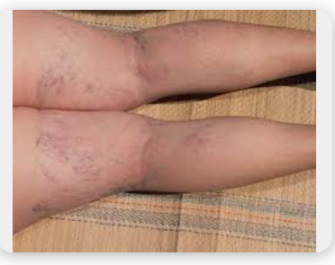
failure of perforating veins to allow bloodflow between superficial and deep veins where they fail and blood is pushed to the superficial veins
20
a blood clot that forms in a deep vein, usually in the leg or pelvis, but it can also occur in the arm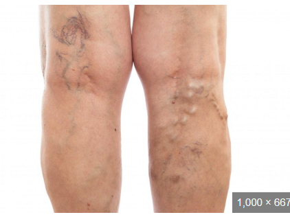
deep vein thrombosis
21
how does lymph drainage work for lateral leg?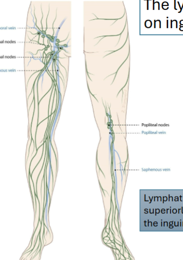
popiteal nodes to inguinal nodes
22
lymph drainage medial thigh?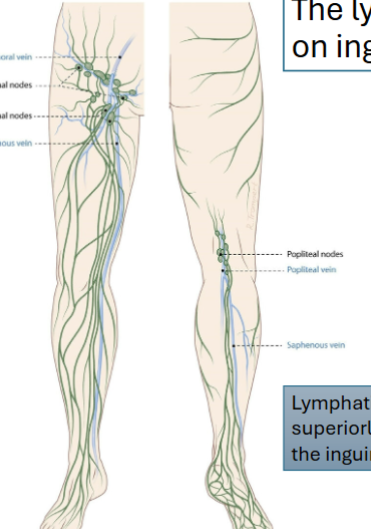
straight to superior inguinal nodes
23
what are the actions of the anterior thigh; innervation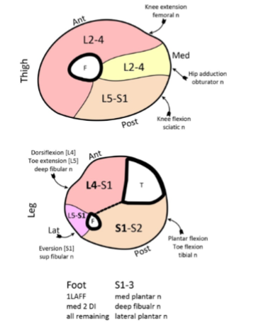
knee extension; femoral n (L2-L4)
24
what is the actions ans innervations medial thigh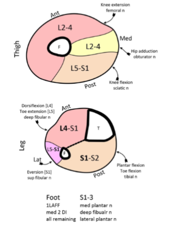
hip adduction; obturator n (L2-4)
25
actions and innervations posterior thigh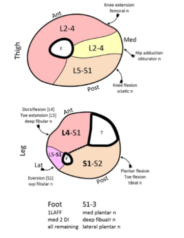
knee flexion; sciatic n (L5-S1)
26
what spinal levels innervate te foot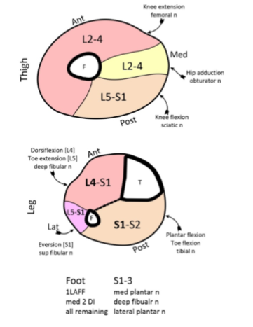
S1-3
27
what actions and innervations anterioir leg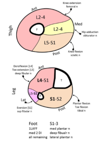
dorsiflexion, toe extension; deep fibular n (L4-S1)
28
actions and innervations lateral leg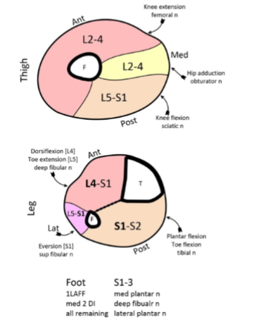
eversion; superficial fibular n (L5-S1)
29
what are the actions and innervations of the posterior leg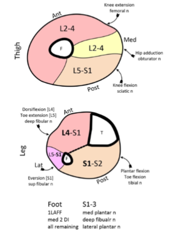
plantar flexion, toe flexion; tibial n (S1-S2)
30
hip abduction level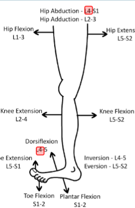
L4-S1
31
hip adduction level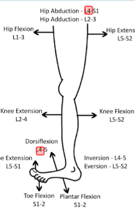
L2-3
32
hip flexion myotome
L1-3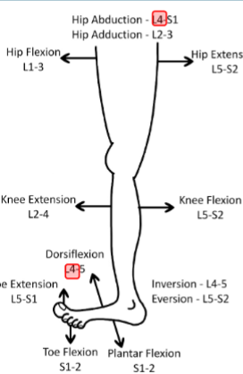
33
hip extension myotome
L5-S2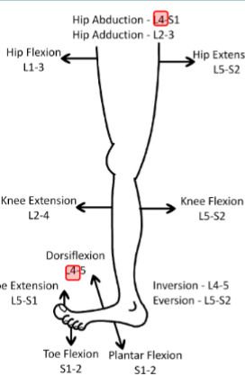
34
knee extension myotome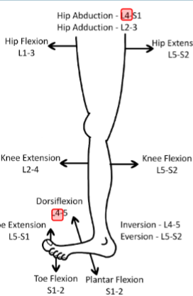
L2-4
35
knee flexion myotome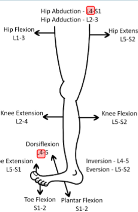
L5-S2
36
dorsiflexion myotome
L4+5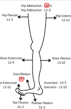
37
toe extension myotome
L5-S1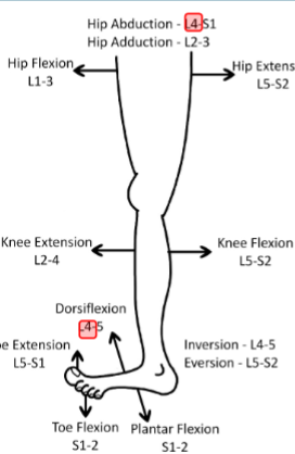
38
inversion myotome
L4-5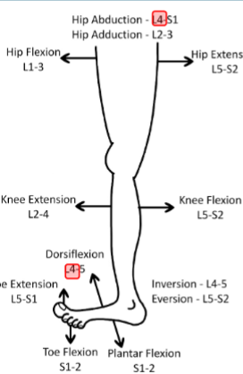
39
eversion myotome
L5-S2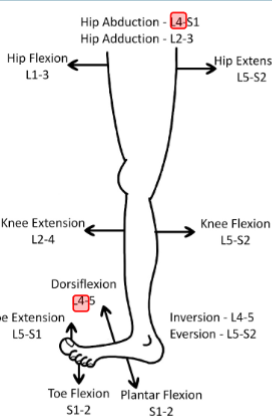
40
plantarflexion myotome
S1-2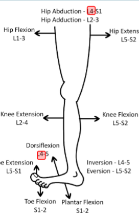
41
iliogastric and ilioinguinal nerve spinal
L1
42
genitofemoral spinal level
L1-L2
43
lateral femoral cutaneos n spinal level
L2-L3
44
obturator n spinal level
L2-4
45
femoral n spinal level
L2-4
46
sup gluteal n spinal level
L4-S1
47
inferior gluteal n spinal level
L5, S1, S2
48
sciatic n spinal level
L4-S2
49
tibial n spinal level
L4-S3
50
what anterior thigh muscles flex the hip/thigh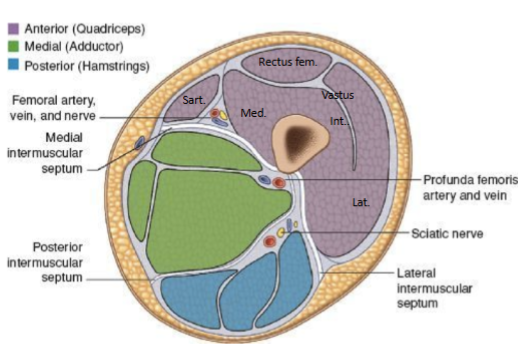
-Iliopsoas
• Sartorius
• Rectus femoris
• Sartorius
• Rectus femoris
51
what anterior thigh muscles extend the knee joint/leg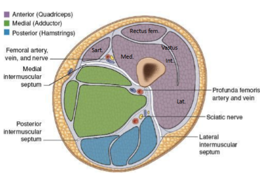
quadriceps
52
what is the blood supply to the anterior thigh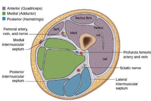
superfiucial femoral a
53
what innervates the anterior thigh muscles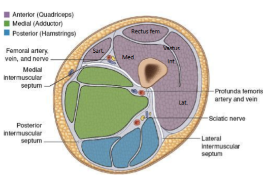
femoral n
54
opening that Allows great saphenous vein to drain
into the femoral vein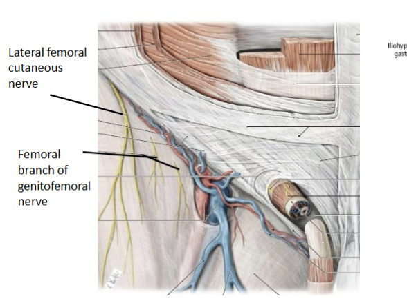
into the femoral vein
saphenous opening
55
what are the borders of the femoral triangle?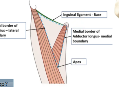
base: inguinal ligament
lat border: medial border of sartorius
med border: medial border of adductor longus
floor: iliopsoas, pectineus, adductor longus
roof: fascia lata
lat border: medial border of sartorius
med border: medial border of adductor longus
floor: iliopsoas, pectineus, adductor longus
roof: fascia lata
56
what lives in the femoral triangle?
femoral n, a, and v
empoty space (femoral canal)
deep inguinal lymph nodes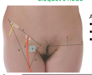
empoty space (femoral canal)
deep inguinal lymph nodes
57
what lives in the lateral femoral sheath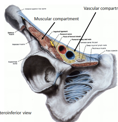
femoral a, femoral branch of gentiofemoral n
58
what lives in the middle femoral sheath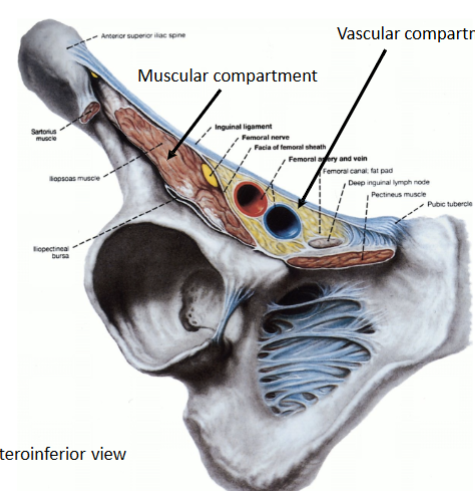
femoral v
59
a type of groin hernia where abdominal contents push through a weakness in the abdominal wall, specifically into the femoral canal. This canal is located in the upper thigh, near the groin, and contains the femoral artery, vein, and nerve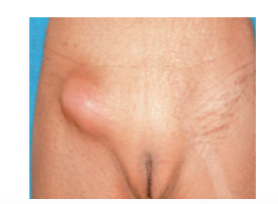
femoral hernia
60
restricted blood flow hernia where tissue may die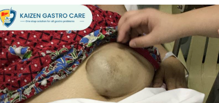
strangulated hernia
61
what can strangulate organs when perforating the femoral triangle in a femoral hernia to cause a strangulated hernia (x2)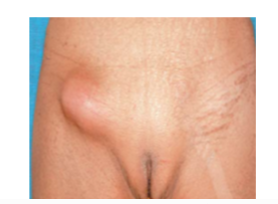
pectineal ligament, lacunar ligmament
62
what lives in the adductor canal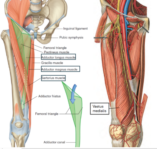
femoral a, femoral v, saphenous n, nerve to vastus medialis
63
o and i iliopsoas m.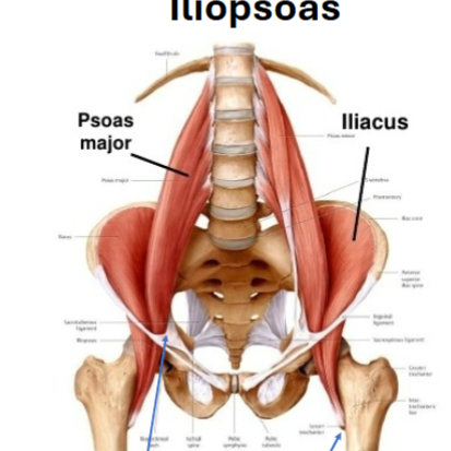
iliacus o: pelvis, psoas major o: lumbar vertebral body
i: lesser trochanter.
i: lesser trochanter.
64
iliopsoas innervation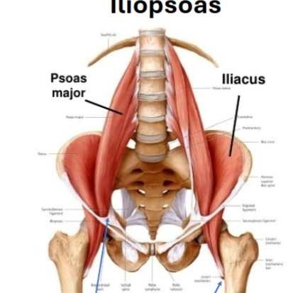
illiacus: femoral n
psoas major: ventral rami L2-L4
psoas major: ventral rami L2-L4
65
a rare and potentially serious infection characterized by a collection of pus in the psoas muscle, located in the lower back and pelvis. It can be categorized as primary, due to hematogenous or lymphatic spread from a distant site, or secondary, resulting from the spread of infection from adjacent structures.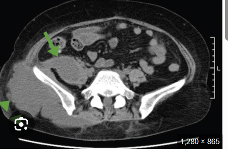
psoas abscess
66
O and sartorius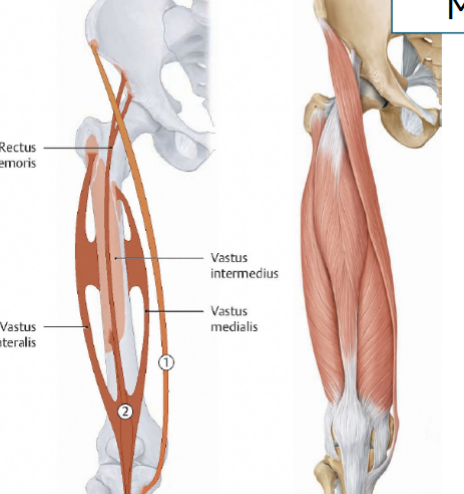
O:ASIS
I: medial tibia, pes anserinus
I: medial tibia, pes anserinus
67
quads muscles (x4)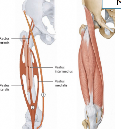
rectus femorus, vastus medialis, vastus lateralis, vastus intermedialis
68
this miscle is the first part of the quads to atrophy in knee joint disease and may result in imporoper leg tracking of the patella if weak (patellofemoral syndrome or patellar dislocation)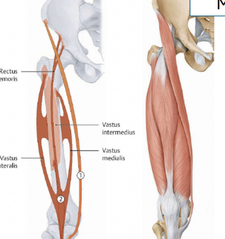
vastus medialis
69
superficial musles of the medial thigh
pectineus, adducyor longus
70
middle muscles of medial thigh
adductor brevis
71
deep muscles of the thigh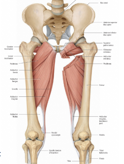
adductor magnus
72
this muscle is the most medial thigh muscle 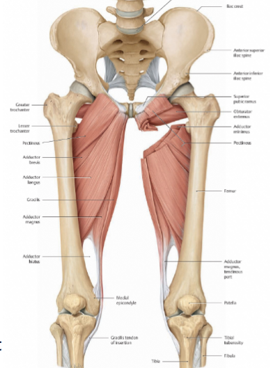
gracilis
73
innervation medial thigh muscles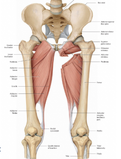
all obturator except pectineus (femoral n), and adductor magnus (sciatic n and obturator n)
74
blood supply to medial thigh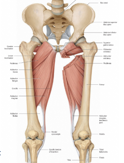
profunda femoris a
75
where is the obturator n located
obturator canal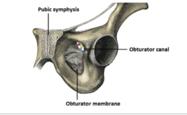
76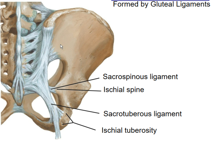
Image occlusion: name the hidden labels
77
Image occlusion: name the hidden labels
78
gluteus maximus o and i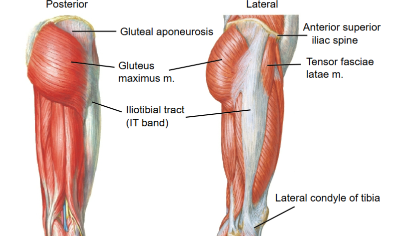
o: illium sacrum sacral tuberous ligament
i: gluteal tuberoscity, IT band
i: gluteal tuberoscity, IT band
79
action of gluteus maximus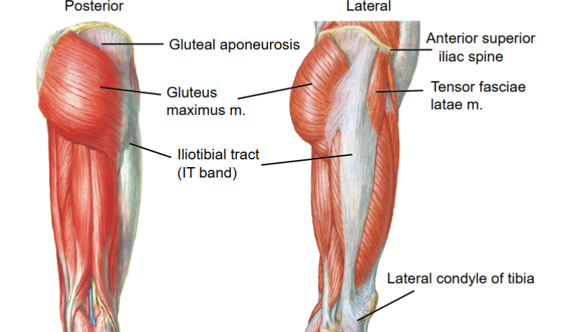
extension and lateral rotation, thigh abduction
80
innervation gluteus maximus m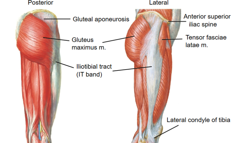
inferior gluteal n
81
o and i gluteus medius and minimus 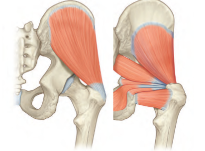
o: ilium
i: greater trochanter
i: greater trochanter
82
actions glut medius and minimus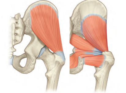
abduct thigh, medially rotate thigh
83
innervation glut medius, glut minimus, and tensor fasciae latae m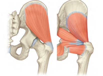
superior gluteal n
84
tensor fasciae latae m o and i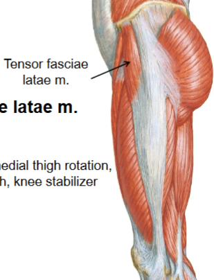
o: ASIS
i: IT band
i: IT band
85
action tensor fasciae latae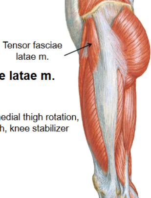
hip flexor, medial thigh rotator, abductor of thigh, knee stabilizer
86
what are the deep gluteal muscles and their action
piriformis, superior gemellus, obturator internus, inferor gemellus, quadratus femoris
lateral rotation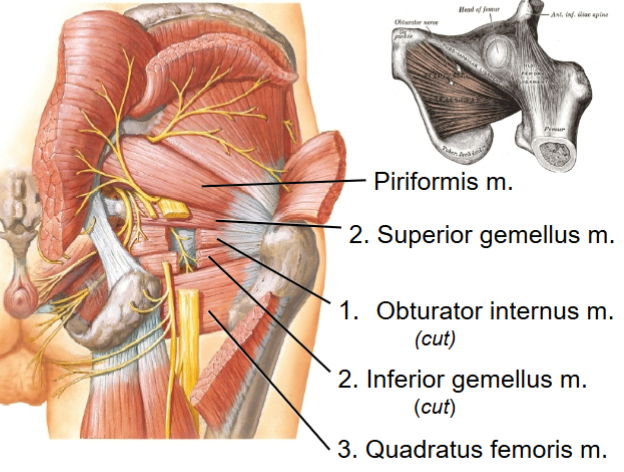
lateral rotation
87
this action of the hip stabilizes the pelvis when controlateral limb is weight bearing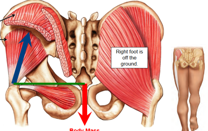
abduction
88
what is a telltale sign to damage to the inferior gluteal n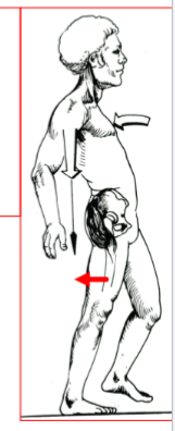
paralysis of gluteus maximus, weakness in extension on thigh, difficulkty waljing up stairs and risinh from seated position
89
telltale signs of superior gluteal n. injury
paralysis of gluteus medius, minimus
(weakness in abduction and medial rotation at hip) and tensor fascia
lata
(weakness in abduction and medial rotation at hip) and tensor fascia
lata
90
weakness in gluteus medius and minimus
demonstrated by inability to maintain a level pelvis when standing on
one foot = pelvis drops to good side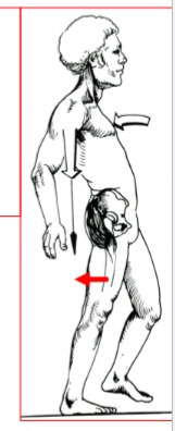
demonstrated by inability to maintain a level pelvis when standing on
one foot = pelvis drops to good side
trendelenburg sign
91
what is the blood supply to the gluteal region?
internal illiac a that branches to sup gluteal, inferior gluteal a, and internal pudendal a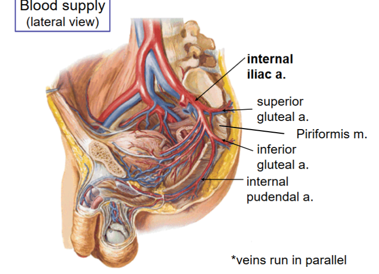
92
innervation of obturator internus and superior gemelis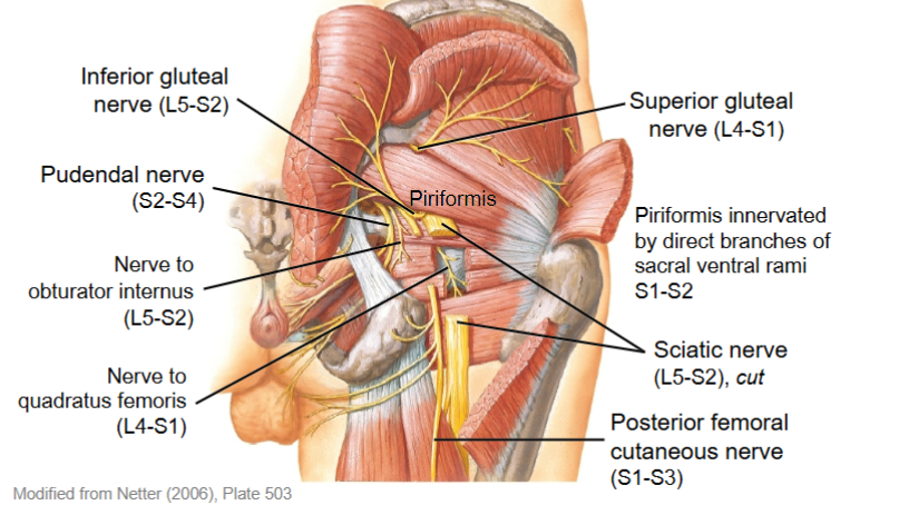
obturator internus n
93
innervation of quadratus femoris and inferior gemelis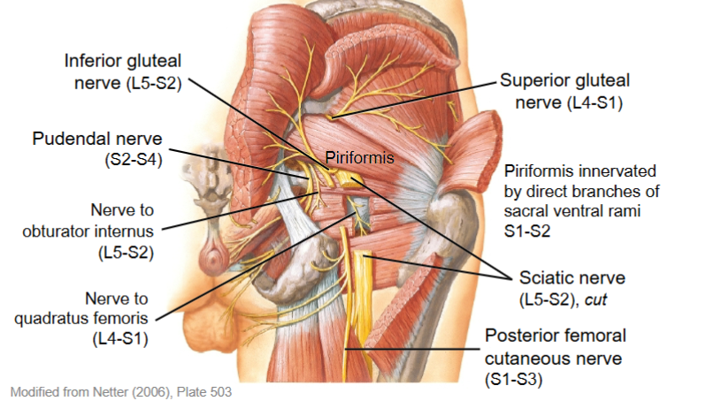
quadratus femoris n
94
compression neuropathy where trhe piriformis muscle compresses the sciatic n, shown via pain when sitting worseninh
piriformis syndrome
95
what region of the glutes does an intramuscular injection occur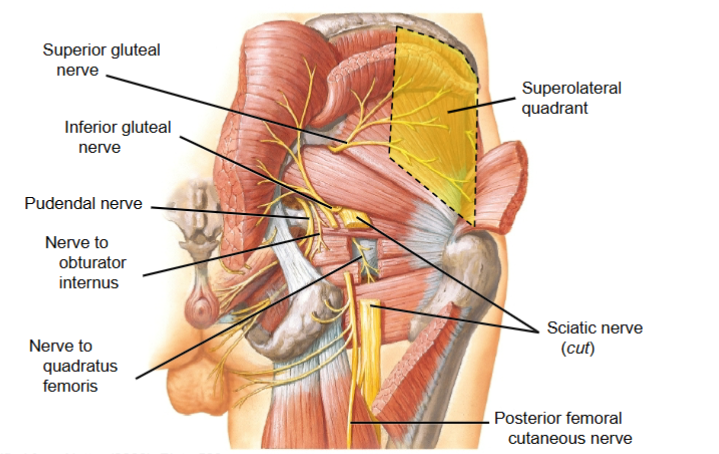
superolateral quadrant
96
o and i semitendinosus and semimembranosus m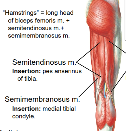
o: ischial tuberocity
i tendinosis: pes anserinus
i membranosus: medial tibial condyle
i tendinosis: pes anserinus
i membranosus: medial tibial condyle
97
hamstrig muscles (x3)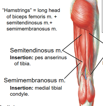
long head biceps femoris, semitendinosus, semimembranosus
98
innervation hamstrings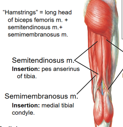
tibial n
99
actions hamstrings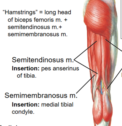
ectens the thigh, flex the leg
100
o and i biceos femoris short head
Origin: lower linea aspera and
supracondylar ridge of femur
Insertion: head of fibula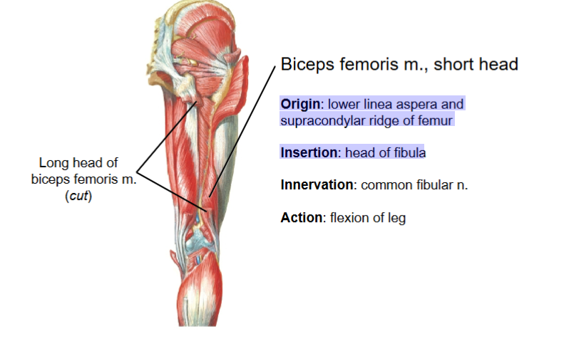
supracondylar ridge of femur
Insertion: head of fibula
101
innervation and action biceps femoris short head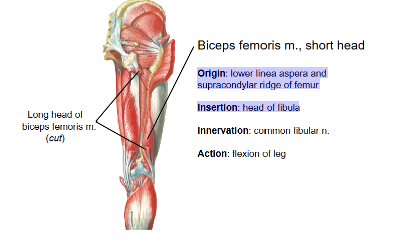
inn: common fibular n
action; flex the leg
action; flex the leg
102
what 3 muscles meet at the pes anserinus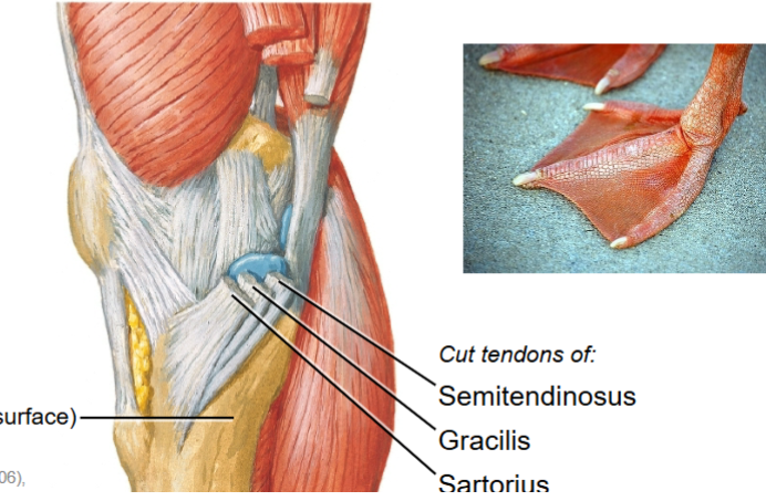
semitendinosus, gracilis, sartorius
103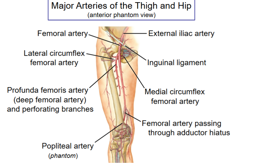
Image occlusion: name the hidden labels
104
Image occlusion: name the hidden labels
105
Image occlusion: name the hidden labels
106
Image occlusion: name the hidden labels
107
Image occlusion: name the hidden labels
108
Image occlusion: name the hidden labels
109
what lives in the popiteal fossa from deep to superior
popiteal a
popiteal v
tibial n
sural n and small saphenous v
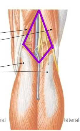
popiteal v
tibial n
sural n and small saphenous v
110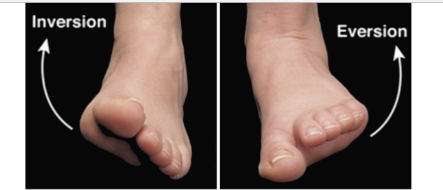
Image occlusion: name the hidden labels
111
Image occlusion: name the hidden labels
112
muscles of the anterior leg
tibialis anterior
extensor hallicis longus
extensor digitorum longus
fibularis tertias
extensor digitorum brevis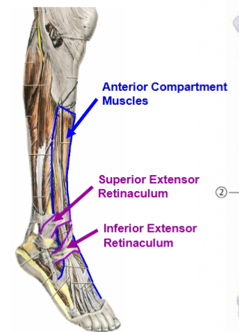
extensor hallicis longus
extensor digitorum longus
fibularis tertias
extensor digitorum brevis
113
what innervates the anterior leg muscles: TA
EHL
EDL
FT
EDB
EHL
EDL
FT
EDB
deep fibular n
114
what is the blood supply of the anterior leg m: TA
EHL
EDL
FT
EDB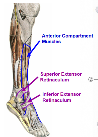
EHL
EDL
FT
EDB
anterior tibial a
115
actions of the anterior leg: TA
EHL
EDL
FT
EDB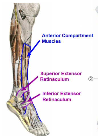
EHL
EDL
FT
EDB
dorsiflexion of ankle
116
muscles of the lat compartment of leg
fibularis longus and fibularis brevis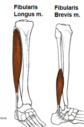
117
innervation lat leg muscles (fib long and brev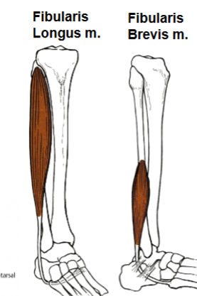
superficial fibular n
118
blood supply of lat leg m (fib long and brev)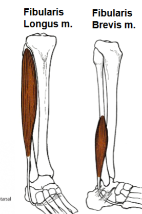
fibular a via perforating branches
119
actions of lat leg m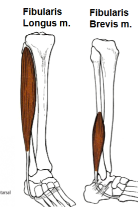
eversion of ankle, plantarflexion
120
posterior superficial leg muscles (x2)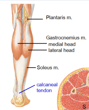
gastrocnemius and soleus
121
where do the posterior superficial leg muscles insert?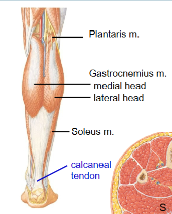
calcaneal tendon
122
what innervates the posterior compartment m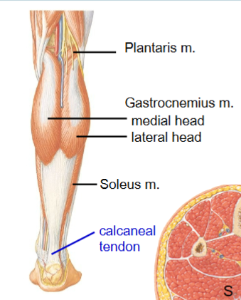
tibial n
123
blood supply to posterior superficial leg musc;es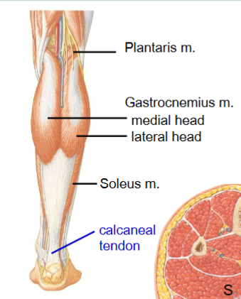
posterior tibial a and fibular a
124
deep posterior leg muscles (x4)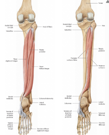
tibialis posterior, flexor digitorum longus, flexor hallucis longus, popliteus m
125
nerve that innervates posterior leg muscles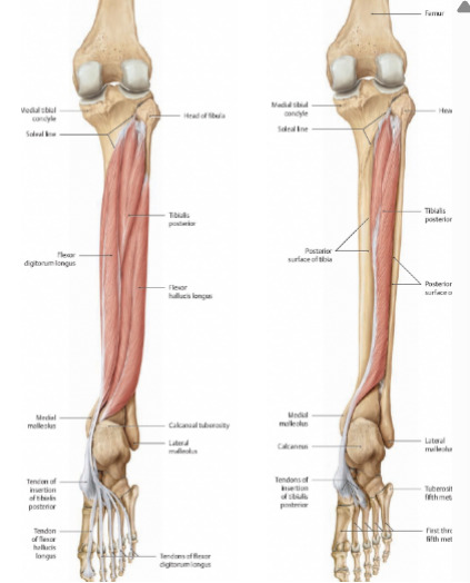
tibial n.
126
vessels of the posterior leg muscles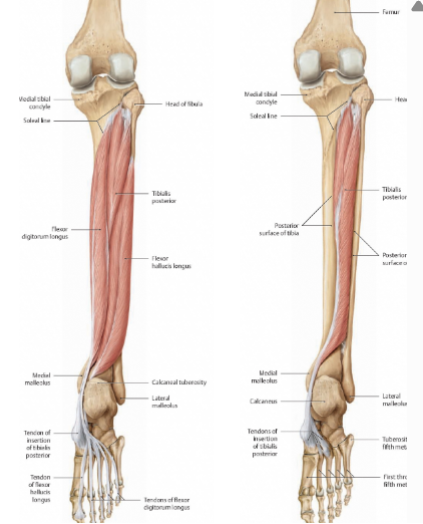
fibular and perneal a and v
posterior tibial a and v
posterior tibial a and v
127
what does the popliteal a divide to bcome?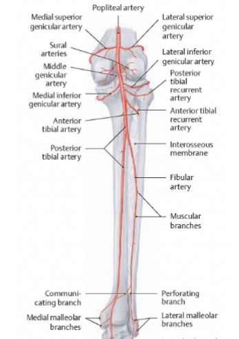
abterior and posterior tibial
128
what does the posterior tibial a divide to become?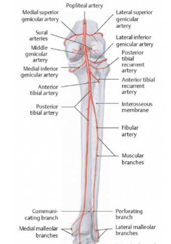
fibular, medial plantar, lateral plantar
129
what runs under the flexor retinaculum? (x6)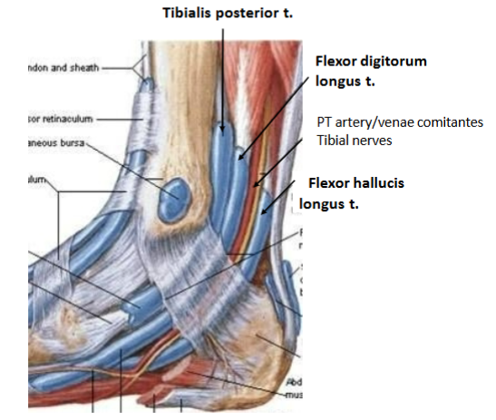
tibialis posterior, flexor digitorum longus, posterior tibial a, venae comitantes, tibial n, flexor hallucis longus
130
what does the inferior extensor retinaculum attach to?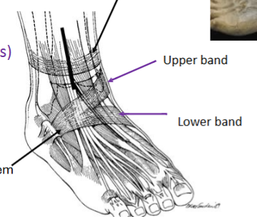
upper band: medial maleolus
lower band: navicular tuberocity
lower band: navicular tuberocity
131
what hppens in tarsal tunnel syndrome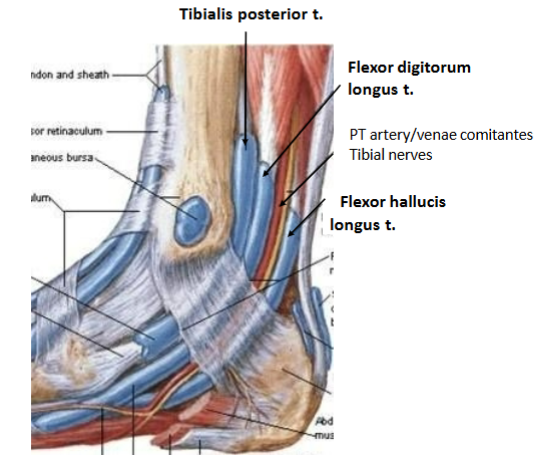
synovial sheaths of flexor retinaculum swell and compress the tibial n.
132
1st layer plantar foot muscles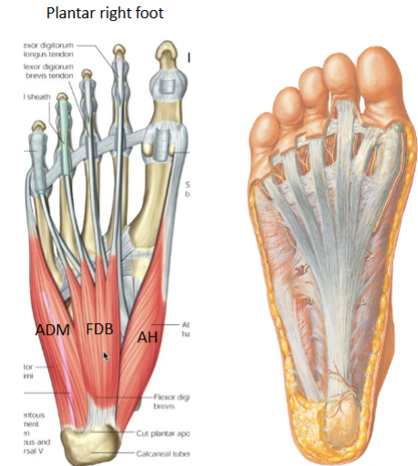
abductor digiti minimi, flexor digitorum brevis, abductor hallucis
133
what innervates the 1st layer of plantar foot m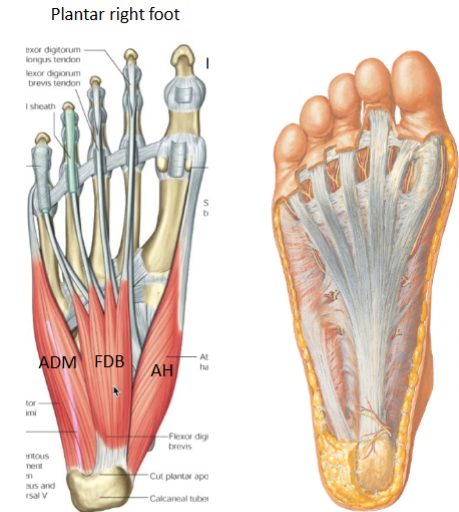
abductor digiti minimi: lateral plantar n
abductor hallucis and flexor digitorum brevis: medial plantar n
abductor hallucis and flexor digitorum brevis: medial plantar n
134
what are the 2nd layer plantar foot muscles (x2)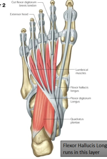
quadratus plantae and 4 lumbricals
135
innervation of 2nd layer of plantar foor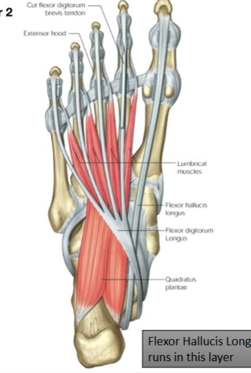
lateral plantar n but 1st lumbrical is medial plantar n
136
what is the knot of henry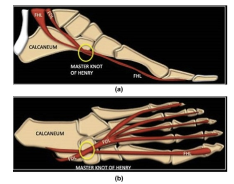
FDL and FHL tendon cross over
137
what is the 3rd layer plantar foot muscles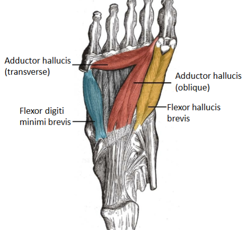
FHB, addutor hallucis, flexor digiti minimi brevis
138
what innercatres the 3rd platar foot muscles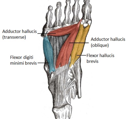
FHB medial plantar
adductor hallucis and flexor digiti minimi brevis lateral plantar n
adductor hallucis and flexor digiti minimi brevis lateral plantar n
139
what are te 4th layer plantar foor muscles 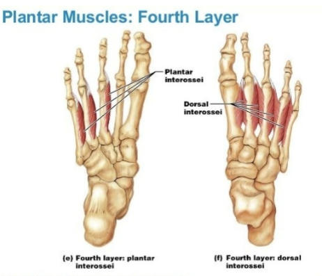
plantar interossei and dporsal interossei
140
action of plantar interossei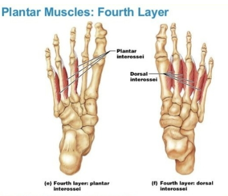
adduct toes 3-5
141
action of drsal interossei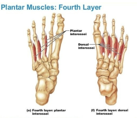
abduct toes 2-4
142
these ligaments maintain the lateral longitudinal arch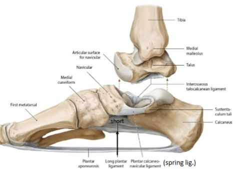
calcaneocuboid ligaments: long and short plantar ligaments
143
this ligament maintains the medial longitudinal arch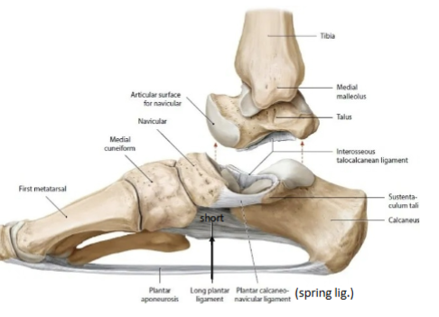
spring ligament (plantar calcaneonavicular)
144
what happens in flatfooted [people?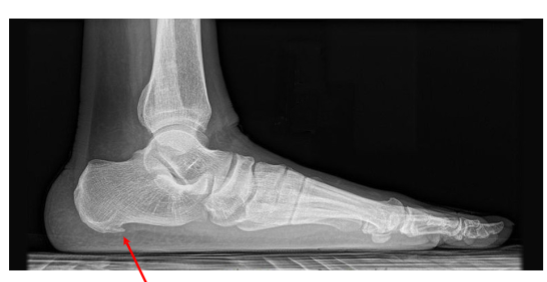
longitudinal ligament is flattened
145
heel spur, boney outgrowth from calcaneal tuberosity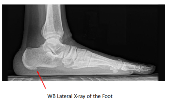
calcaneal spur
146
Patients present with symptoms
in first/second decade
• muscle atrophy causes
inverted champagne bottle
leg shape
• pes cavus; hammertoe, high
arches
• Tinglingurning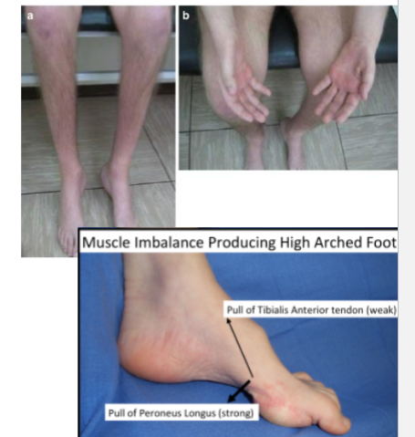
in first/second decade
• muscle atrophy causes
inverted champagne bottle
leg shape
• pes cavus; hammertoe, high
arches
• Tinglingurning
charcot marie tooth disease
147
these arteries anastosome with each other and provide blood to the talus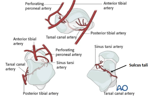
posterior tibial a, sinus tarsi a
148
what is most likely to damage the femoral a
distal femur fracture
bakers cyst
bakers cyst
149
where is the pars in the cervical region
pedicle
150
where is the pars in the thoracic and lumbar region
lamina
151
what type of joint is the hip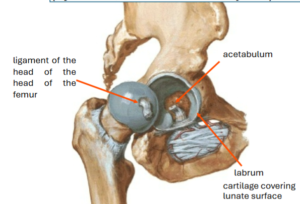
synovial ball and socket
152
this ligament supports the head of the femur so it stayts in the joinmt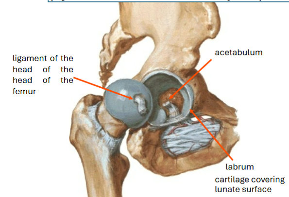
ligament of the head of the femur
153
this ligament helps support the joint capsule/hip joint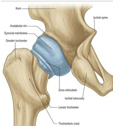
zona orbicularis
154
what arteries supply blood to the hip joint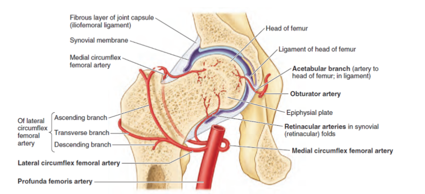
acetablar branch of obturator a: disappears into adulthood
medial circumflex femoral a with retinacular a: main blood supply to the femur head
lateral circumflex frmoral a
medial circumflex femoral a with retinacular a: main blood supply to the femur head
lateral circumflex frmoral a
155
this hip joint ligament prevemts hyperextension
iliofemoral ligament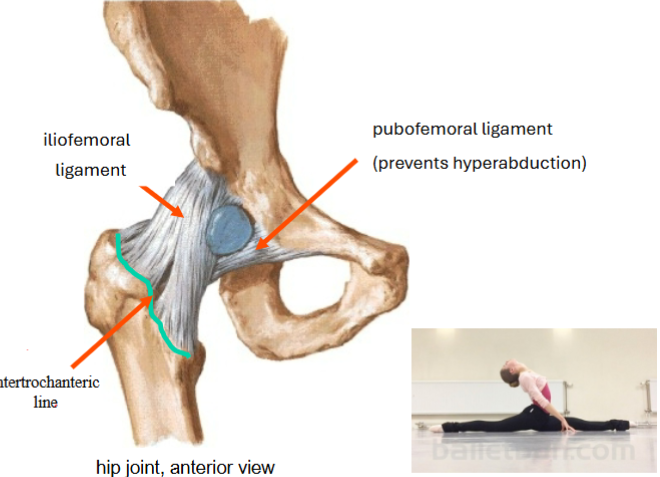
156
this hip joint ligamennt prevents hyperabduction
pubofemoral ligament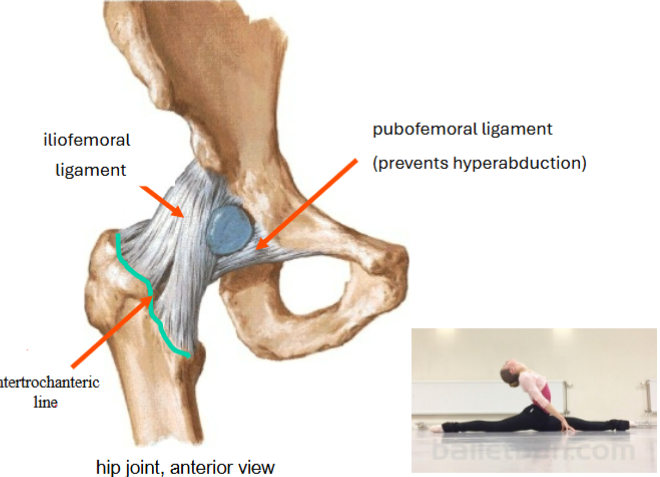
157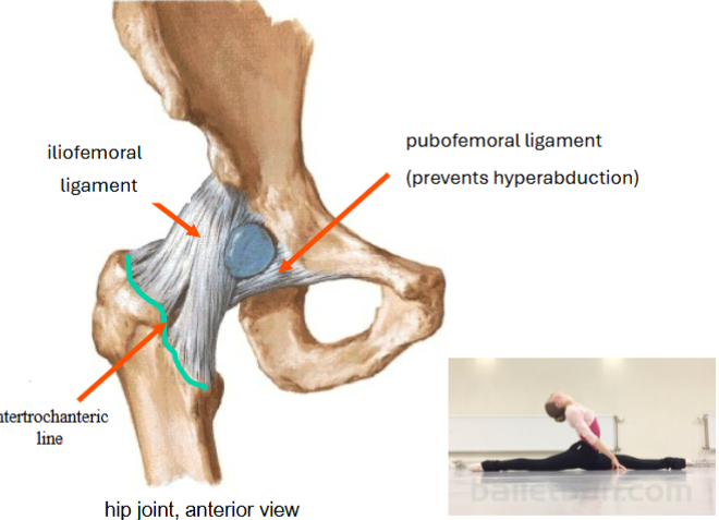
Image occlusion: name the hidden labels
158
Image occlusion: name the hidden labels
159
this hip ligament prevents hyper medial rotation of the hip
ischiofemoral ligament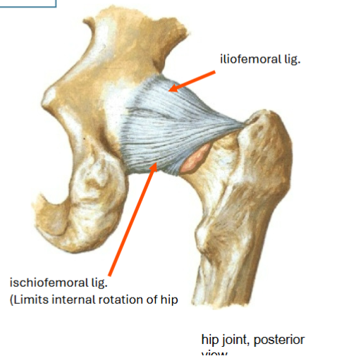
160
what is a posterior femur injury most likely to damage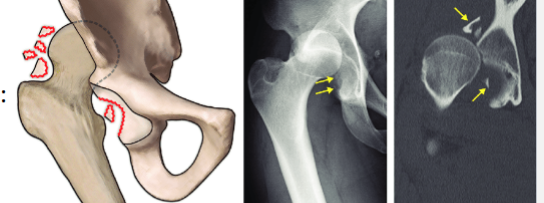
sciatic n
161
what is an anterior femoral disloaction most likely to damage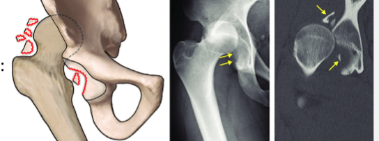
structures in the femoral traingle
162
where wll a posterior femoral disloaction leave the hipjoint?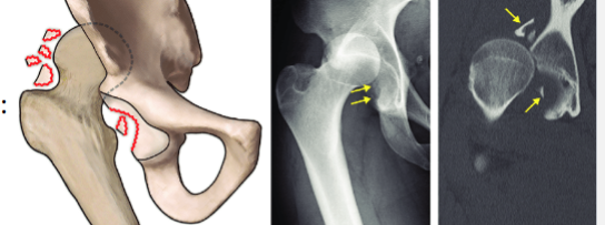
shortened limb shown
internally rotated (medial)
posterior adducted
(ShIN PAd)
internally rotated (medial)
posterior adducted
(ShIN PAd)
163
what type of joint is the knee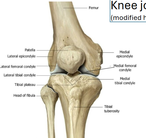
synovial hinge
164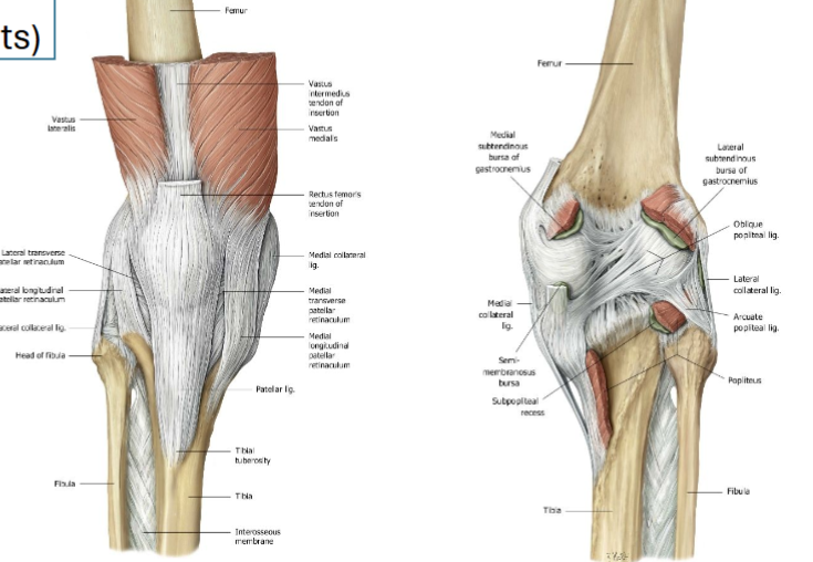
Image occlusion: name the hidden labels
165
Image occlusion: name the hidden labels
166
Image occlusion: name the hidden labels
167
Image occlusion: name the hidden labels
168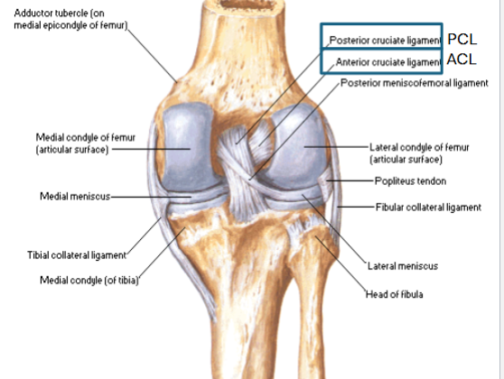
Image occlusion: name the hidden labels
169
Image occlusion: name the hidden labels
170
ligament prevents excessive anterior tibial movement 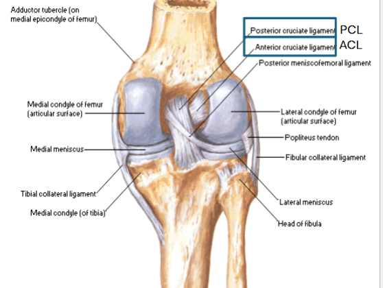
ACL
171
ligament prevents excessive posterior tibial movement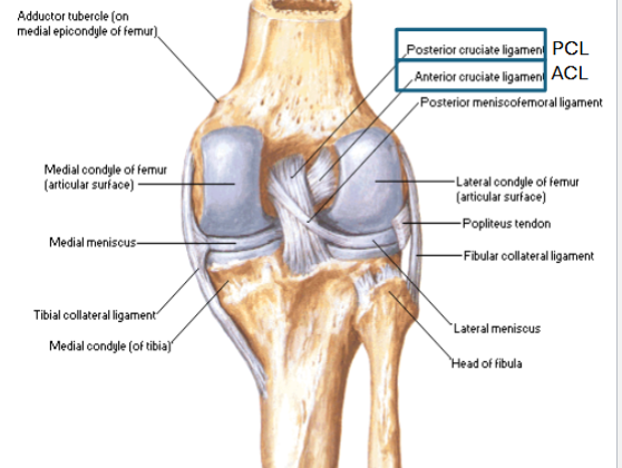
PCL
172
what can an abnormal increase of Q-angle cause?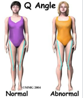
patellofemoral syndrome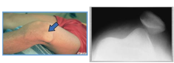
173
how do you treat patellofemral syndrome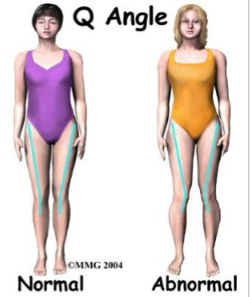
strengthen vastus mediallis
174
synovial hinge joint between talus and leg; site of dorsiflexion and plantarflexion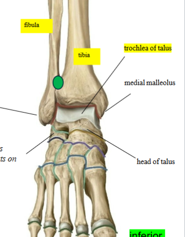
talocrural joint
175
stabilizing ligaments of the ankle (x2)
deltoid igament (MCL): strongest and most extensive
LCL: weaker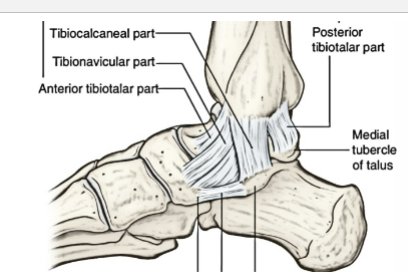
LCL: weaker
176
inversion injury to the ankle damages this ligament
calcaneo fibular and talo fibular ligaments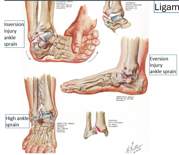
177
eversion injury to the ankle can damage these ligaments
deltoid ligament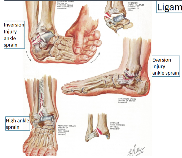
178
high ankle sprain or abduction sprain damages these ligaents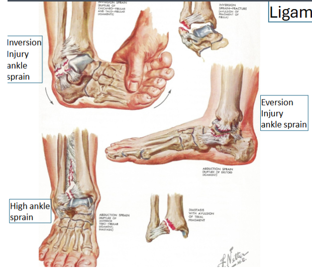
anterior tibio fibular ligament
179
what are the 7 p's of compartment syndrome
pain
parastesia
paralysis
palor
pulselessness
poikilothermia (coolness)
pressure
parastesia
paralysis
palor
pulselessness
poikilothermia (coolness)
pressure
180
an aneurysm of the popliteal artery can compress this nerve
tibial n
181
distal fractures of the femur and knee disloactions can damage this
popliteal a
182
Bursitis in the semimembranosus bursa can lead to a swelling 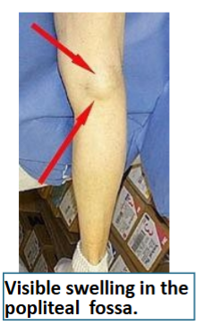
baker's cyst
183
the knee angles outward, “bowleg”; the distal tibia is angled inward (varum)
genu varum
184
the knee angles inward, “knock-knee”; the distal tibia is angled outward (valga)
genu valgum
185
Can be injured by trauma to the sacral ala, bony changes in the SI joint, local metastases or
diabetic amyotrophy. The axons from L4 in the lumbosacral trunk are destined for the sciatic
nerve, as are the axons from L5. L5 deficits will appear as an L5 radiculopathy, but will spare
the deep back muscles. L4 deficits will be restricted to the distribution of the tibal, superficial and
deep fibular nerves (sparing structures innervated by the femoral nerve)
diabetic amyotrophy. The axons from L4 in the lumbosacral trunk are destined for the sciatic
nerve, as are the axons from L5. L5 deficits will appear as an L5 radiculopathy, but will spare
the deep back muscles. L4 deficits will be restricted to the distribution of the tibal, superficial and
deep fibular nerves (sparing structures innervated by the femoral nerve)
lumbosacral trunk
186
where is sympathetic innervation from for the LL
T10-L2
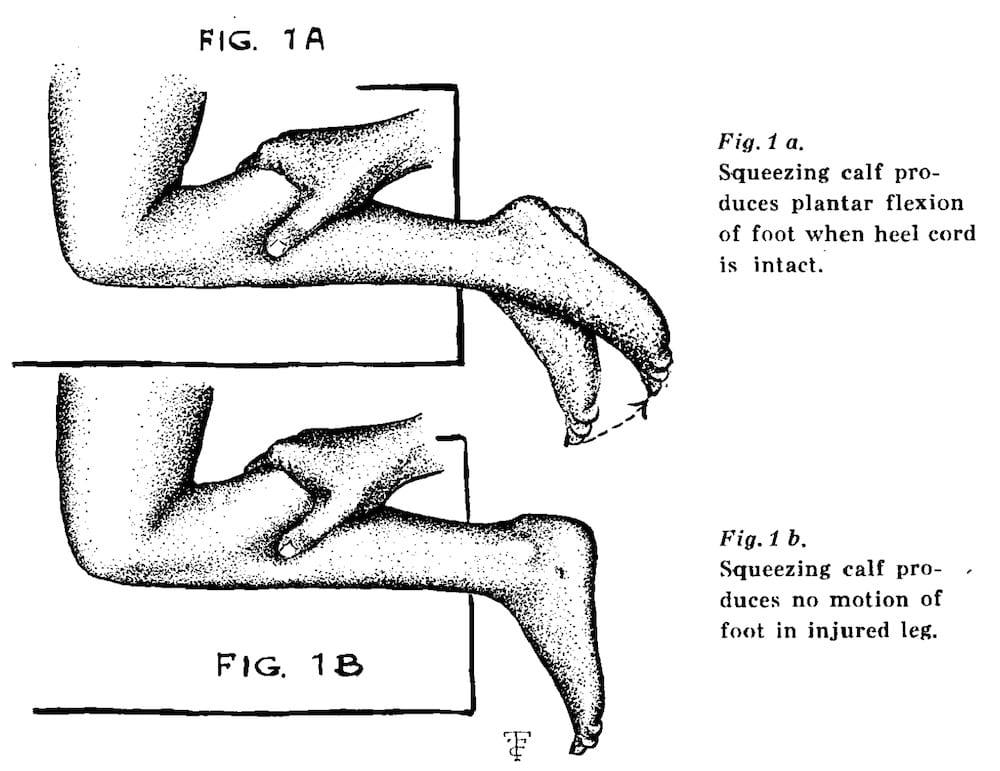
187
used to assess the integrity of the Achilles tendon; with the pt laying face down and
the foot hanging off the edge of the table, squeeze the belly of the gastrocnemius muscle – with an
intact muscle and tendon, the foot will plantarflex; if the Achilles tendon is torn, the foot will not move
the foot hanging off the edge of the table, squeeze the belly of the gastrocnemius muscle – with an
intact muscle and tendon, the foot will plantarflex; if the Achilles tendon is torn, the foot will not move
thompson test