Embryo exam 2
Study these flashcardsFree · spaced repetition · iPhone, Android, Mac & web
First 300 of 330 cards
1
what system is the first to develop and function
circulatory
2
oxygenated blood for the fetus comes from here
placenta
3
cardiac precurspr cells lie in the ________- and migrate to the __________ to form the _____________
epiblastl splabchnic mesoderm; primative heart field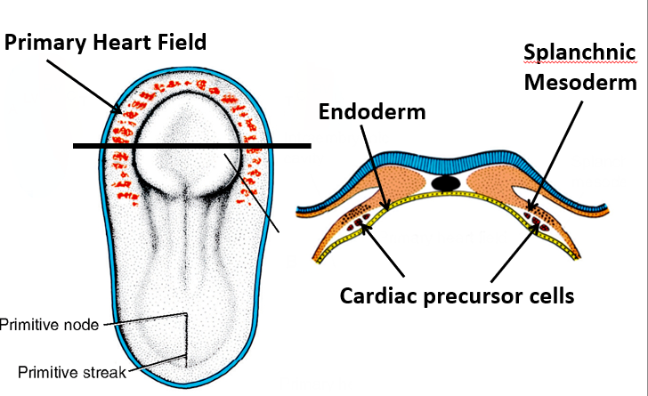
4
the secondary heart field resides in ________ and lengthens the arterial and veinous poles
visceral mesoderm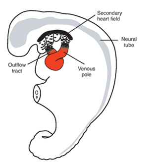
5
what brings the heart from the head to the chest cavity
lateral and cephallic-cxaudal folding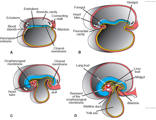
6
this if taken in pregnancy can lead to heart defects because sertatonin is essential in the embryonic folding that creates the primative heart
SSRIs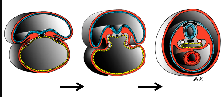
7
this suspends the transverse heart tube and later forms the transverse pericardial sinus
dorsal mesocardium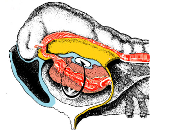
8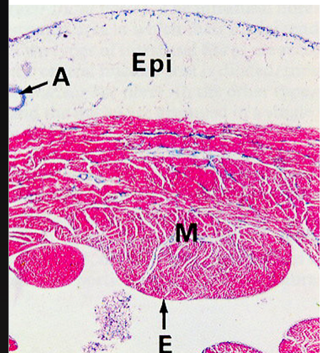
Image occlusion: name the hidden labels
9
Image occlusion: name the hidden labels
10
what does the bulbus cordis give rise to?
TA (roots of the great vessels) conus cordis (outflow ventricles), and trabeculated part of the right ventricle
11
what are the first 4 parts of the heart tube?
outflow tract
ventricle
atrium
sinus venosus
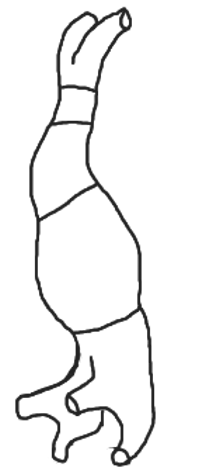
ventricle
atrium
sinus venosus
12
what day does the cardiac tube begin to bend?
day 23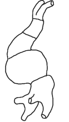
13
what makes up the cardiac tube on day 23
truncus arteriosus
bulbus cordis
ventricle
atrium
sinus venosus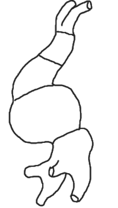
bulbus cordis
ventricle
atrium
sinus venosus
14
what day does cardiac looping complete?
day 28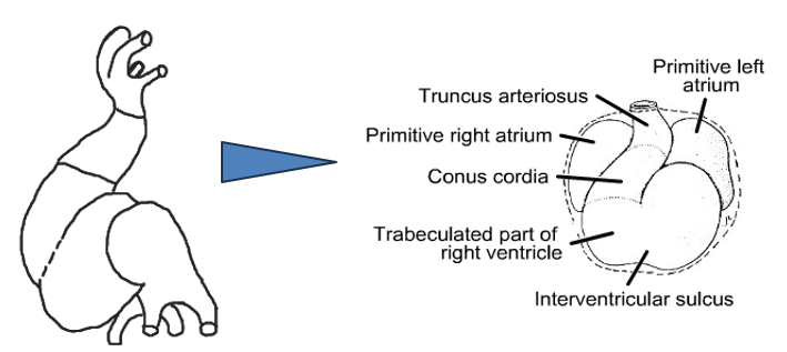
15
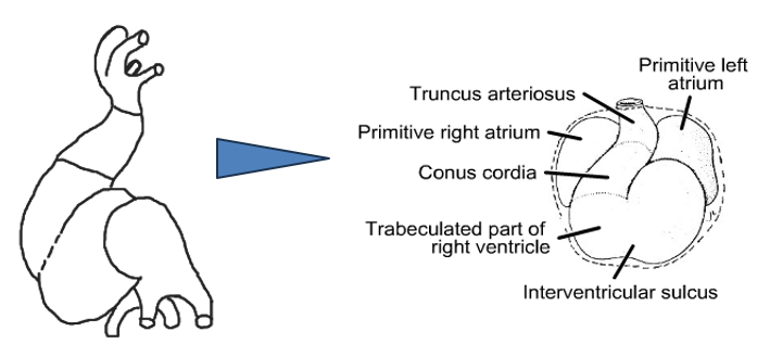
where does everything end up at the end of cardiac looping?
Bulbus cordis moves anteriorly, inferiorly and to the right
The ventricle moves anteriorly, inferiorly, and to the left
The atrium shifts posteriorly and superiorly
The ventricle moves anteriorly, inferiorly, and to the left
The atrium shifts posteriorly and superiorly
16
where does the sinus venosus receive blood from bilaterallu (x3)
right and left sinus horns that receiuve blood from the vitelline, umbillical and common cardinal vein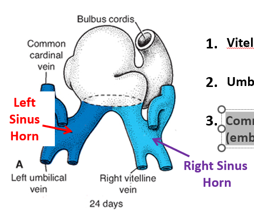
17
where does the vitelline v get its blood from
yolk sac
18
where does trhe umbilical v get its blood from
placenta
19
where does the common cardinal v get ts blood from
embryo
20
what happens at wks 4 and 5 of venous shift in the heart
right umbilical v and left viteline v obliterate, left common cardinal obliterates later making the left sinys horn less important and regressing
21
what does the right sinus horn become
sinus venarum of the right atrium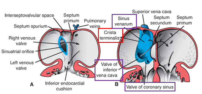
22
what does the incorporation of the right sinus horn result in
creation of 2 valves, IVC and coronary sinus valves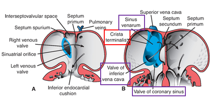
23
endocardial cushions from _____________ serve to partition rhe truncus
neural crest
24
endocardial cushions from __________ partition atria, ventricals, and AV canals
splanchnic mesoderm
25
this creates oblique blood flow once the septum primum has formed
septum secundum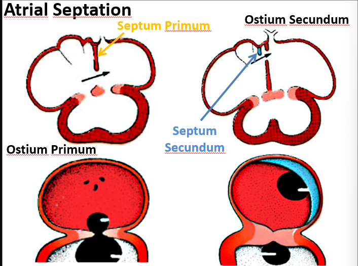
26
this allows blood flow once the septum primum forms
ostium secundum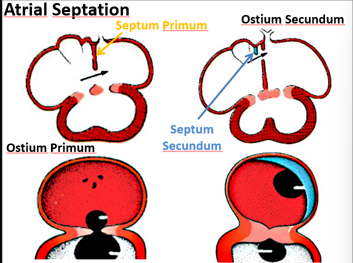
27
the septum secundum becomes this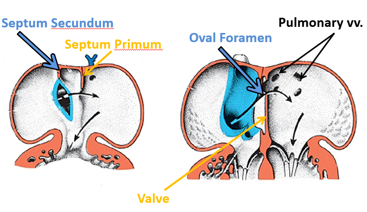
oval foramen
28
this gives rise to the pulmonary vein in the left atrium
dorsal mesocardium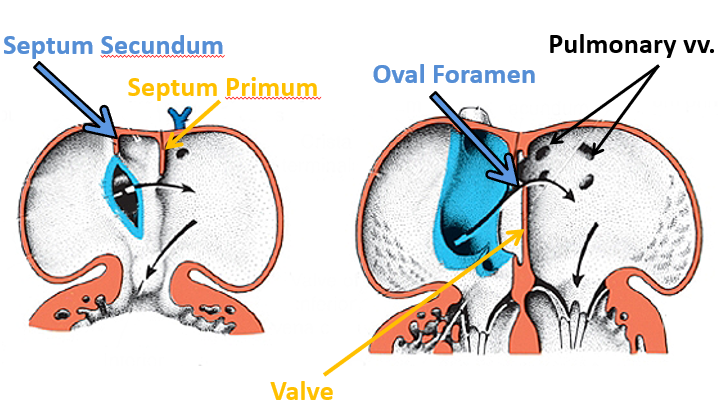
29
this forms the AV valves
mesenchymal tissue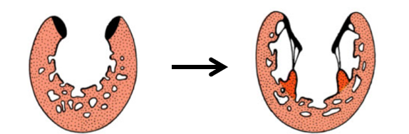
30
the primary ventricke forms (x2)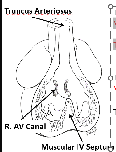
Most of the left ventricle and part of the right
The muscular interventricular septum
The muscular interventricular septum
31
the truncus arteriosus forms (x2) 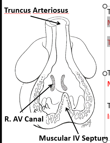
infundibulum and aortic vestibule
32
what forms the IV septum
primative ventricle and conotruncal ridges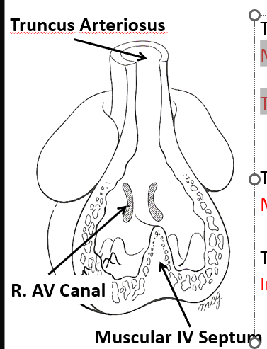
33
what are the 4 abnirmalities of teralogy of fallot
pulmonary stenosis
right hypertrophy
overlying aorta
interVentricular septal defect
(PROVe)
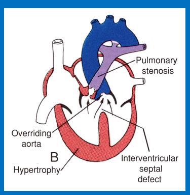
right hypertrophy
overlying aorta
interVentricular septal defect
(PROVe)
34
conducting system of the heart (x3) steps
Initially pacemaker is located in caudal left cardiac tube
Sinus venosus soon takes over its role (Sinuatrial node)
Cells from the sinus venosus and the AV canal form the AV node and bundle
Sinus venosus soon takes over its role (Sinuatrial node)
Cells from the sinus venosus and the AV canal form the AV node and bundle
35
1 atrium 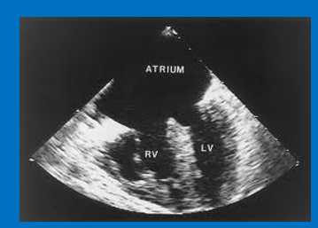
triloculare
36
the ductus arteriosus persists as this
ligamentum arteriosus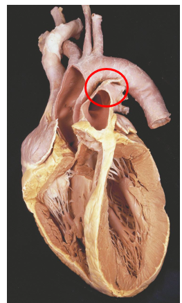
37
all arteries and lymphatics develop from this germ layer
mesoderm
38
extraembryonic mesoderm vessels form initially from _____
yolk sac
39
big 3 arterial networks and what they supply
aortic arches: head and neck, etc
vitelline: guts
umbilical: placenta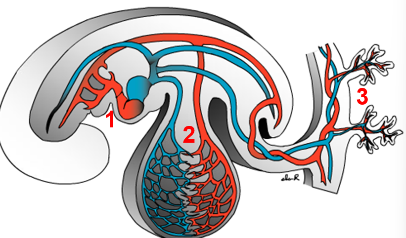
vitelline: guts
umbilical: placenta
40
what does arch IV become on the right
subclavian A
41
what does arch IV become on the left
arch of aorta
42
what does arch V become
NOTHING IT DIED
43
what does arch VI become on the right
Rught pulmonary A
44
what does arch VI become on the left
left pulmonary arch
45
what dpes the dorsal aorta become on the right
DEAD
46
what dpoes the dorsal aorta become on the left
descending aorta
47
this connects the pulmonary a to left dorsal a
ductus arteriosus
48
what does the ductus arteriosus become
ligamentum arteriosus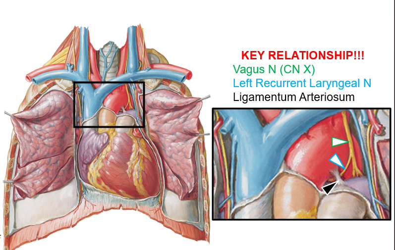
49
what runs under the ligamentum arteriosum
left recurrent laryngeal n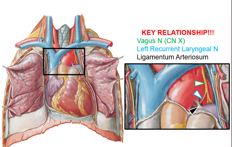
50
produces a continuous murmur head best at L sternal border
permits flow from the aorta into the pulmonary artery (PA) recirculation through lungs = L overload and enlargement
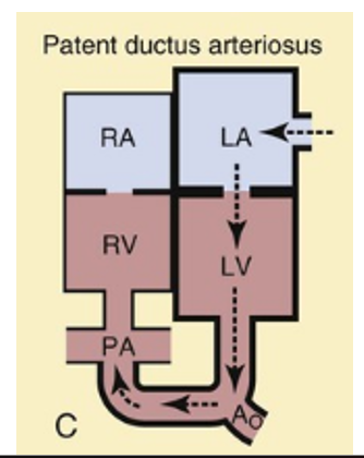
permits flow from the aorta into the pulmonary artery (PA) recirculation through lungs = L overload and enlargement
patent ductus arteriosus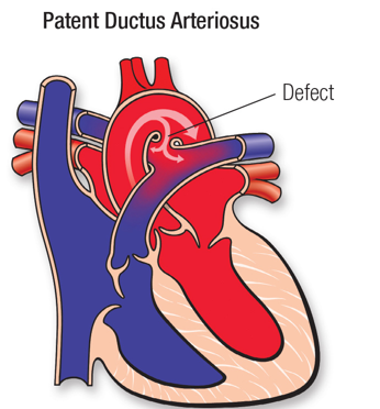
51
narrowing of the aorta
coarctation of the aorta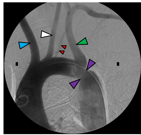
52
where does narrowing of the aorta usually occur in coarctation cases
immediately distal to left subclavian a; usually there is also a bicuspic aortic valve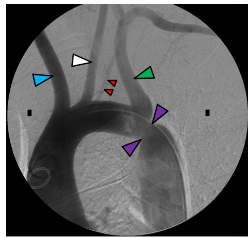
53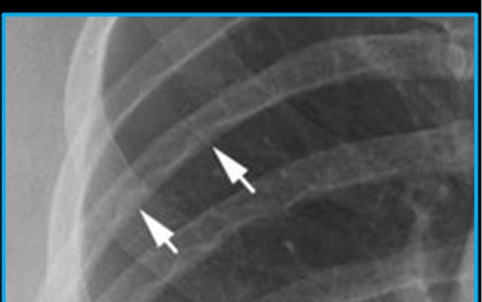
key signs and symptoms of coarctation of the aorta
54
condition where right subclavuan a hooks esophagus and makes swallowing difficult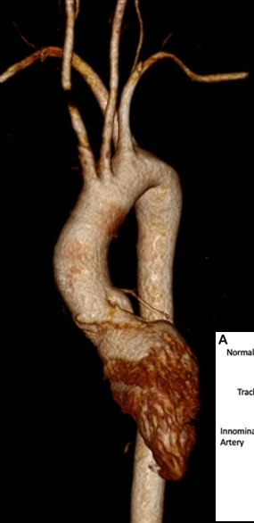
abbuut right subclavian a
55
symetric system of veins present at 5th week of development
vitelline
umbilical
cardinal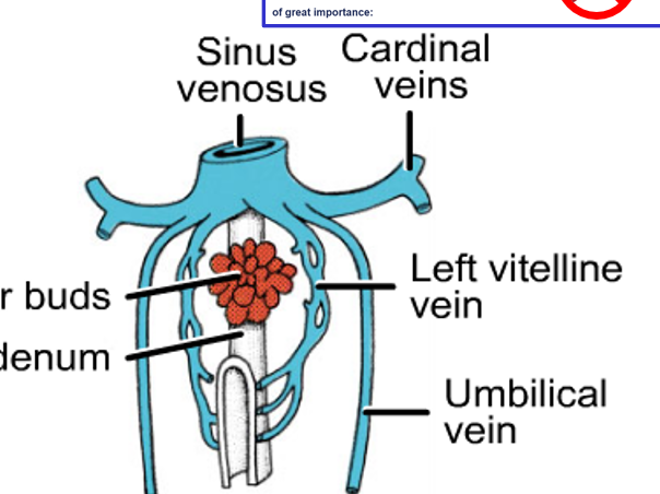
umbilical
cardinal
56
what hap[pens during shiftin of vitelline and umbilical veins
right umbilical vein and left vitellinevein are lost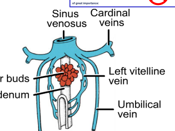
57
what do the cardinal veins becpme (x3)
SVC, azygous, IVC
58
what does the testicukar or ovarian v (R) and (L) drain into
R: inferior vena cava
L: left renal v
L: left renal v
59
what does this show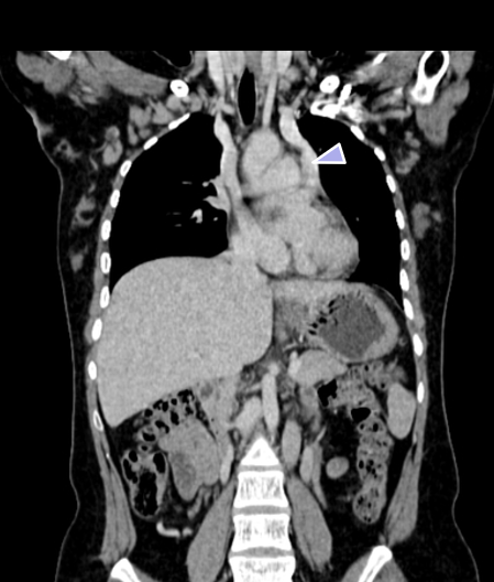
double superior vena cava
60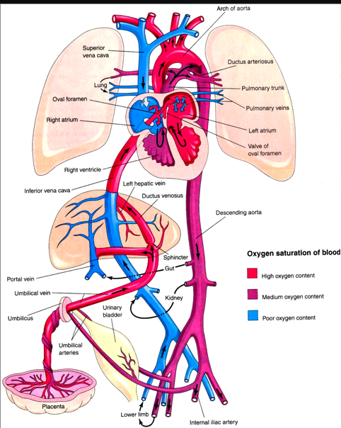
Image occlusion: name the hidden labels
61
Image occlusion: name the hidden labels
62
Image occlusion: name the hidden labels
63
Image occlusion: name the hidden labels
64
Image occlusion: name the hidden labels
65
Image occlusion: name the hidden labels
66
Image occlusion: name the hidden labels
67
Image occlusion: name the hidden labels
68
Image occlusion: name the hidden labels
69
Image occlusion: name the hidden labels
70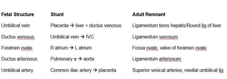
Image occlusion: name the hidden labels
71
Image occlusion: name the hidden labels
72
Image occlusion: name the hidden labels
73
Image occlusion: name the hidden labels
74
Image occlusion: name the hidden labels
75
Image occlusion: name the hidden labels
76
Image occlusion: name the hidden labels
77
Image occlusion: name the hidden labels
78
Image occlusion: name the hidden labels
79
Image occlusion: name the hidden labels
80
Image occlusion: name the hidden labels
81
what are the two important shunts that develop in the embryonic heart?
foramen ovale and ductus arteriosus
82
defects in endocardial cushions
atrial septal defects (ASD)
83
excessive absorption of septum primum or poor
development of septum secundum
development of septum secundum
ostium secundum ASD
84
lack of closure of ostium primum; occurs near AV valves
and commonly associated with valvular problems or VSD
and commonly associated with valvular problems or VSD
ostium primum ASD
85
defects in the muscular part are common but usually spontaneously resolve (not
clinical)
• defects in the membranous part are much more serious (70-80% of clinical VSDs) -
the clinical consequences depend on the size of the defect: a large defect can cause
left to right shunting of blood while a small defect may close spontaneously
clinical)
• defects in the membranous part are much more serious (70-80% of clinical VSDs) -
the clinical consequences depend on the size of the defect: a large defect can cause
left to right shunting of blood while a small defect may close spontaneously
Ventricular septal defects
86
absence of a atrial septum resulting in a three chambered
heart
heart
cor triloculare biventrivulare
87
this leads to hypertrophy of the right atrium and
ventricle and hypotrophy of the left chambers; patient typically dies shortly after birth
ventricle and hypotrophy of the left chambers; patient typically dies shortly after birth
premature closure of the oval foramen
88
the septum that normally divides the aorta and pulmonary trunk does not form properly
transposition of the great vessels
89
always have patency of the foramen ovale, VSD, underdeveloped
right ventricle and hypertrophy of the left ventricle
right ventricle and hypertrophy of the left ventricle
tricuspid valve atresia
90
improper formation of the tricuspid valve where the valves are partially
fused to the ventricular wall; part of the right ventricle becomes “atrialized”; accompanied by
tricuspid regurgitation; indicated by cyanosis and heart failure; usually accompanied by a
ASD
fused to the ventricular wall; part of the right ventricle becomes “atrialized”; accompanied by
tricuspid regurgitation; indicated by cyanosis and heart failure; usually accompanied by a
ASD
ebstein anomaly
91
poorly developed left ventricle; as blood returns from the
lungs it must pass through an ASD to the right atrium, into the right ventricle and then
through a patent ductus arterious into the systemic circulation; without surgical correction
this condition is fatal
lungs it must pass through an ASD to the right atrium, into the right ventricle and then
through a patent ductus arterious into the systemic circulation; without surgical correction
this condition is fatal
hypoblastic left heart syndrome
92
• conotruncal ridges do not fuse
• accompanied by IV septal defect
• the truncus gets blood from both ventricles
• accompanied by IV septal defect
• the truncus gets blood from both ventricles
persistent truncus arteriosus
93
dilation of lymphatic channels or congenital hypoplasia of
lymphatic channels
lymphatic channels
congenital lymphedema
94
large swellings composed of fluid filled cavities; involve jugular lymph
sacs that fail to connect lymphatic vessels
sacs that fail to connect lymphatic vessels
cystic hygroma
95
what do neural crest cells give rise to in the respiratory system (x2)
laryngeal cartilages and intramural ganglia
96
what does the splanchnic mesoderm give rise to respiratory (x3)
tracheal cartilages, smooth muscle of trachea and bronchi
97
what does the paraxial mesoderm give rise to (respiratpory)
somitomeres-skeletal muscle ofd larynx
98
what does the enfdoderm give rise to in the respiratory system
epithelial lining and glands
99
this is the name of the initial outgrowth of the respiratory system from the foregut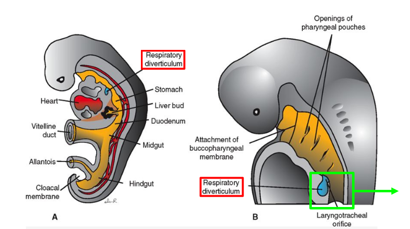
respiratory diverticulum
100
development of lungbuds: these ridges seperate the lung buds from the foregut
tracheoesophageal ridges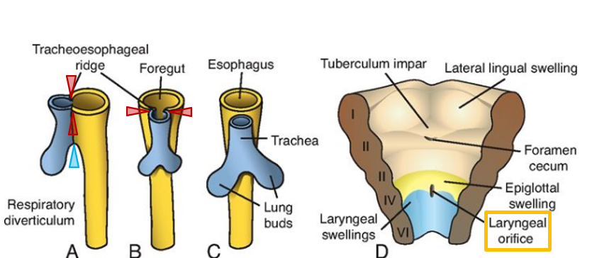
101
development of lungbuds: these seperate the trachea and the lungbuds from esophagus
esophagotracheal septum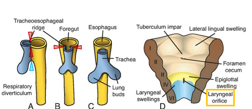
102
what is the 4th pharyngeal arch innervated by
external laryngeal n. (from superior laryngeal)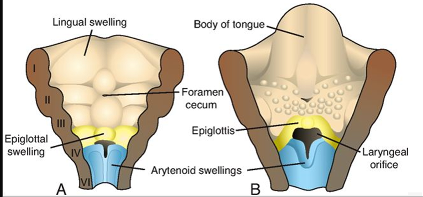
103
what is the 6th pharygeal arch innervated by?
recurrent laryngeal n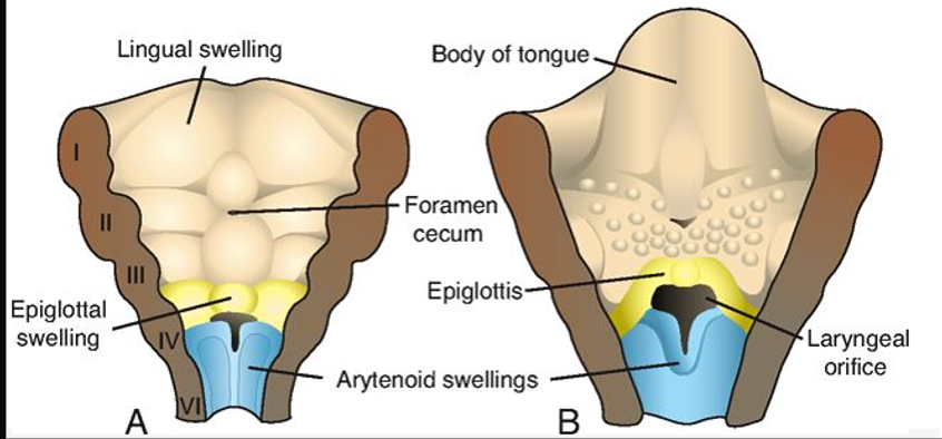
104
what are the fxs of amniotic fluid in lung development (x3)
Together with fetal (airway) secretions, keeps fetal airway patent/distended
Stimulates growth and maturation (necessary for epithelial maturation and surfactant production)
Breathing & swallowing movements (swallowing, inhale, exhale)
Stimulates growth and maturation (necessary for epithelial maturation and surfactant production)
Breathing & swallowing movements (swallowing, inhale, exhale)
105
inhalation in eutero of fetal feces
meconium aspiration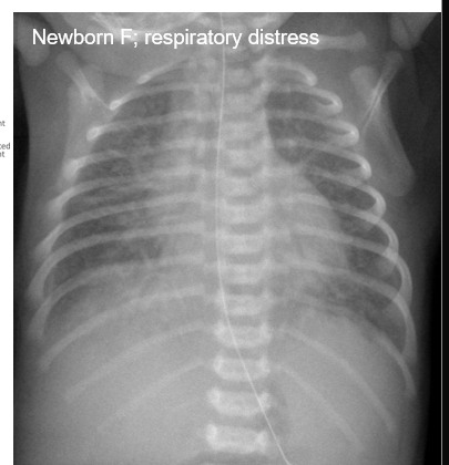
106
during weeks ~4-7
the lungs grow into the ceolom; pleural cavity
the lungs grow into the ceolom; pleural cavity
embryonic stage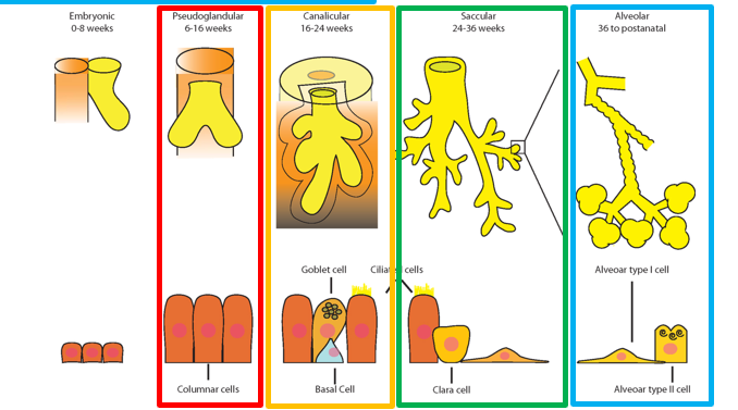
107
during weeks ~5-16
the lungs grow into the pleural cavities
terminal bronchioles but no gas exchange structures yet
the lungs grow into the pleural cavities
terminal bronchioles but no gas exchange structures yet
pseudoglandular stage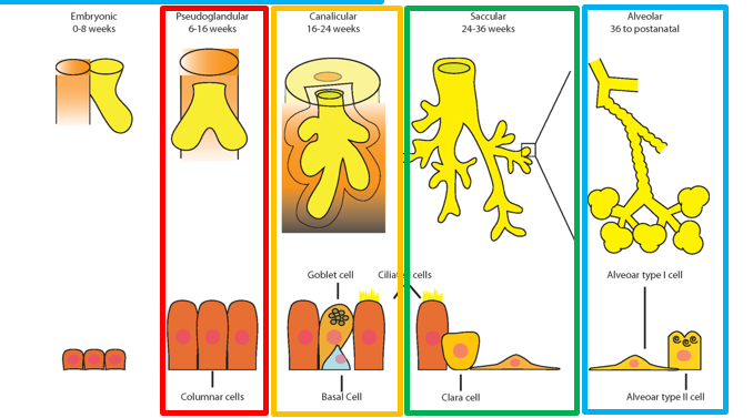
108
during weeks ~16-26
respiratory bronchioles and alveolar ducts are present
gas exchange is possible towards the end of this stage
respiratory bronchioles and alveolar ducts are present
gas exchange is possible towards the end of this stage
canalicular stage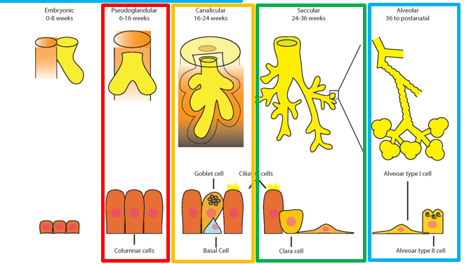
109
during weeks ~26-birth
the epithelium becomes thin to form Type I alveolar cells
capillaries bulge into alveoli
near the beginning of this stage, type II alveolar cells begin to produce surfactant
the epithelium becomes thin to form Type I alveolar cells
capillaries bulge into alveoli
near the beginning of this stage, type II alveolar cells begin to produce surfactant
terminal sac stage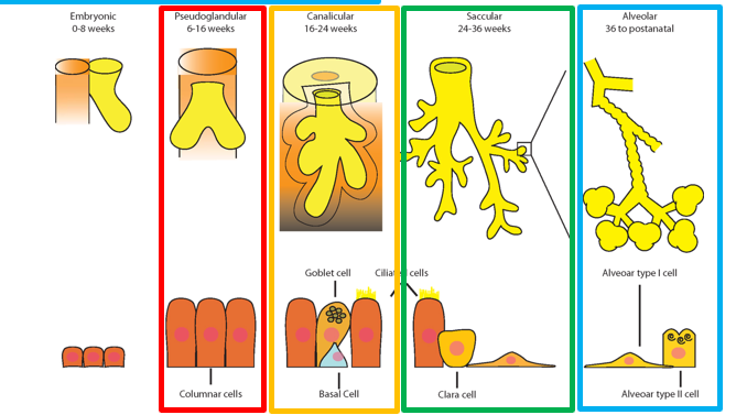
110
begins near birth and continues until approximately 8 years of age
mature alveoli form only after birth
during this period, the number and size of alveoli increases
mature alveoli form only after birth
during this period, the number and size of alveoli increases
alveolar stage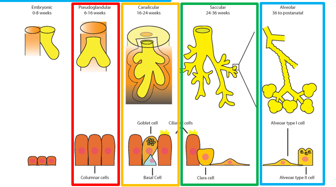
111
what is CHAOS syndrome?
congenital high airway obstruction syndrome
112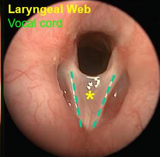
laryngeal web
113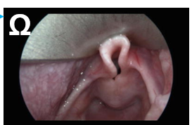
laryngomalacia
114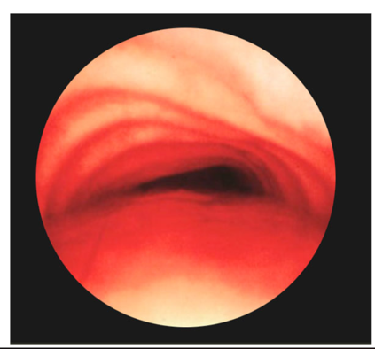
tracheomalacia
115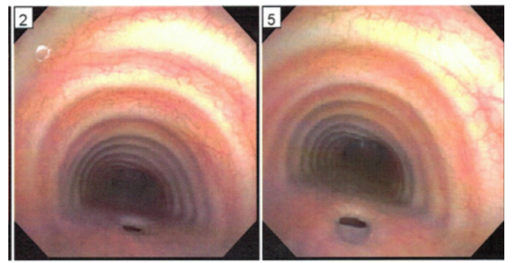
tracheoesophageal fistula
116
extra superior lobe of rught lung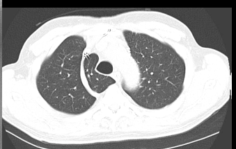
azygos lobe
117
Results from insufficient surfactant – this causes alveoli to collapse during expiration
respiratory distress syndrome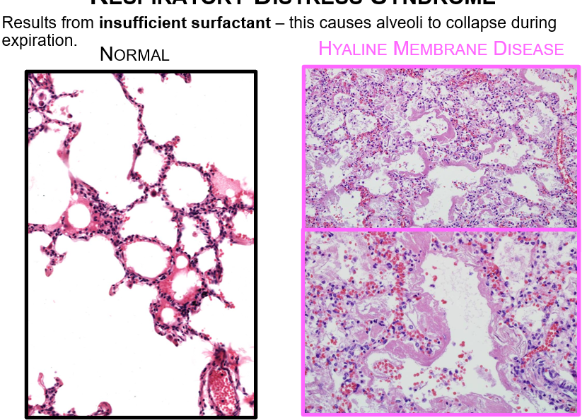
118
lobes of lung tissue arising from the trachea or esophagus
ectopic lung tissue
119
agenesis, absence of one or both lungs; congenital absence of a lung has an incidence of 0.007% (2/30,000) of autopsies (Ellis,1917)
aplasia, formation of rudimentary bronchi
hypoplasia, incomplete development of the lung
aplasia, formation of rudimentary bronchi
hypoplasia, incomplete development of the lung
hypoplasia
120
what is a VACTERL association
Vertebral defect
Anorectal malformation
Cardiac defect
Tracheal esophageal fistula
Renal defects
Limb defects
Anorectal malformation
Cardiac defect
Tracheal esophageal fistula
Renal defects
Limb defects
121
what does the endoderm form in the gut tube?
Epithelial lining from pharynx to upper 2/3 anal canal
Epithelium of accessory GI organs (liver, pancreas, etc.)
Epithelium of accessory GI organs (liver, pancreas, etc.)
122
what does the mesoderm form in the gut tube
Connective tissue (includes peritoneum) and smooth muscle
123
what does the ectoderm form in the gut tube
Epithelium at rostral and caudal ends of tube (mouth, lower 1/3 anal canal)
Neural crest (neuroectoderm) contributes to ganglia
Neural crest (neuroectoderm) contributes to ganglia
124
this provides the communucation between the gut and yolk sac while the gut tube is forming
vitelline duct (yolk stalk)
125
this artery supplies the foregut
celliac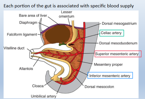
126
this artery supplies the midgut
superior mesenteric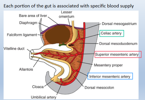
127
this artery supplies the hijdgut
inferior mesenteric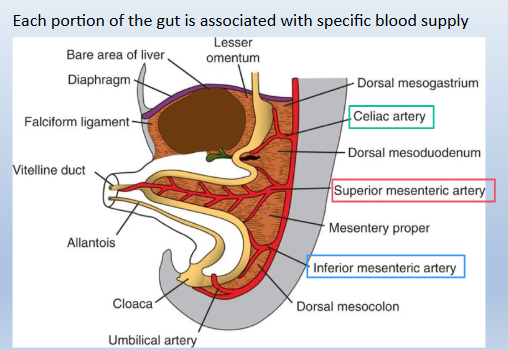
128
what happens in a tracheoesophageal fistula?
blind end of esophagus is seperate from distal end because distal end never seperated frpm trachea (85% of cases)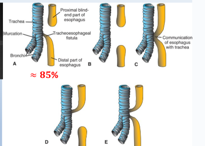
129
this causes the stomach to rotate when being formed
differential growth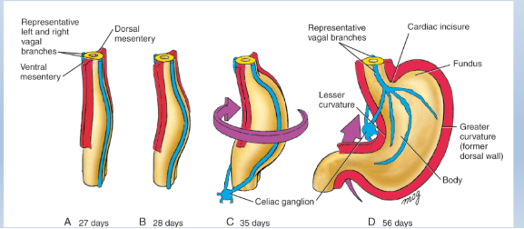
130
what innervates the stomach
vagus nerve
L vagus=anterior aspect
R vagus posterior stomach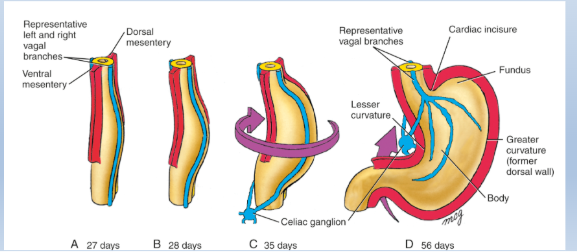
L vagus=anterior aspect
R vagus posterior stomach
131
the stomach pulls the duodenum _____________________ (c shape); it receives blood from these arteries
superiorly and to the right
celiac and mesenteric
celiac and mesenteric
132
the duodenum has a solid stage before going through thus where cavitiues are formed
recanalizing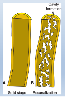
133
what is this case? what caused it possibly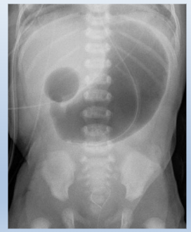
duodenal atresia
failure to recanalize
failure to recanalize
134
what outgrows from the foregut (x3)
liver bud: drom duodenum
gallbladder: from bile duct: solidify and recanalize
pancreas: ventral and dorsal buds; drom duodenum
gallbladder: from bile duct: solidify and recanalize
pancreas: ventral and dorsal buds; drom duodenum
135
this causes the posterior movement of the ventral bud of the pancreas and causes the pancreatic buds ad the ducts to fuse
growthotation of the stomach
136
a rare, congenital condition where a ring of pancreatic tissue encircles the duodenum, the first part of the small intestine. It is caused by an embryonic developmental anomaly, potentially linked to trisomy 21 (Down syndrome). Symptoms, which can appear in infancy or adulthood, include vomiting, abdominal pain, and duodenal abstructoion
annular pancreas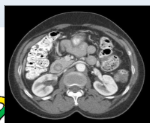
137
what forms the primary intestinal loop
rapid growth of gut tibe and mesenteru
138
what forms from the cephallic limb of midgut
distal duodenum, jejunum, part of ileum
139
what forms from the caudal limb of midgut
lowqer ileum through proximal 2/3 transverse colon
140
explain the process of umbilical herniation at 6wks
intestoinal loop rotates outward at 90* counterclockwise around SMA, retracts around 10wks at 180* counterclockwise around SMA where small intestne comes back in first than large intestine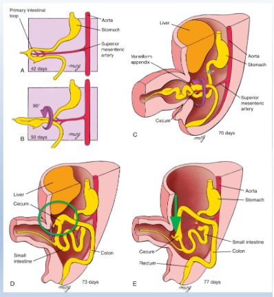
141
what is this? how does this occur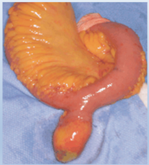
meckel's diverticulum; persistance of vitelline duct
142
what is the rule of 2s with meckel's diverticulum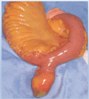
2% of population has Meckel’s diverticulum
2% of them will have sx
Typically 2 inches long
Typically 2 feet from ileocecal valve
2/3 of people who have it will have ectopic mucosa => 2 types-> gastric mucosa and pancreatic mucosa
Most commonly diagnosed at 2 y/o
2% of them will have sx
Typically 2 inches long
Typically 2 feet from ileocecal valve
2/3 of people who have it will have ectopic mucosa => 2 types-> gastric mucosa and pancreatic mucosa
Most commonly diagnosed at 2 y/o
143
what does this show; what cause sthis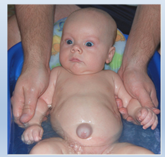
umbilical hetrnia; failure to fully return large intestine to abdominal cavity
144
what is this? what is an earrly sign of this?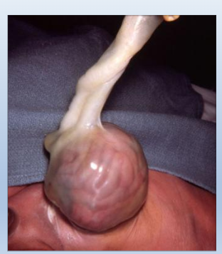
gastroschisis: a birth defect where there is a hole in the abdominal wall through which the intestines protrude into the amniotic fluid
No known cause
Cause elevated AFP
No known cause
Cause elevated AFP
145
what does this show; what are some complications of this?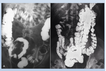
malrotation of the gut
can cause volvulus (surgically correctable) and blood restrictions
can cause volvulus (surgically correctable) and blood restrictions
146
the hindgut terminates here; where the endoderm meets ectoiderm here is referred to as this
cloaca; cloacal membrane
147
this divides the cloaca into urogenital sinus and anorectal canal; it also distinguishes urogenital membrane and anal membrane
urorectal septum
148
Lower 1/3 of anal canal derived from ectoderm at proctodeum – invaginates to form _______
anal pit
149
the degeneration of this estabilishes continuity between parts of the anal canal
anal membrane
150
rectum forms fistulka with vagina; from a blind nonfunctional anal pit
rectovaginal fistula
151
an abnormal connection between the rectum (the last part of the large intestine) and the perineum (the area between the anus and the genitals)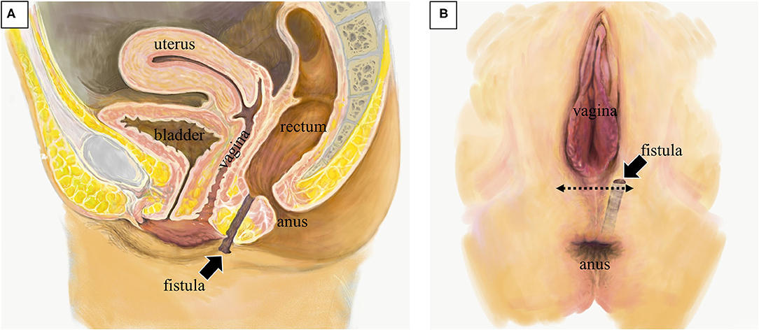
rectoperineal fistula
152
what does this show; what can cause this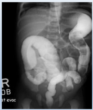
Hischprung's disease
failure of neural crest cells to migrate or imperforated anus
failure of neural crest cells to migrate or imperforated anus
153
somatic layer of lateral plate mesoderm (________ (Parietal)
splanchnic layer of lateral plate mesoderm (________) (Visceral)
splanchnic layer of lateral plate mesoderm (________) (Visceral)
GSA; GVA
154
the visceral and parietal layers of lateral plate mesoderm are continuous as this when the coelom is forming
dorsal mesentery: supports the gut
155
external heart. Skin/membrane may cover heart. Ventral wall defect. Sometimes w other midline anomalies, very low survival rate ~10%. Improper somatopleure folding.
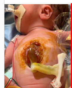
ectopic cordis
156
a severe congenital defect characterized by five central abdominal and thoracic abnormalities: a midline abdominal wall defect (often an omphalocele), a defect of the lower sternum, a deficient anterior diaphragm, a defect in the diaphragmatic pericardium, and a malformed heart, often found outside the chest (ectopia cordis).
pantology of Cantrell
157
herniation of guts through an enlarged umbilical ring; covered by amnion; high rate of mortality (25%), children also usually have cardiac and neural tube defects
-Organs within sac protrube into umbilicus. Seen often w chromosomal disorders. HIGH mortality. Midline defect!!!!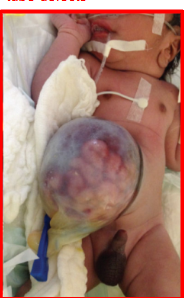
-Organs within sac protrube into umbilicus. Seen often w chromosomal disorders. HIGH mortality. Midline defect!!!!
omphalocele
158
improper closing of body wall; herniation of guts through a body wall defect INTO the amniotic cavity – viscera can be damaged by exposure to amniotic fluid, can be complicated by volvulus
- wall did not close, organs herniate into amniotic fluid (caustic to intestine!) Usually right of midline defect. High/low maternal age, substance exposure. No clear genetic association. Volvulos common. Try to fix w Silo operation= plastic sleeve around intestine (still outside cavity) and slowly return into cavity. Still high mortality.
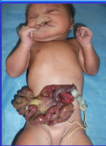
- wall did not close, organs herniate into amniotic fluid (caustic to intestine!) Usually right of midline defect. High/low maternal age, substance exposure. No clear genetic association. Volvulos common. Try to fix w Silo operation= plastic sleeve around intestine (still outside cavity) and slowly return into cavity. Still high mortality.
gastroschisis
159
what is this? what causes it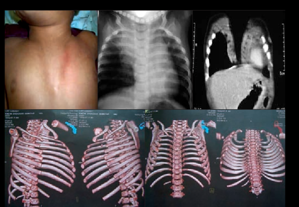
chest wall deformuty; failure of sternal bar to fuse durin gestation
160
partitioning of the coelom: this derived from splanchnic mesoderm is part of the developong diaphragm and seperates the heart and liver
septum transversum
161
what does the septum transversum become
central tendon of diaphragm
162
these are opening that conect with the abdominal cavity on either side of the foregut. If they do not close, a certain type of diaphragmatic hernia can occur
pericardoperitoneal canals
163
the growth of the lungs form these; the fusion of them forms three coimparents (x3)
pleuropericardial membranes
com[parments: pericardial sac, R pleureal cavity, L pleural cavity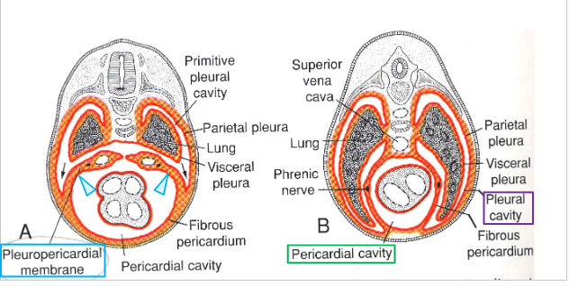
com[parments: pericardial sac, R pleureal cavity, L pleural cavity
164
the pleuropericardial membranes contain these which are covered in somatic mesoderm
phrenic nerve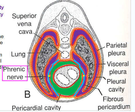
165
this permits comunication from the pleural cavities to the peritoneal cavity
pericardioperitoneal canals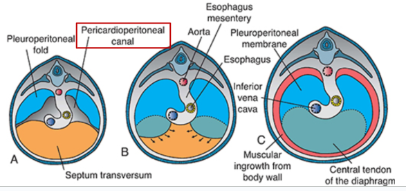
166
common; defect in the right crus and/or short esophagus
the upper part of the stomach is retained in the thorax and the stomach is constricted at the level of the diaphragm
the upper part of the stomach is retained in the thorax and the stomach is constricted at the level of the diaphragm
esophageal hernia
167
rare; opening at costosternal junction (foramen of Morgagni: between septum transversum and costal margin of diaphragm)
90% on the right side
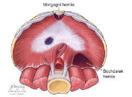
90% on the right side
hernia of morgagni
168
a developmental defect in the posterolateral diaphragm that permits herniation of abdominal contents into the thorax.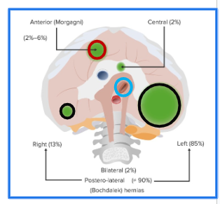
bockdeleck hernia
169
dorsal mesentery is these types of mesoderm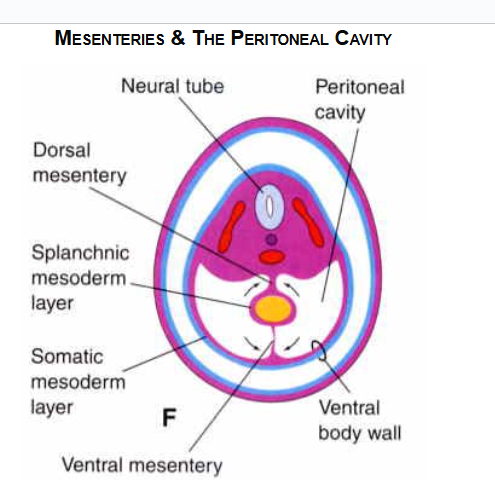
splanchic and somatic.
170
grwoth of the liver divides the mesentery into...(x2)
Lesser omentum: hepatogastric, hepatoduodenal ligament
Falciform ligament: carries the left umbilical vein towards liver
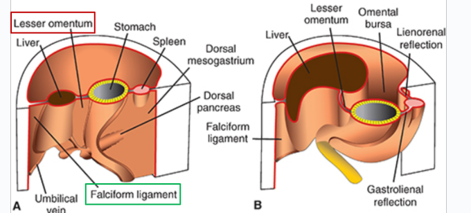
Falciform ligament: carries the left umbilical vein towards liver
171
these ligaments guard bare area of liver
coronary and triangular
172
these mesenteries are the splenic vessels and tail of pancreas
lineorenal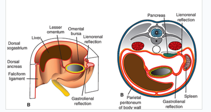
173
these mesenteries are the short gastroic arteries and left gastropiplooic a
gastrolineal 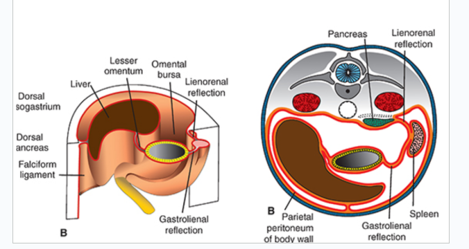
174
the greater omentum is divided (x3)
gastrophrenic
gastrosplenic
gastrocolic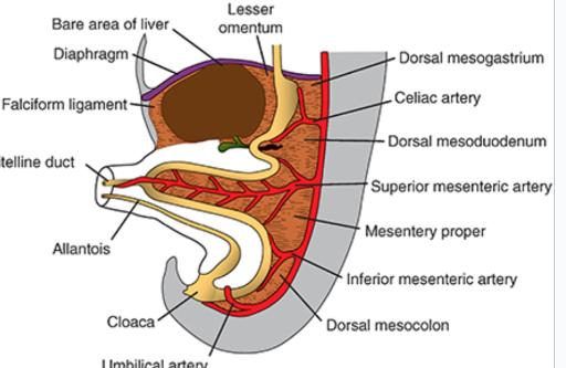
gastrosplenic
gastrocolic
175
this is the bursa behind the stomach border (greater omentum)
lesser sac
lesser sac
omental bursa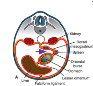
176
a medical condition where a part of the intestine twists around itself, obstructing the flow of food and waste
-Twists around mesentery or d/t lack of mesentery. Gangrenous bowel, perforated bowel. Can see intermittent pain from twist and untrist 🡪harder to diagnose.
-Twists around mesentery or d/t lack of mesentery. Gangrenous bowel, perforated bowel. Can see intermittent pain from twist and untrist 🡪harder to diagnose.
volvulus
177
multiple small splenic nodules without a main body
polysplenia.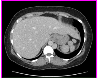
178
what does this show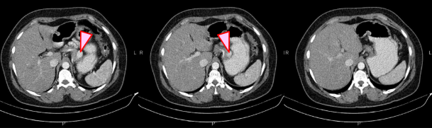
accessory spleen
179
what organs are peritoneal
stomacj, spleen, jejunum, ileum, transverse and sigmoid colon. part of f=duodenu,m
180
what organs are primary retroperitoneal
kidneys, ureters, bladder, aorta
181
what organs are secondary retroperitoneal
duodenum (parts 2-4)
ascending + descending colon
rectum
pancreas
gallbladder and liver
ascending + descending colon
rectum
pancreas
gallbladder and liver
182
has a mesenteru
peritoneal
183
never had a mesentery
primary retroperitoneal
184
had a mesentery in embryo but gone
secondary retroperitoneal
185
this is the longitudinal elevation of intermediate mesoderm that later develkops parts of the urinary and reproductive system
urogenital ridge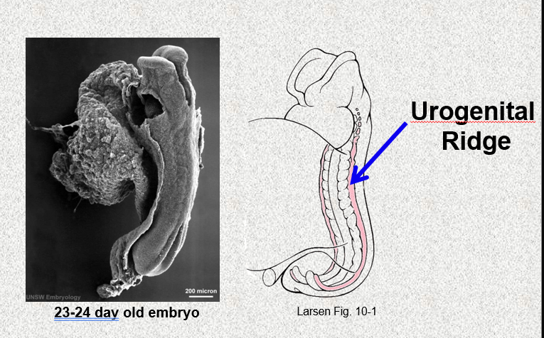
186
what does the urogenital ridge give rise to (x2)
nephrogenic ccord
gonadal ridge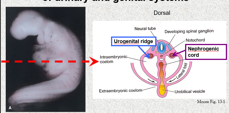
gonadal ridge
187
what is the timeline of kidney development (x3)
pronephros: ridimentary and nonfunctional
mesonephros: function for a short time in early fetal period
metanephros: permanant kidney
mesonephros: function for a short time in early fetal period
metanephros: permanant kidney
188
what are the first set of kidneys called? wjere do they forst appear?
pronephrol; nephrogenic cord of cervocal region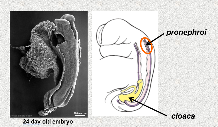
189
what do the mesonephroi develop from? where do they develop?
intermediate mesoderm; upper thpracic to upper lumbar region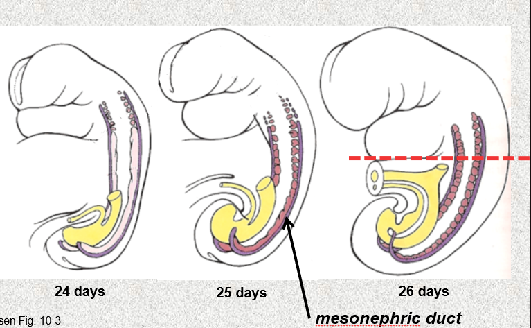
190
what makes up the promordium of the permanent kidney
ureteric bud and metanephric blastema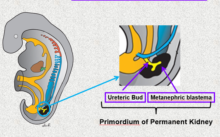
191
where do the metanephroi develop? what sprouts from it?
caudal region of the embryo; mesophrenic duct that becomes parts of the adult kidney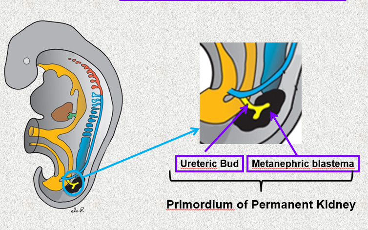
192
what does the ureteric bud give rise to? (5)
urteter, renal pelvis, minor and major xalyces, collecting tubles
193
the metanephric blastema will give rise to this...
nephrons
194
how do the kidneys change position during development
ascend due to the grpwth pf the vaudal embryo
rotate 90* medially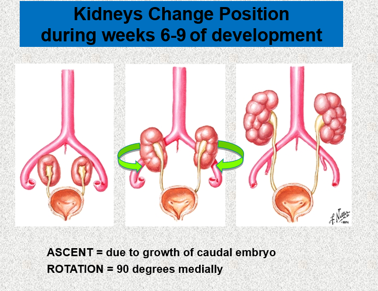
rotate 90* medially
195
what are all the potential blood supplies to the kidney as it develops
internal illiac a
common iliac a
distal aorta
abdominal a
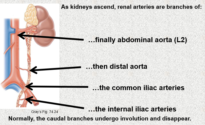
common iliac a
distal aorta
abdominal a
196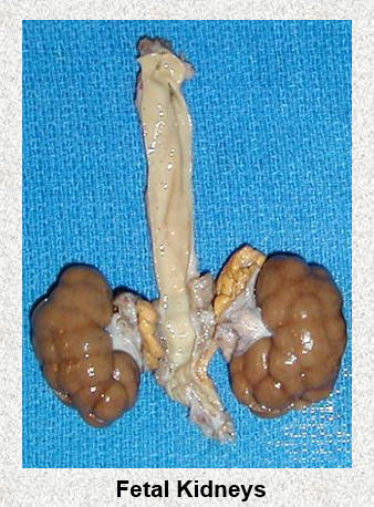
Image occlusion: name the hidden labels
197
If a kidney does not ascend (ectopic) where will it most likely be found?
pelvis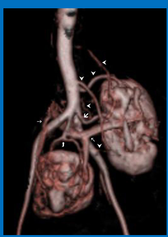
198
. If there is a failure of rotation, the hilum faces _____________, which is the embryonic position. If over rotation occurs, then the hilum will face _________-
anteriorly; posteriorly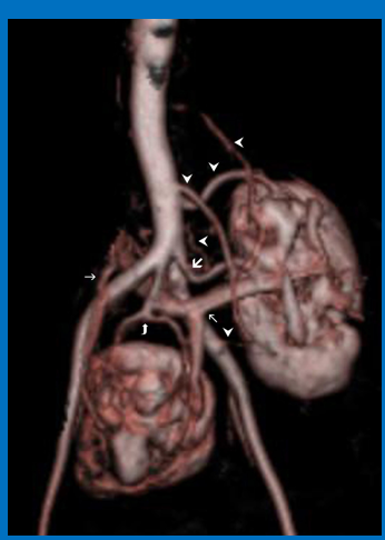
199
missing kidney
renal agenesis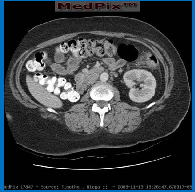
200
an additional, fully developed kidney that is present in addition to the two normal kidneys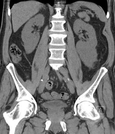
supernymerary kidney
201
a condition where a ureter, the tube that carries urine from the kidney to the bladder, is abnormally wide and cannot function properly.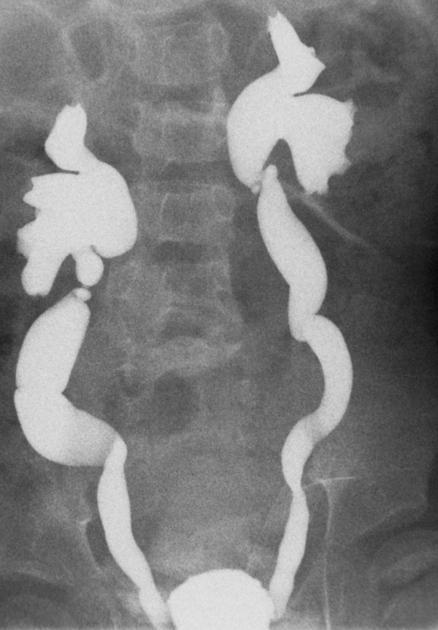
megaloureter
202
a congenital anomaly where the ureter, the tube that carries urine from the kidney to the bladder, splits into two separate branches before reaching the bladder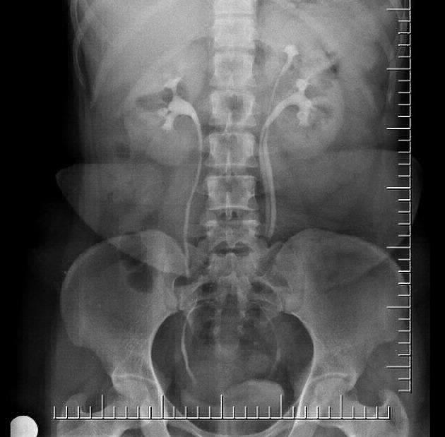
bifid ureter
203
what is this? what did they get stuck on causing this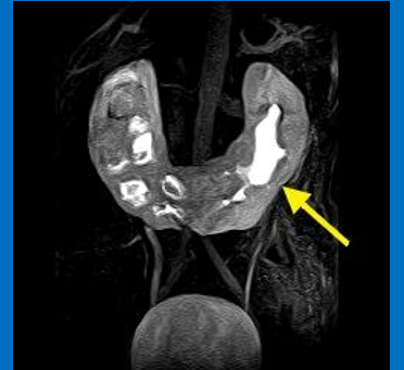
horseshoe kidney; inferior mesenteric a
204
this divides the cloaca; what segments is the cloaca divided into?
urorectal septum; anal canal and urogenital sinus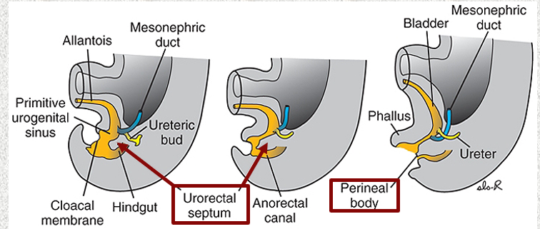
205
what are the 3 parts of the urogenital sinus
vesicle part, pelvic part, phallic part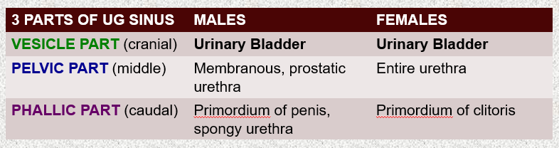
206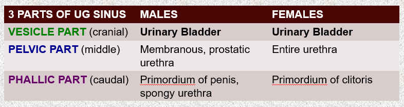
Image occlusion: name the hidden labels
207
Image occlusion: name the hidden labels
208
Image occlusion: name the hidden labels
209
Image occlusion: name the hidden labels
210
Image occlusion: name the hidden labels
211
Image occlusion: name the hidden labels
212
Confluence of urethra, vagina, and rectum
into a common channel
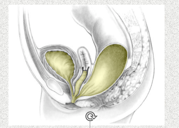
into a common channel
persistent cloaca
213
what does the allantois become in the yrorectal region?
urachus
214
a rare, fluid-filled sac in the midline of the lower abdomen, formed from a remnant of a fetal structure called the urachus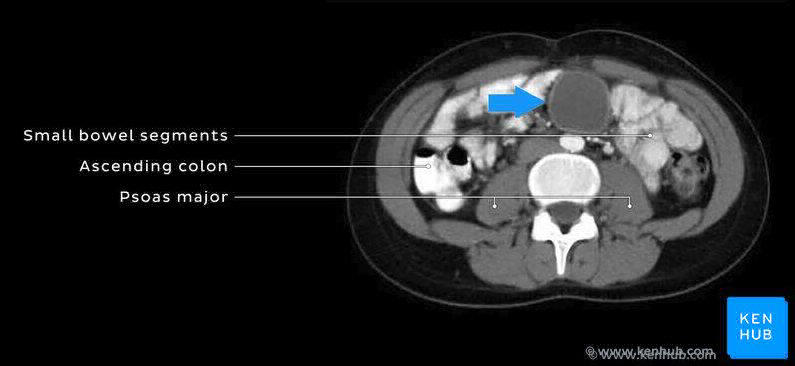
urachal cyst
215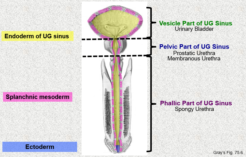
Image occlusion: name the hidden labels
216
Image occlusion: name the hidden labels
217
Image occlusion: name the hidden labels
218
Image occlusion: name the hidden labels
219
Image occlusion: name the hidden labels
220
Image occlusion: name the hidden labels
221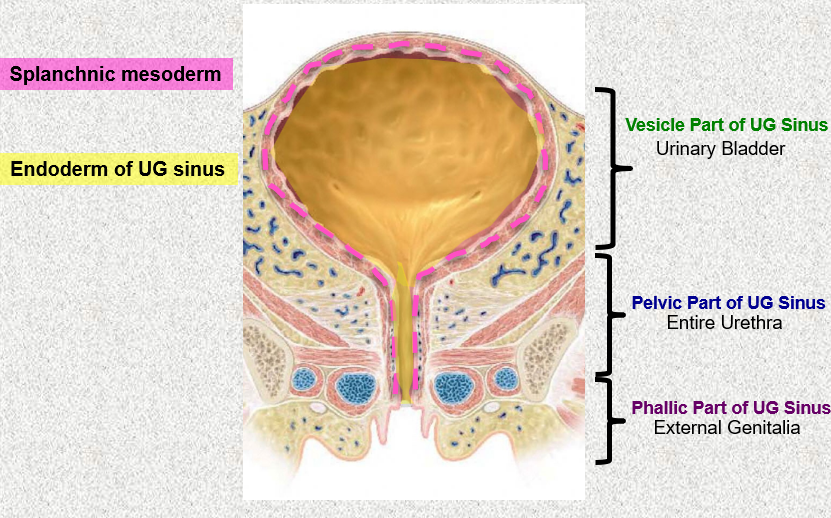
Image occlusion: name the hidden labels
222
Image occlusion: name the hidden labels
223
Image occlusion: name the hidden labels
224
Image occlusion: name the hidden labels
225
Image occlusion: name the hidden labels
226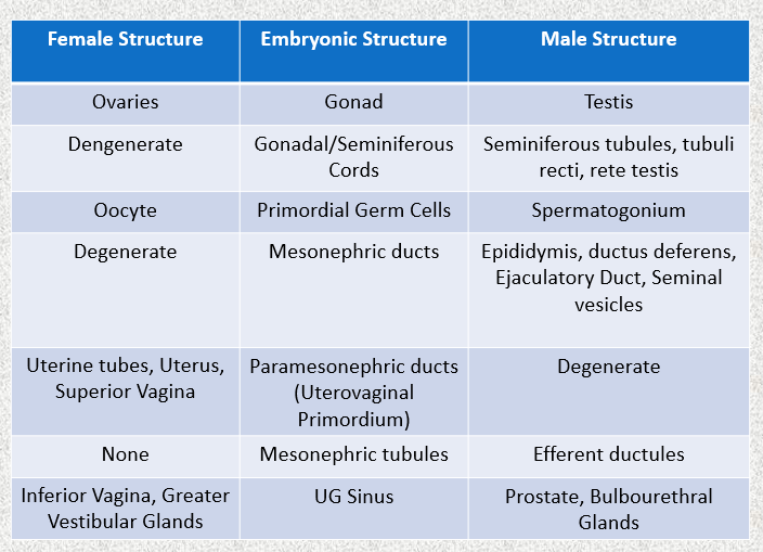
Image occlusion: name the hidden labels
227
Image occlusion: name the hidden labels
228
Image occlusion: name the hidden labels
229
Image occlusion: name the hidden labels
230
Image occlusion: name the hidden labels
231
Image occlusion: name the hidden labels
232
Image occlusion: name the hidden labels
233
Image occlusion: name the hidden labels
234
Image occlusion: name the hidden labels
235
Image occlusion: name the hidden labels
236
Image occlusion: name the hidden labels
237
Image occlusion: name the hidden labels
238
Image occlusion: name the hidden labels
239
Image occlusion: name the hidden labels
240
where does differentiation of the early genital systems begin?
dorsal body wall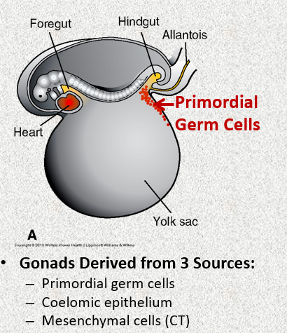
241
Early genital system development: primordial germ cells migrate from _______ through dorsal mesentery to reach ________-
umbilical vesicles; posterior body wall (gonadal ridges)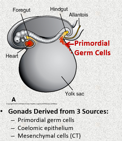
242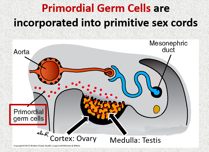
Image occlusion: name the hidden labels
243
Image occlusion: name the hidden labels
244
Image occlusion: name the hidden labels
245
The _- gene on the short arm of the Y chromosome acts as a switch that dorects production o the testes determining factor (TDF)
SRY 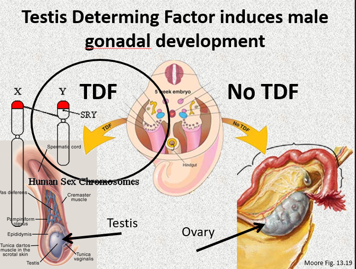
246
in males, primitive sex cords become ________- and give rise to the _______
testis cords; seminiferous cords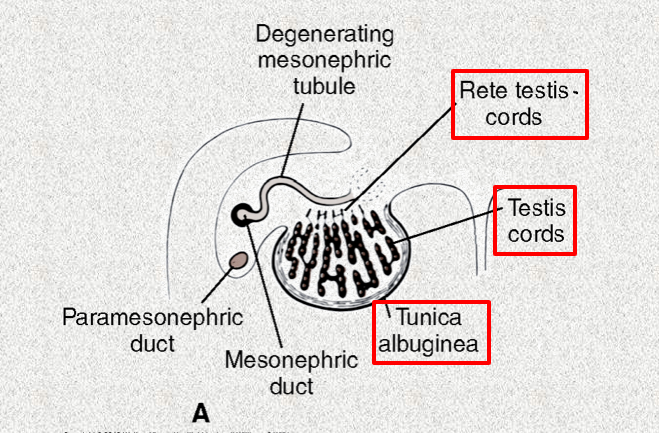
247
what do the seminiferous cords become (x3)
seminiferous tubules, tubuli recti, rete testis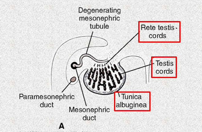
248
These derived fro the epitheliym secrete MIS
sertoli cells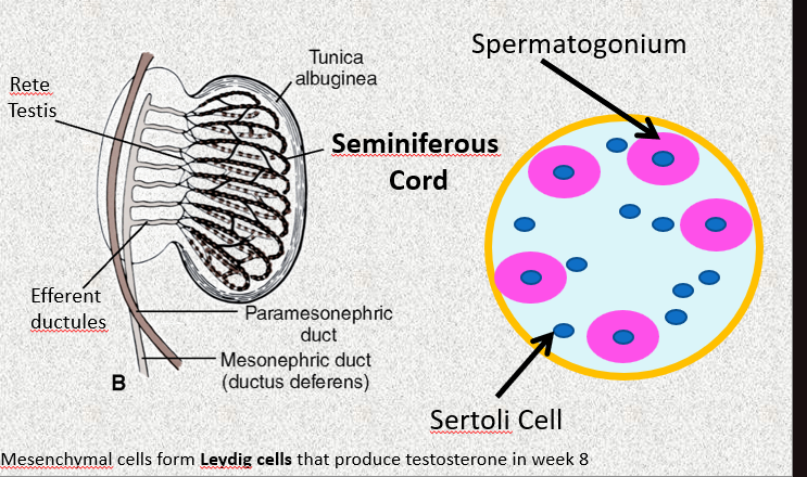
249
this leads the indifferent gonads to differentiate into the ovary
no TDF present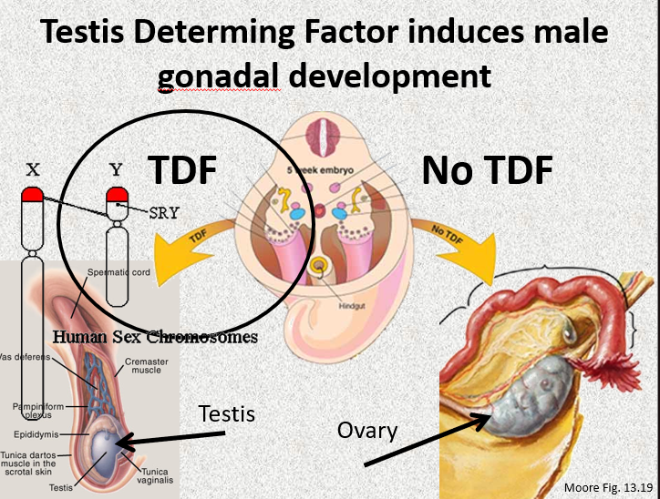
250
females: the primordial follicles are formed by __________ breaking up into clusters and surrounding germ cells
cortical cords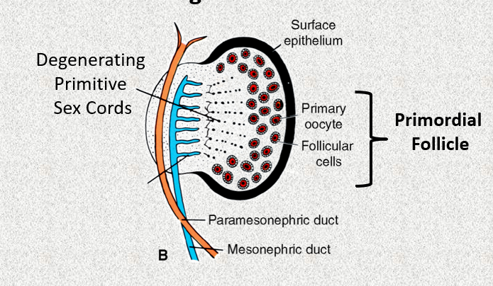
251
how do the testes descend
through the inguinal canal down to the scrotom
-vaginal process forms the scrotal lining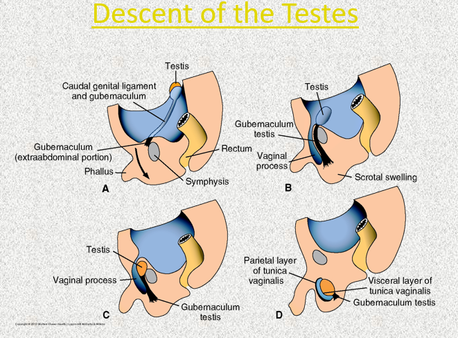
-vaginal process forms the scrotal lining
252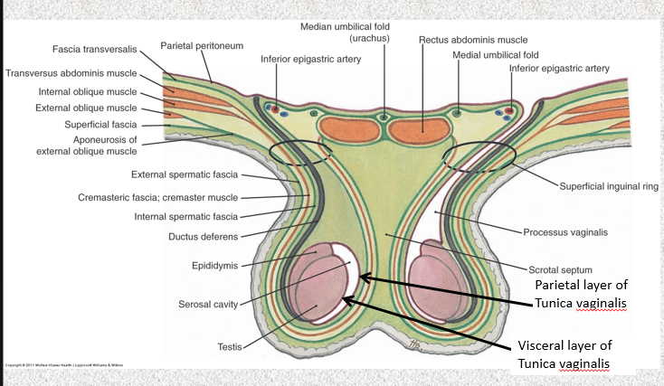
Image occlusion: name the hidden labels
253
Image occlusion: name the hidden labels
254
Image occlusion: name the hidden labels
255
Image occlusion: name the hidden labels
256
Image occlusion: name the hidden labels
257
Image occlusion: name the hidden labels
258
these ducts persist in males
mesonephrenic
259
these ducts persist in females
paramesonephrenic
260
these secrete the testosterine that induces masculine differentiate of the mesonephrenic ducts
leydig cells (interstitial cells)
261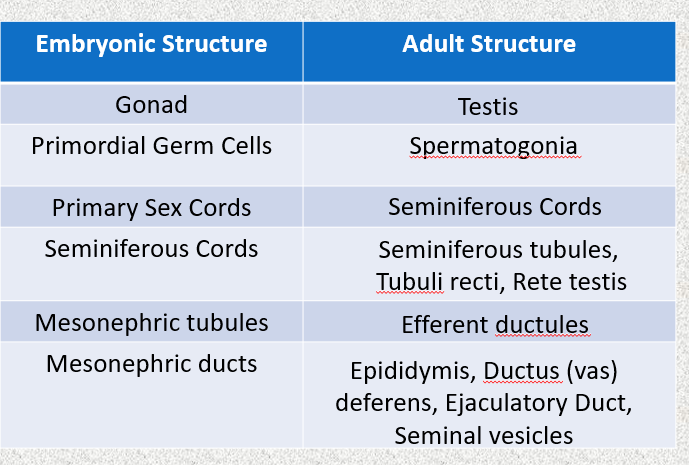
Image occlusion: name the hidden labels
262
Image occlusion: name the hidden labels
263
Image occlusion: name the hidden labels
264
Image occlusion: name the hidden labels
265
Image occlusion: name the hidden labels
266
Image occlusion: name the hidden labels
267
female: what do the urethral and paraurethral glands develop from
develop as buds from te urethrainto the surrounding mesenchyme
268
female: what do the greater vestibular glands develop from
outgrowths from the UG sinus
269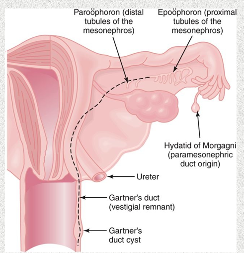
Image occlusion: name the hidden labels
270
Image occlusion: name the hidden labels
271
Image occlusion: name the hidden labels
272
Image occlusion: name the hidden labels
273
Image occlusion: name the hidden labels
274
what is the vagina formed from? (x2)
paramesonephric ducts and UG sinus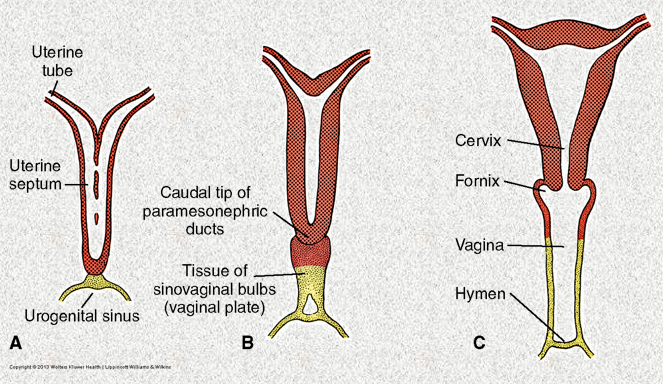
275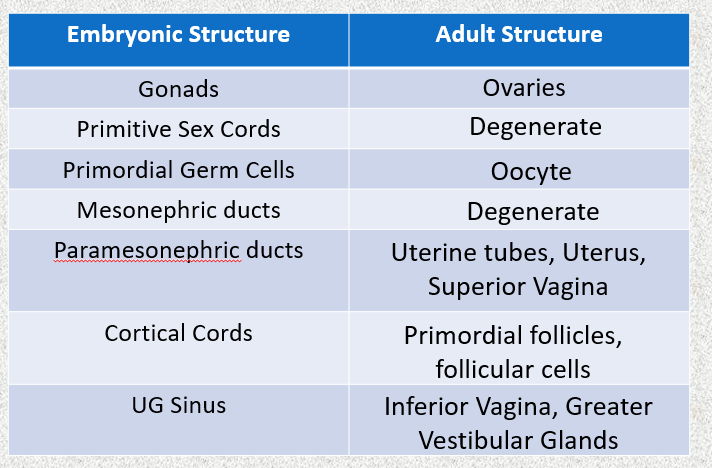
Image occlusion: name the hidden labels
276
Image occlusion: name the hidden labels
277
Image occlusion: name the hidden labels
278
Image occlusion: name the hidden labels
279
Image occlusion: name the hidden labels
280
Image occlusion: name the hidden labels
281
Image occlusion: name the hidden labels
282
what is this? which structure formed improperlu
1 sided uterus; paramesonephric ducts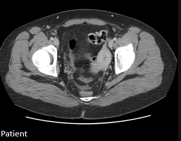
283
what is the test called that shows thie dye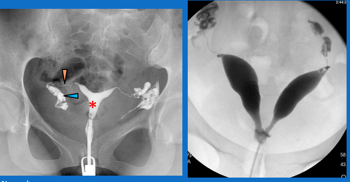
hysterosalpingogram
284
double uterus, is a rare congenital condition where a person is born with two separate uteruses, often accompanied by two cervices and a double vagina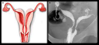
uterus didelphys
285
congenital uterine anomaly where the top of the uterus has a small, saddle-shaped indentation.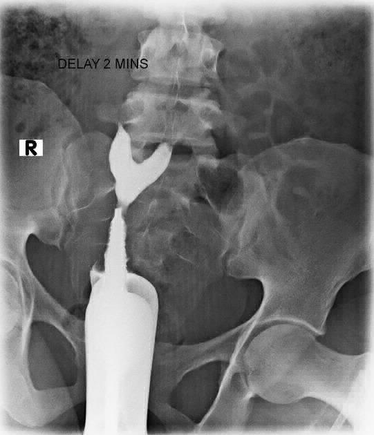
arcuate uterus
286
rare congenital uterine malformation where the uterus is divided into two separate horns (bicornuate) but has a single cervix (unicollis); 2 cervix would be bicollis (add after bicornis)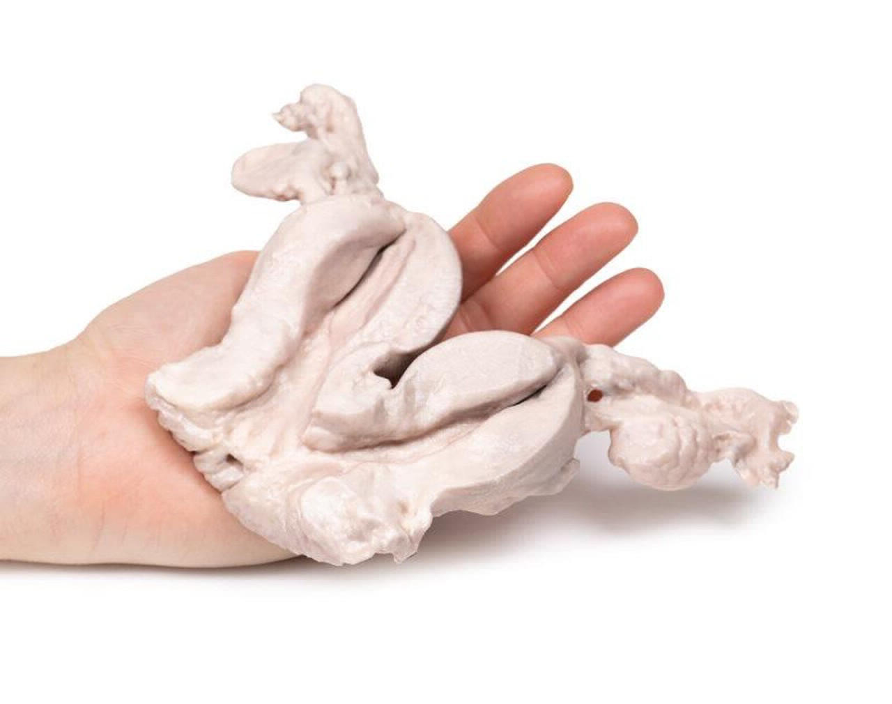
uterus bicornis
287
males: the primordial phallus enlarges to form the _____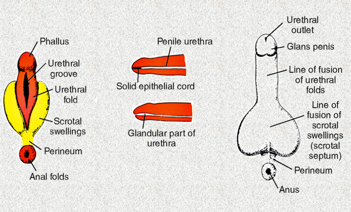
penis
288
males: the urethral folds form the ;ateral walls of the ______-- on the ventral surface of the penis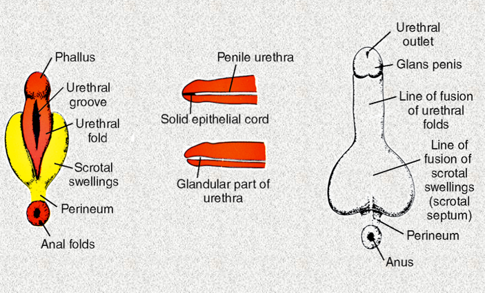
urethral groove which become the spongy urethra
289
what is the glans of the penis formed from
ectodermal ingrowth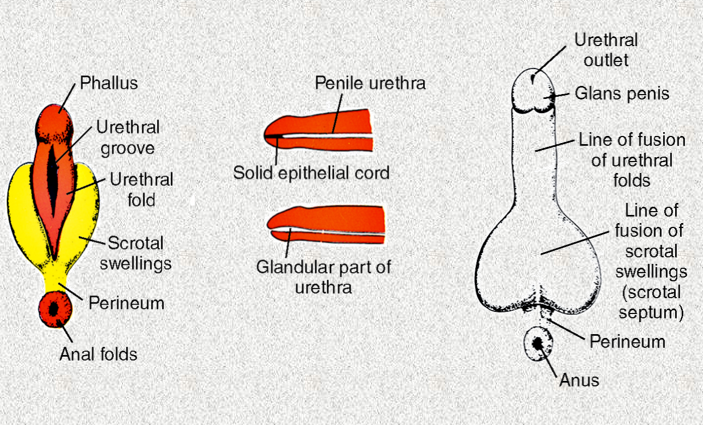
290
the periphery of the glans penis breaks down to form this
prepuce (foreskin)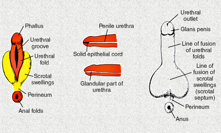
291
the labioscrotal swellings fuse to form ...
scrotum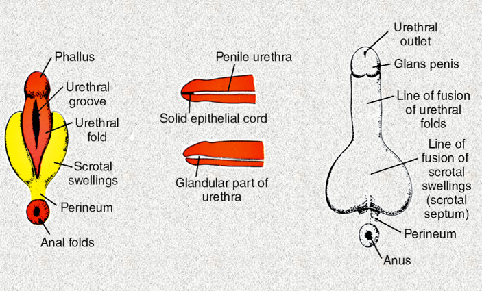
292
when fusion of the urethral folds are incomplete
hypospadias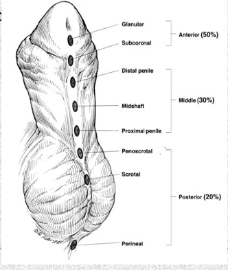
293
female: this decreases in sixe to become the clitoris
primordial phallus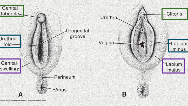
294
female: the urogenital fold fuse posteriorly to form the ______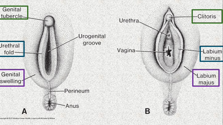
frenulum
295
female: the unfused portions of the urogenita folds form the _____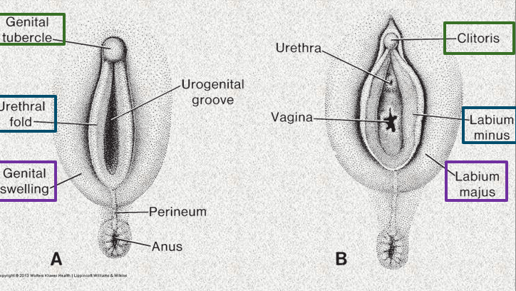
labia minora
296
the labiosacral folds fuse posteriorly to form ____ and anteriorly to form _________
posterior labial commissure; anterior labial commissure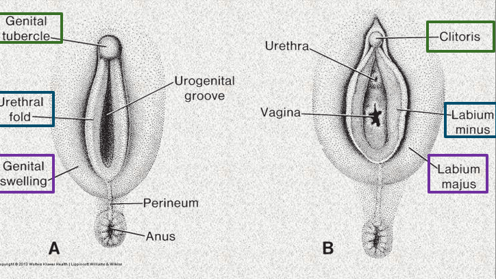
297
the urethral folds fuse posteriorly to form the _______- of the labia minora
frenulum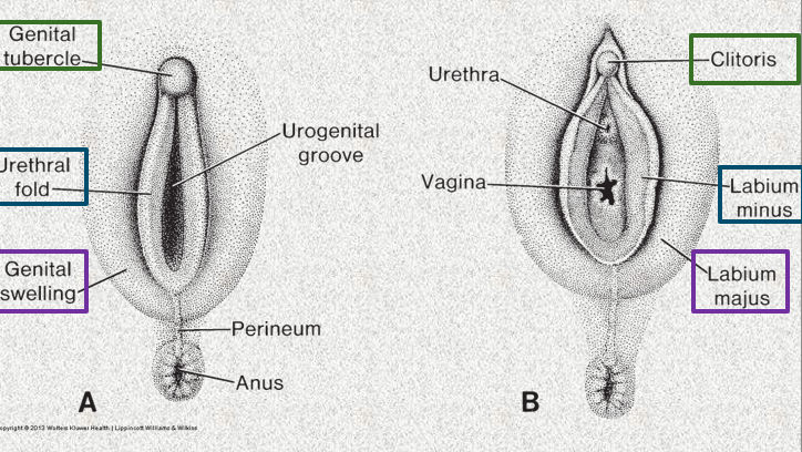
298
a rare genetic disorder where the body is unable to respond to male hormones (androgens), such as testosterone.
androgen insensitivity syndrome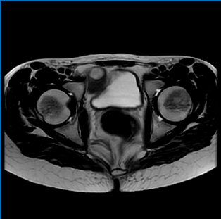
299
what is the maternal part of the placenta derived from 9germ layer)
endoderm
300
what is the maternal part of the placenta called?
decidua
30 more cards in OutLearn. Open the full deck