First 300 of 479 cards
1
top of the skull
calvarium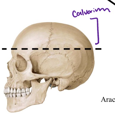
2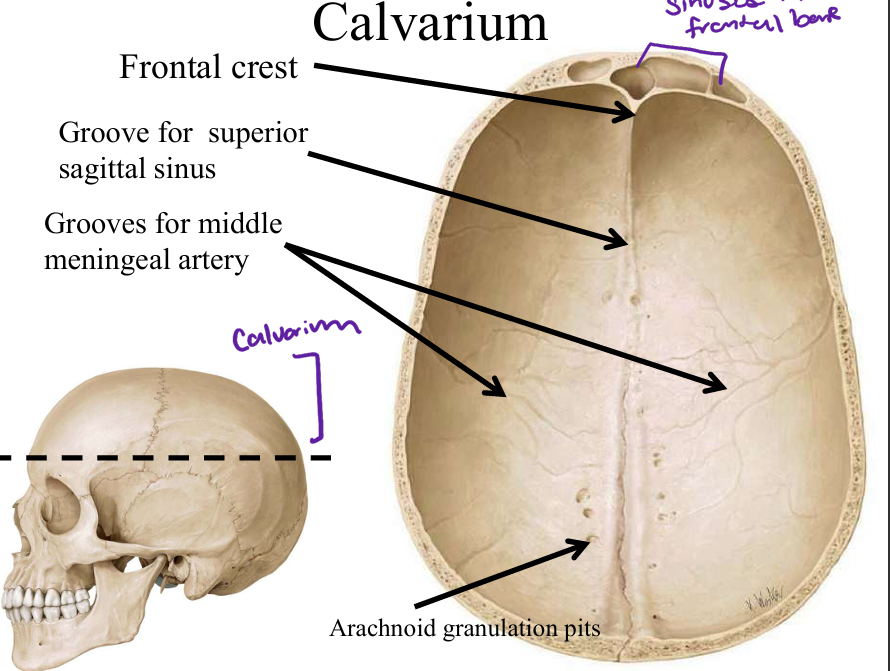
Image occlusion: name the hidden labels
3
Image occlusion: name the hidden labels
4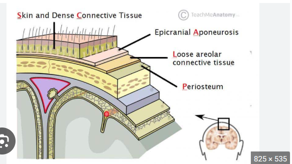
Image occlusion: name the hidden labels
5
Image occlusion: name the hidden labels
6
Image occlusion: name the hidden labels
7
Image occlusion: name the hidden labels
8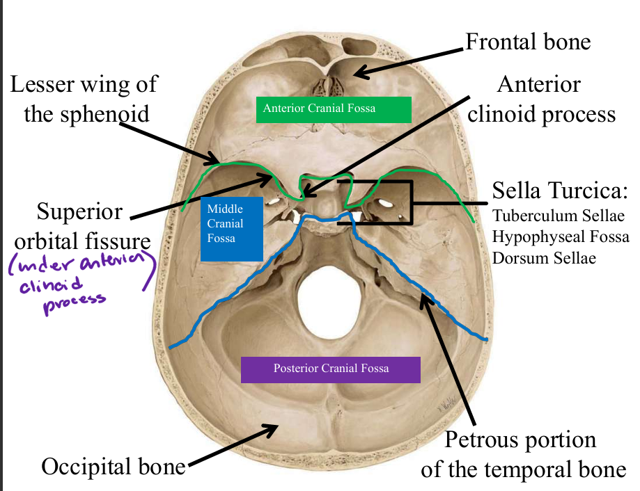
Image occlusion: name the hidden labels
9
Image occlusion: name the hidden labels
10
Image occlusion: name the hidden labels
11
Image occlusion: name the hidden labels
12
Image occlusion: name the hidden labels
13
Image occlusion: name the hidden labels
14
Image occlusion: name the hidden labels
15
Image occlusion: name the hidden labels
16
channels between the layers of dura
dural venous sinuses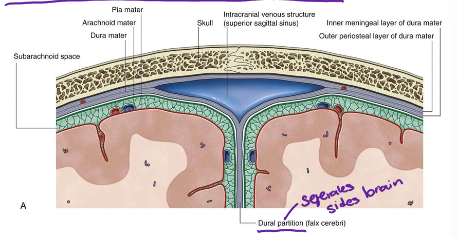
17
a potential space located between the dura mater (the outermost layer of the meninges) and the inner surface of the vertebral column
epidural space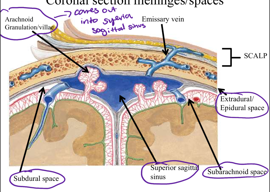
18
a narrow, fluid-filled area located between the brain and spinal cord and the innermost layer of the meninges, called the pia mater
subarachnoid space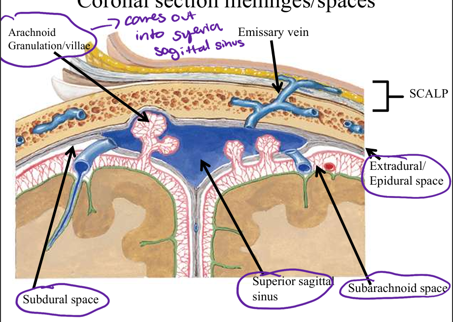
19
a thin, potential space located between the dura mater (outermost layer of the meninges) and the arachnoid mater (middle layer of the meninges) of the brain
subdural space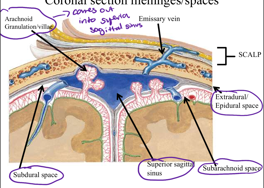
20
dural fold between cerebrum R and L
falx cerebri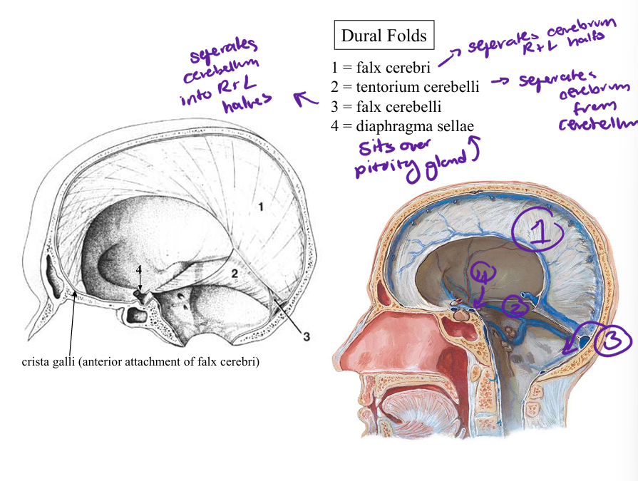
21
dual fold between cerebellum and cerebrum
tetorium cerebelli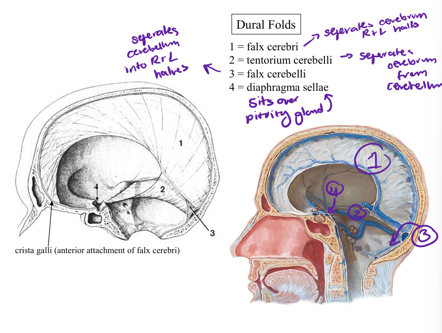
22
dural fold
falx cerebelli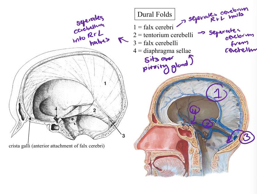
23
dural fold over pituitary gland
diaphragma stellae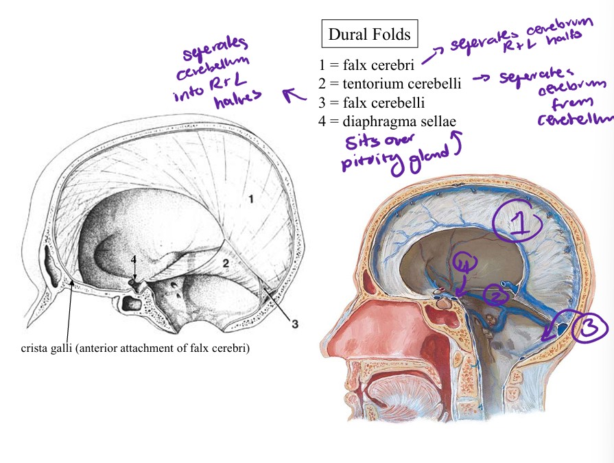
24
venous channels formed between 2 dural layers
dural venous sinuses
25
what drains into the dural venous sinus
rain & meninges-
diploe-
vertebral column
-deep face, orbit, nasal cavity-parietal, mastoid, occipital areas via
emissary vv.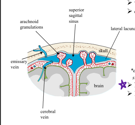
diploe-
vertebral column
-deep face, orbit, nasal cavity-parietal, mastoid, occipital areas via
emissary vv.
26
what drains the dural sinus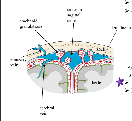
internal jugular v
27
does the dural venous sinuses have valves?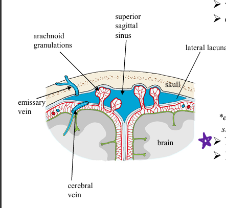
no
28
the dural venous sinuses run between layers of _____-
dura mater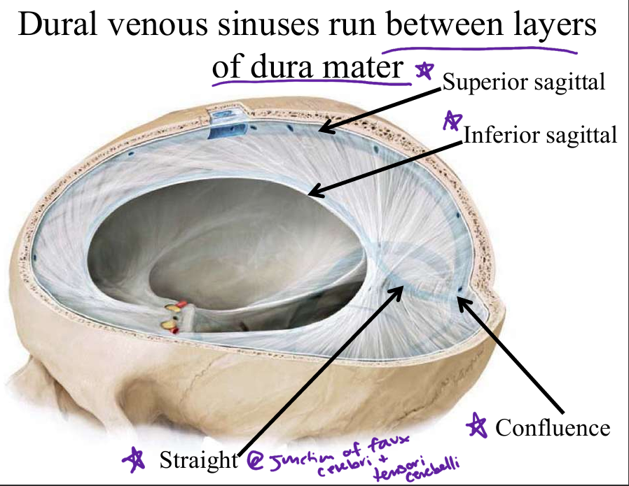
29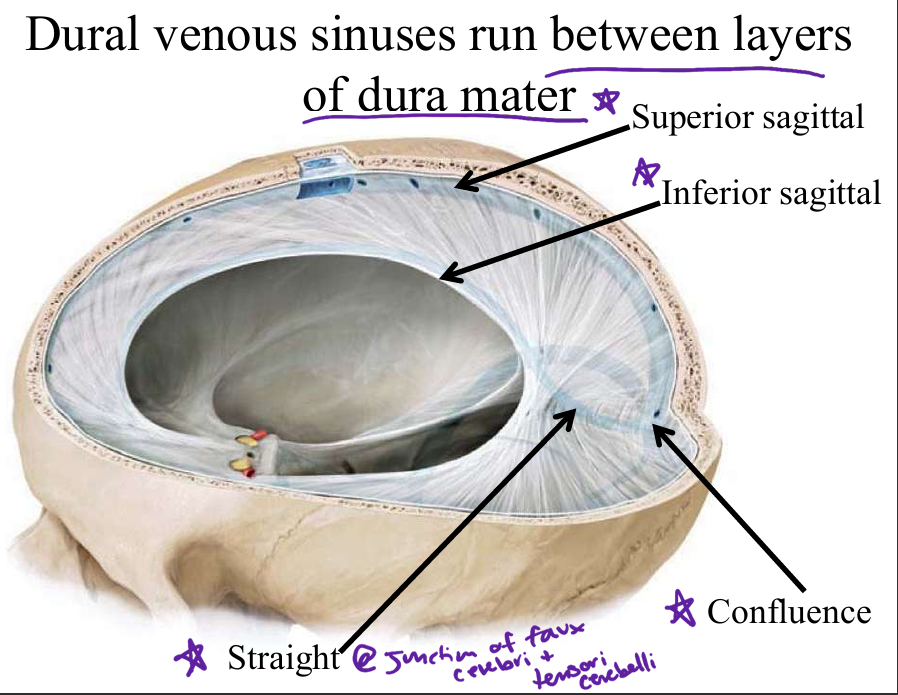
Image occlusion: name the hidden labels
30
Image occlusion: name the hidden labels
31
Image occlusion: name the hidden labels
32
Image occlusion: name the hidden labels
33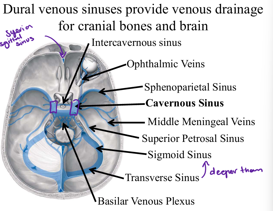
Image occlusion: name the hidden labels
34
Image occlusion: name the hidden labels
35
Image occlusion: name the hidden labels
36
Image occlusion: name the hidden labels
37
Image occlusion: name the hidden labels
38
Image occlusion: name the hidden labels
39
Image occlusion: name the hidden labels
40
Image occlusion: name the hidden labels
41
Image occlusion: name the hidden labels
42
how do wound from the superficial scalp transfer to the brain (infection)
internal cranial venous sinuses
43
what passes through the outer wall of the cavernous sinus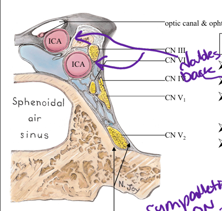
CN III, IV, V1, V2
44
what passe through the cavernous sinus
interbal carotid a and CN VI
45
what does the cavernous sinus drain to?
superiuor and inferior petrosal sinyses
46
explain the danger zone of the face?
infections, thrombophlebitis (inflamed vein with clots)
from facial vein can travel intracranially via superior
ophthamic v. to cavernous sinus or inferior ophthalmic v. to
cavernous sinus or pterygoid venous plexus…this would lead
to thrombophlebitis of cavernous sinus which is fatal in more
than 1/3 of cases
from facial vein can travel intracranially via superior
ophthamic v. to cavernous sinus or inferior ophthalmic v. to
cavernous sinus or pterygoid venous plexus…this would lead
to thrombophlebitis of cavernous sinus which is fatal in more
than 1/3 of cases
47
dural innervation
C1,C2,C3, vagus m
48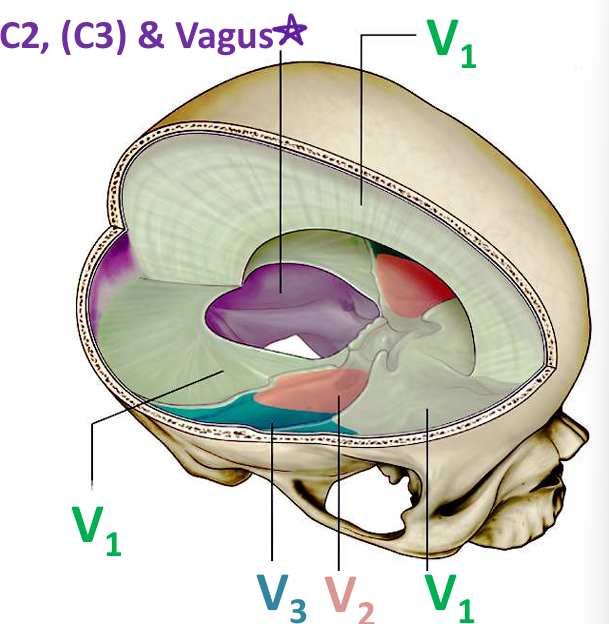
Image occlusion: name the hidden labels
49
Image occlusion: name the hidden labels
50
what artery supplies the ACF floor and branch of anterior ethmoidal a
anterior meningeal a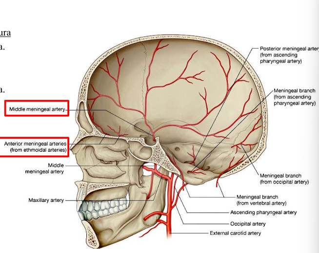
51
what artery suplies MCF, most of dura, brach of maxilary a
middle meningeal a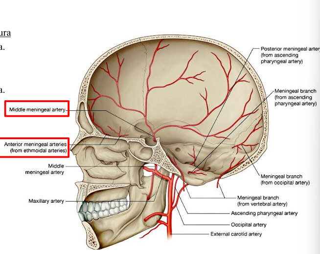
52
what artery supplies MCf and branch of maxillary a
accessory meningeal a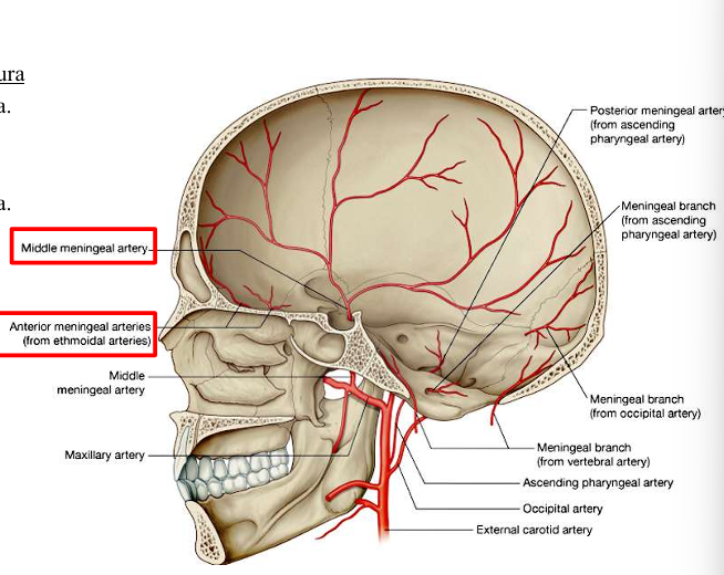
53
what artery supplies the PCF floor and branch of occipital and vertebral a
posterior meningeal aa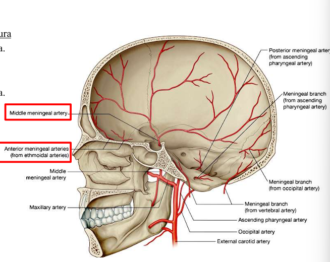
54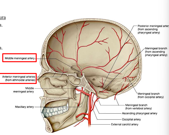
Image occlusion: name the hidden labels
55
Image occlusion: name the hidden labels
56
Image occlusion: name the hidden labels
57
Image occlusion: name the hidden labels
58
Image occlusion: name the hidden labels
59
Image occlusion: name the hidden labels
60
Acute/Chronic
Venous
old age; minor fall/injury
infants/children abuse; shaken baby syndrome
-cresceny t shapped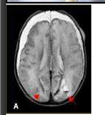
Venous
old age; minor fall/injury
infants/children abuse; shaken baby syndrome
-cresceny t shapped
subdural hematoma
61
where does tearing occur in subdural hematoma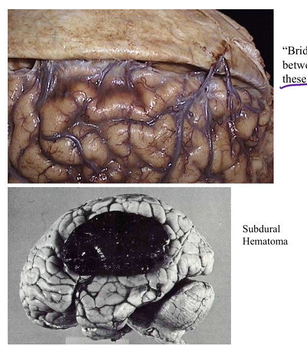
bridging between cerevral veinsbetween arachnoid and dura
62
-Symptoms due to pressure-Arterial
Headache
Confusion/disorientation
Lethargy
Unresponsiveness
Can have initial LUCID
interva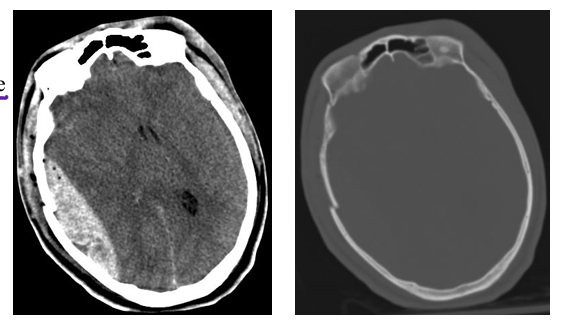
Headache
Confusion/disorientation
Lethargy
Unresponsiveness
Can have initial LUCID
interva
epidural hematoma
63
usually arterial, can be venous
Traumauptured aneurysm
Sudden, painful headache
Neck stiffness
Vomiting/nausea
Feel like head will explode
Loss of consciousness
Aversion to light (photophobia)
Bloody CSF on CT or LP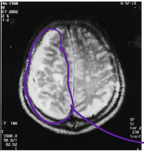
Traumauptured aneurysm
Sudden, painful headache
Neck stiffness
Vomiting/nausea
Feel like head will explode
Loss of consciousness
Aversion to light (photophobia)
Bloody CSF on CT or LP
subarachnoid hemorrhage
64
Sensory ganglion of CN V
Houses cell bodies of pseudounipolar sensory
neurons
a.k.a.; Gasserian, Semilunar
Sensory ganglion of CN V
Houses cell bodies of pseudounipolar sensory
neurons
a.k.a.; Gasserian, Semilunar
trigeminal ganglion
65
-Sensory disorder in CN V-Described as stabbing, lightning bolt-like pain
that may be very temporary or lengthy in duration -More likely in V
2
, V
3
than in V
1-May be due to blood vessel constricting
sensory root of CN V
(middle meningeal a.)
that may be very temporary or lengthy in duration -More likely in V
2
, V
3
than in V
1-May be due to blood vessel constricting
sensory root of CN V
(middle meningeal a.)
trigeminal neuralgia
66
parasympathetic supply to yhe parotid gland is frpm
CN IX
67
fx masseter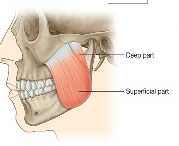
elevate and close mandible
68
innervation masseter m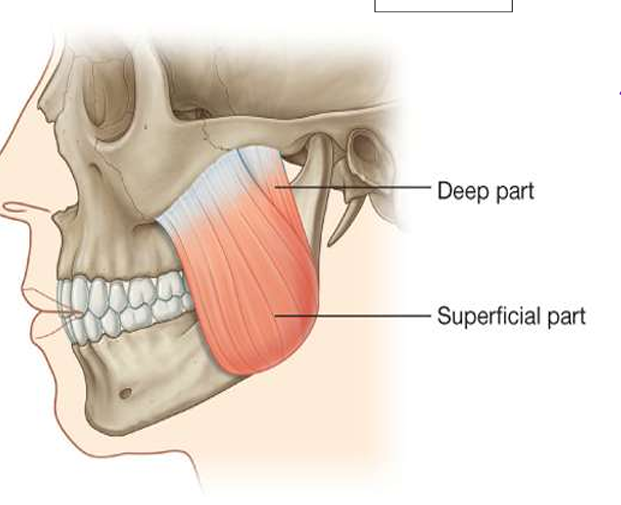
mandibular n from CN V3
69
innervation temporalis m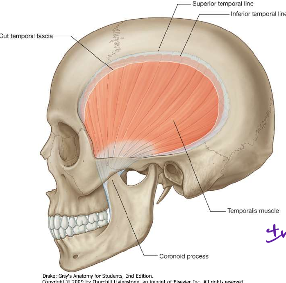
CN V3 mandibular n
70
fx temporalis m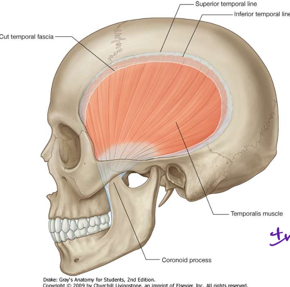
elevation and retavyiom of jaw
71
fx lateral pterygoid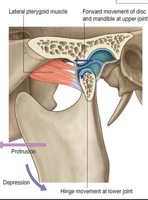
Protrusion
Side-to-side movements
of the mandible
Side-to-side movements
of the mandible
72
innervation lateral pterygoid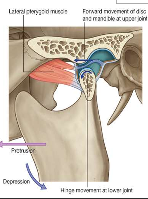
Lateral pterygoid nerve
from the anterior trunk of
V3
from the anterior trunk of
V3
73
fx medial pterygoid m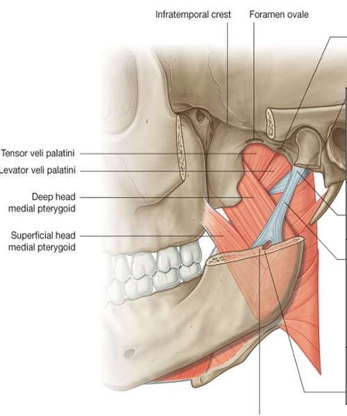
Elevation of the
mandible
Protrusion
Side-to-side
movements of the
mandible
mandible
Protrusion
Side-to-side
movements of the
mandible
74
innervation medial pterygoid m
medial pterygoud n from anterior trunk of V3
75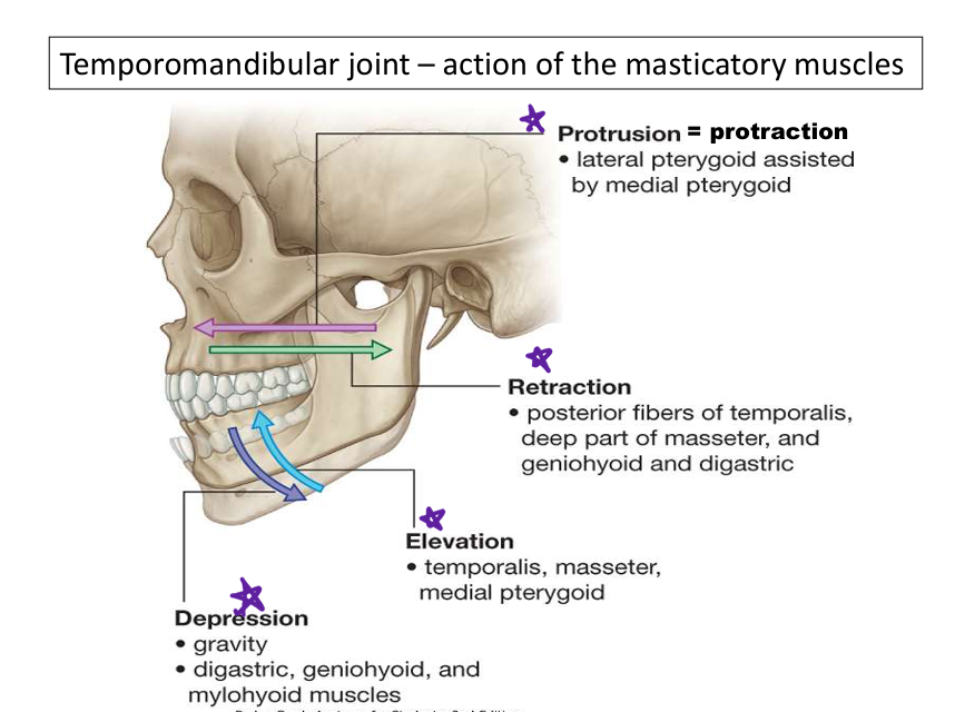
Image occlusion: name the hidden labels
76
Image occlusion: name the hidden labels
77
Image occlusion: name the hidden labels
78
Image occlusion: name the hidden labels
79
responsible for hindge/elevation depression of the jaw
mandibular fossa
80
responsible for the gliding movemet of the jaw
articular tubercle
81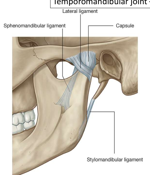
Image occlusion: name the hidden labels
82
Image occlusion: name the hidden labels
83
Image occlusion: name the hidden labels
84
what does this show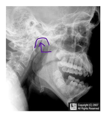
dislpocated jaw
85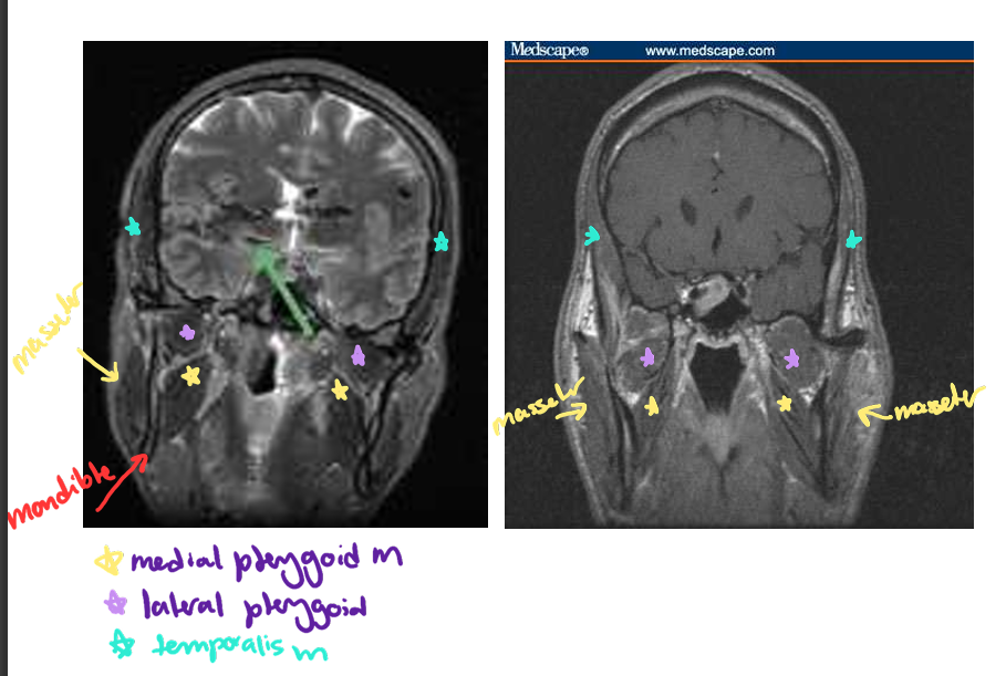
Image occlusion: name the hidden labels
86
Image occlusion: name the hidden labels
87
Image occlusion: name the hidden labels
88
branches of infratemporal fdossa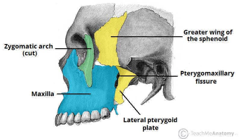
•Sphenomandibular ligament, medial and lateral pterygoid
•Maxillary artery and its branches
•Pterygoid venous plexus
•Mandibular nerve (CNV
3
) and its branches
•One branch of the facial nerve (CN VII): chorda tympani
•One branch of the glossopharyngeal nerve (CN IX): lesser
petrosal nerve
•One branch of Maxillary nerve (CNV
): post. sup. alveolar n.
•Maxillary artery and its branches
•Pterygoid venous plexus
•Mandibular nerve (CNV
3
) and its branches
•One branch of the facial nerve (CN VII): chorda tympani
•One branch of the glossopharyngeal nerve (CN IX): lesser
petrosal nerve
•One branch of Maxillary nerve (CNV
): post. sup. alveolar n.
89
where do the parts of the maxilarry artery run
First part (between mandibular neck and the sphenomandibular ligament)
Second part (related to the lateral pterygoid muscle)
Third part (in the sphenopalatine fossa)
Second part (related to the lateral pterygoid muscle)
Third part (in the sphenopalatine fossa)
90
artery that supplies the foramen spinosum; fro first part maxulkary a
middle meninheal a
91
artery that supplies the mandibular canal fro first part of maxilary a
inferior alveolar a
92
artery that supplies the temporalis m; from 2nd oart of maxillary a
deep temporal a
93
artery thjat runs with the buccal n and supplies the cheek and buccinator m
buccal a
94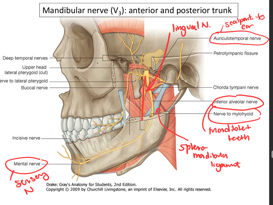
Image occlusion: name the hidden labels
95
Image occlusion: name the hidden labels
96
Image occlusion: name the hidden labels
97
Image occlusion: name the hidden labels
98
Image occlusion: name the hidden labels
99
Image occlusion: name the hidden labels
100
Image occlusion: name the hidden labels
101
• Sensory innervation of the dura mater mainly in the middle cranial
fossa
• Supply the mastoid cells that communicate with the middle ear
fossa
• Supply the mastoid cells that communicate with the middle ear
reccurent meningeal branch of V3
102
It also gives innervation to the tensor veli palatini
• It enters into the temporal bone above the pharyngotympanic canal
(superior part of the canalis musculotubarius) innervating the tensor
tympani muscle located in the canal
• It enters into the temporal bone above the pharyngotympanic canal
(superior part of the canalis musculotubarius) innervating the tensor
tympani muscle located in the canal
medial pterygoid n fro mandibular CN V3
103
• Usually 2 nerves ascending to the infratemporal fossa
• Supply the temporalis muscle from its deep surface
• Supply the temporalis muscle from its deep surface
deep temporal n from mandibular CN V3
104
• Passes laterally over the lateral pterygoid muscle
• Runs through the mandibular notch innervating the masseter muscle
• Runs through the mandibular notch innervating the masseter muscle
masseteric n from CN V3
105
Runs into the medial surface of the lateral pterygoid, innervating the muscle
lateral pterygoid n from CN V3
106
• Predominantly sensory nerve
• May carry motor fibers to the lateral pterygoid and temporalis muscle
• Passes laterally between the upper and lower heads of the lateral pterygoid
• Descends anterior to the mandibular ramus
• Often pierces the tendon of the temporalis muscle
• Runs on the lateral surface of the buccinator
• Innervates the oral mucosa and the skin of the cheek
• May carry motor fibers to the lateral pterygoid and temporalis muscle
• Passes laterally between the upper and lower heads of the lateral pterygoid
• Descends anterior to the mandibular ramus
• Often pierces the tendon of the temporalis muscle
• Runs on the lateral surface of the buccinator
• Innervates the oral mucosa and the skin of the cheek
buccal n from CN V3
107
• Sensory innervation of the anterior two-third of the tongue, oral mucosa on the floor of
the oral cavity and lower lingual gingiva
• Chorda tympani merges with the lingual nerve carrying taste fibers from the anterior
two-third of the tongue and preganglionic parasympathetic fibers for the
submandibular and sublingual glands (ggl: submandibular)
• Descends on the lateral surface of the medial pterygoid muscle
• Runs on the superior surface of the mylohyoid
the oral cavity and lower lingual gingiva
• Chorda tympani merges with the lingual nerve carrying taste fibers from the anterior
two-third of the tongue and preganglionic parasympathetic fibers for the
submandibular and sublingual glands (ggl: submandibular)
• Descends on the lateral surface of the medial pterygoid muscle
• Runs on the superior surface of the mylohyoid
lingual n from CN V3
108
•Inferior alveolar nerve
• Descends on the lateral surface of the medial pterygoid muscle
• Passes between the sphenomandibular ligament and the mandibular ramus
• Enters into the mandibular canal where provides sensory innervation of the lower teeth
and the associated gingiva
• Terminal branch exits the mandible through the mental foramen as mental nerve
providing sensory innervation of the mucosa and skin of the lower lip and chin
• Before entering in the mandibular canal inferior alveolar nerve gives a motor branch:
the nerve to the mylohyoid that runs in the mylohyoid groove and innervates the
mylohyoid and anterior belly of digastric muscles
• Descends on the lateral surface of the medial pterygoid muscle
• Passes between the sphenomandibular ligament and the mandibular ramus
• Enters into the mandibular canal where provides sensory innervation of the lower teeth
and the associated gingiva
• Terminal branch exits the mandible through the mental foramen as mental nerve
providing sensory innervation of the mucosa and skin of the lower lip and chin
• Before entering in the mandibular canal inferior alveolar nerve gives a motor branch:
the nerve to the mylohyoid that runs in the mylohyoid groove and innervates the
mylohyoid and anterior belly of digastric muscles
inferior alveolar n from CN V3
109
• Originates with two roots that surround the middle meningeal artery
• Passes between the tensor veli palatini and the upper head of lateral pterygoid
• Then passes between the sphenomandibular ligament and the mandibular neck
• Curves around the mandibular neck and ascends between it and the ear
• Sensory innervation of the temple, external ear, external acoustic meatus, tympanic
membrane and the temporomandibular joint
• Also delivers postganglionic parasympathetic fibers from the otic ggl (medial side of the
mandibular nerve just under the foramen ovale) to the parotid gland (preganglionic
fibers from CN IX via lesser petrosal nerve)
• Passes between the tensor veli palatini and the upper head of lateral pterygoid
• Then passes between the sphenomandibular ligament and the mandibular neck
• Curves around the mandibular neck and ascends between it and the ear
• Sensory innervation of the temple, external ear, external acoustic meatus, tympanic
membrane and the temporomandibular joint
• Also delivers postganglionic parasympathetic fibers from the otic ggl (medial side of the
mandibular nerve just under the foramen ovale) to the parotid gland (preganglionic
fibers from CN IX via lesser petrosal nerve)
auriulotemporal n from CN V3
110
structures that pass through the parotid gland
Transverse facial a/v (superficial temporal)
CN VII
Retromandibular v
External carotid
Auriculotemporal n. (V3)
Superficial and deep parotid nodes
CN VII
Retromandibular v
External carotid
Auriculotemporal n. (V3)
Superficial and deep parotid nodes
111
innervation to parotid gland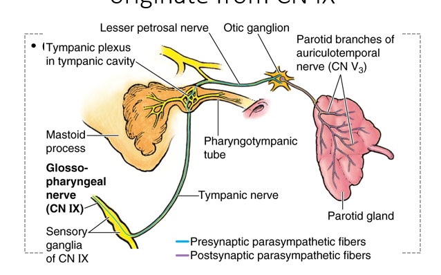
Parasympathetic innveration from CN IX.
Exits jugular foramen. Tympanic nerves goes up and back into tympanic cavity. Then gives off lesser petrosal nerve that goes thru middle cranial fossa then typical exits foramen ovale to get to infratempral fossa. Synapses otic ganglion in infratemporal fossa. Then uses auricotemporal n to get to parotid gland
Exits jugular foramen. Tympanic nerves goes up and back into tympanic cavity. Then gives off lesser petrosal nerve that goes thru middle cranial fossa then typical exits foramen ovale to get to infratempral fossa. Synapses otic ganglion in infratemporal fossa. Then uses auricotemporal n to get to parotid gland
112
major blood vessel that supplies the face
facial a
113
this vein drains much of the strictures of the face`
facial v
114
what does the facial v drain into
retromandibular v trhan IJV
115
infections from here can travel thrpug what to the braon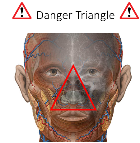
ophthalmic vv
116
Separates the oral cavity from the
nasal cavity
• Formed by the palatine processes of
the maxillae (anterior ¾) and the
horizontal processes of the palatine
bones (posterior ¼)
• Superiorly covered my respiratory
mucosa
• Inferiorly covered by oral mucosa
forming
nasal cavity
• Formed by the palatine processes of
the maxillae (anterior ¾) and the
horizontal processes of the palatine
bones (posterior ¼)
• Superiorly covered my respiratory
mucosa
• Inferiorly covered by oral mucosa
forming
hard palate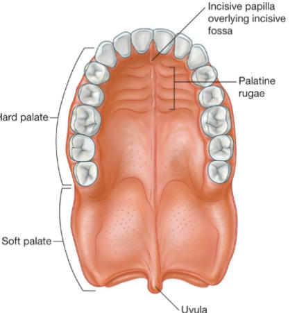
117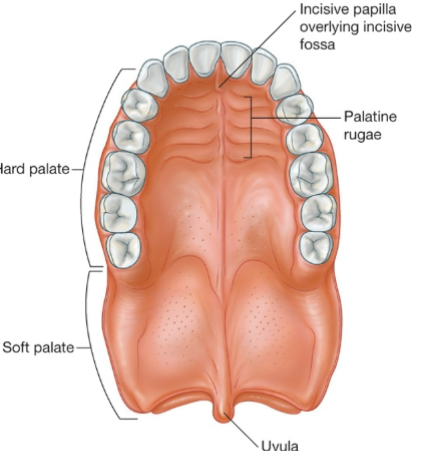
Image occlusion: name the hidden labels
118
Image occlusion: name the hidden labels
119
Image occlusion: name the hidden labels
120
Image occlusion: name the hidden labels
121
Image occlusion: name the hidden labels
122
Continues posteriorly from the hard
palate
• Superiorly covered my respiratory
mucosa
• Inferiorly covered by oral mucosa
• Can be depressed and elevated
• Uvula – small projection hanging
from the posterior free margin
palate
• Superiorly covered my respiratory
mucosa
• Inferiorly covered by oral mucosa
• Can be depressed and elevated
• Uvula – small projection hanging
from the posterior free margin
soft palate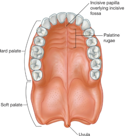
123
5 muscles associated wth the soft palate
Tensor veli palatini
• Levator veli palatini
• Palatopharyngeus
• Palatoglossus
• Musculus uvulae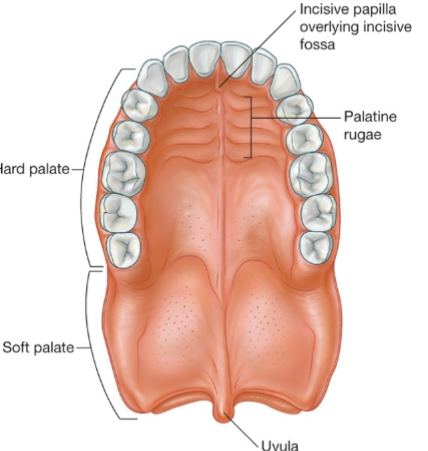
• Levator veli palatini
• Palatopharyngeus
• Palatoglossus
• Musculus uvulae
124
actions of tensor veli palatini
• Tenses the soft palate
• Opens the
pharyngotympanic tube
(yawning, swallowing)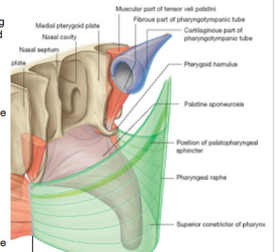
• Opens the
pharyngotympanic tube
(yawning, swallowing)
125
innervation tensor velu palatini
medial pterygoid nerve of the
mandibular nerve (V3)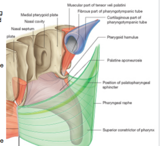
mandibular nerve (V3)
126
actions levator veli palatini
• Elevate the palate (close the
pharyngeal isthmus between the
nasopharynx and oropharynx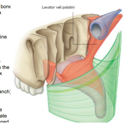
pharyngeal isthmus between the
nasopharynx and oropharynx
127
innervation levaor velu palatini
vagus n (pharyngeal branch)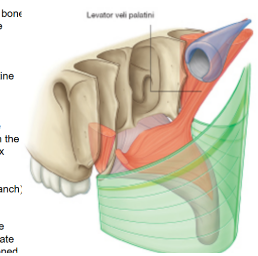
128
clinical testing for the levator veli palatini
Patient say ‘ah’, if the muscle
function is abnormal, the palate
deviates away from the lesioned
site (normal: elevates evenly)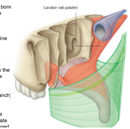
function is abnormal, the palate
deviates away from the lesioned
site (normal: elevates evenly)
129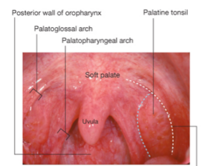
Image occlusion: name the hidden labels
130
Image occlusion: name the hidden labels
131
Image occlusion: name the hidden labels
132
Image occlusion: name the hidden labels
133
Image occlusion: name the hidden labels
134
Image occlusion: name the hidden labels
135
palatopharyngeus m action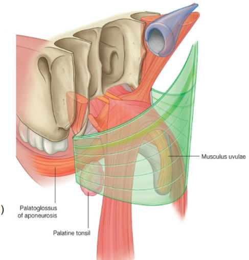
• Depresses soft palate,
• Narrows the isthmus faucium
• Elevates pharynx
• Narrows the isthmus faucium
• Elevates pharynx
136
innervation palatopharyngeus m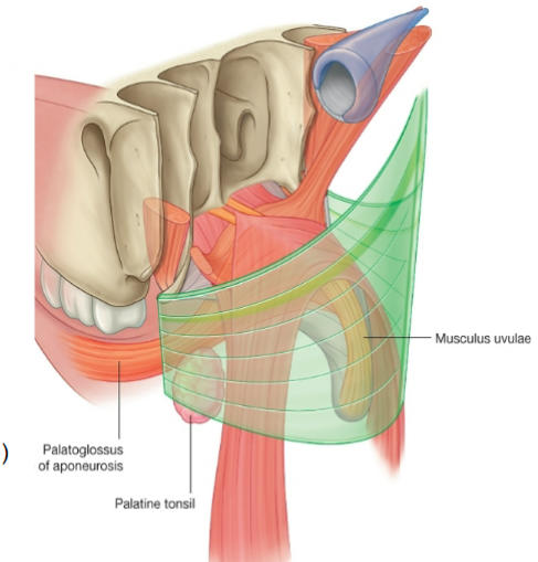
vagus n pharyngeal branch
137
actions of the palatoglossus m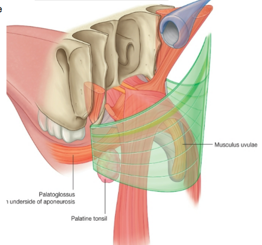
•Depresses soft palate
•Narrows the isthmus faucium
•Elevates back of the tongue
•Narrows the isthmus faucium
•Elevates back of the tongue
138
innervation of the palatoglossus m 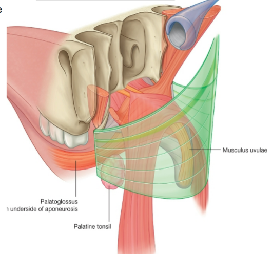
vagus n pharyngeal branch
139
action musculus uvulae
elevate and retract uvula
thickens centtral region of soft palate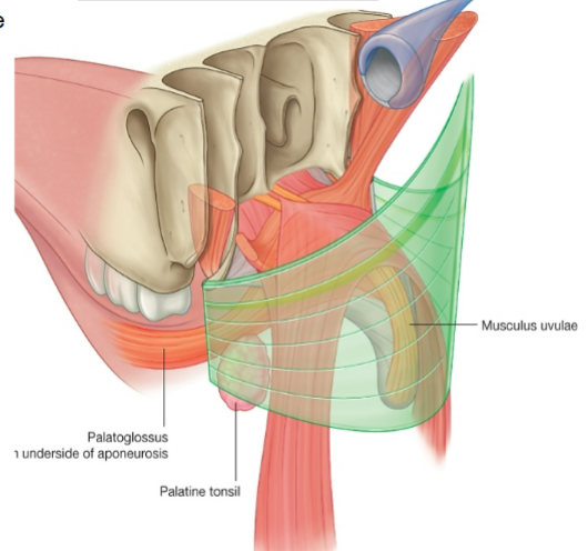
thickens centtral region of soft palate
140
innervation musculus uvulae
vagus n pharngeal branch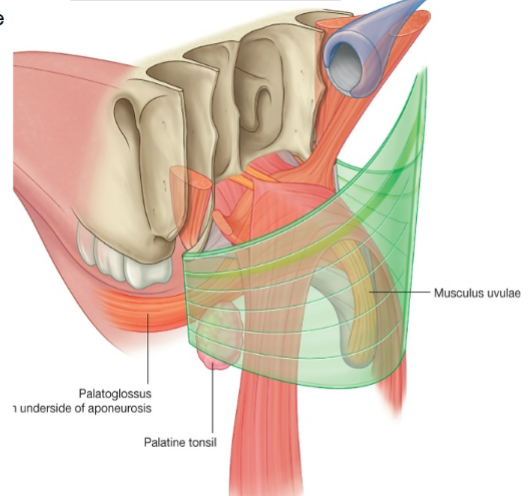
141
the nasopalatine n leaves through the ___________ foramen to reac the nasal cavity and leaves through the ___- to supply the anterior hard palare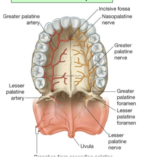
sphenopalatine; incisive canal
142
what does the nasopalatine nerve innervate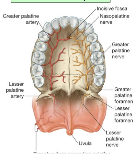
anterior hard palate
143
the greater and lessser palatine n pass throh the ________-- and enter the oral surface of the palate through the ______ foramina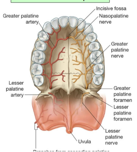
palatine canal; greater and lesser palatine foramine
144
the greater palatine n innervate ________________ 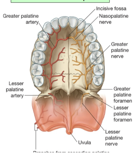
mucosa and glands of the hard
palate, gingiva until the first
premolar, and give the posterior
inferior nasal nerves that enter
nasal cavity through small foramina
of the hard palate
palate, gingiva until the first
premolar, and give the posterior
inferior nasal nerves that enter
nasal cavity through small foramina
of the hard palate
145
the lesser palatine n innervates the ...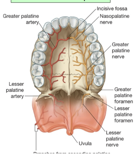
mucosa and glands of the soft palate
146
the ascendin palatine a is a branch of this a; it loops ocer this muscle and penetrates this fascia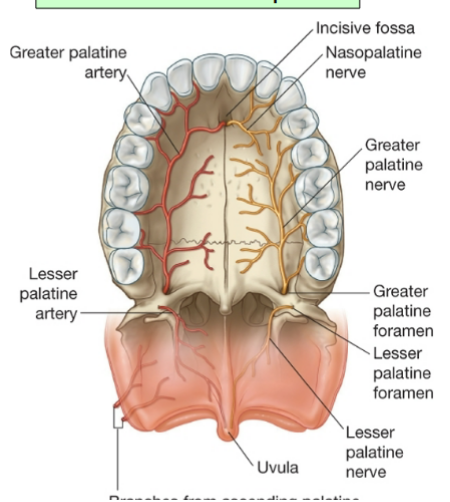
facial a; superior constroctor m; pharyngeal fascia
147
the ascending palatine a follows the _____-- to the soft palate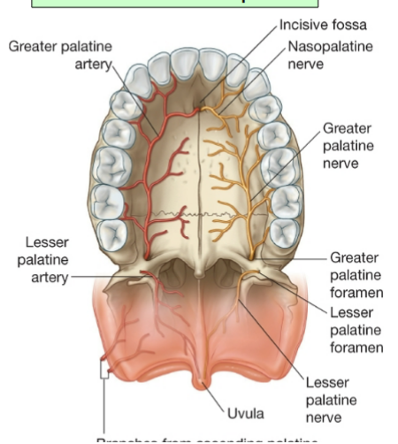
levator veli palatini
148
this a has the same course as the ascending palatine a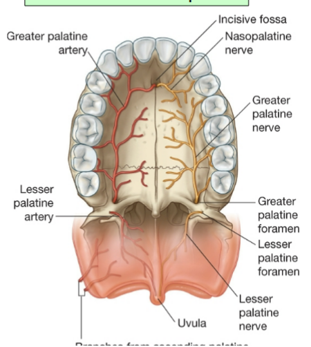
palatine branch of ascending palatine a
149
the descending palatine a passes through the _________- with the palatine n; it enters theoral surface of the palate through the ______________-- foramina as these arteries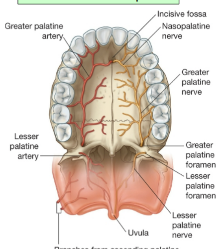
palatine canal; greater and lesser palatine foramina; greater and lesser palatine a
150
the descending palatine a supplies this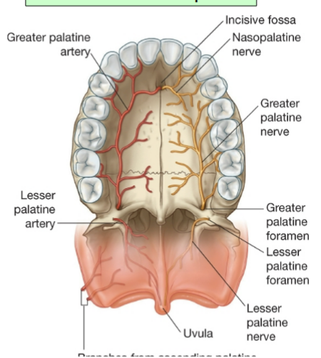
hard and soft palate
151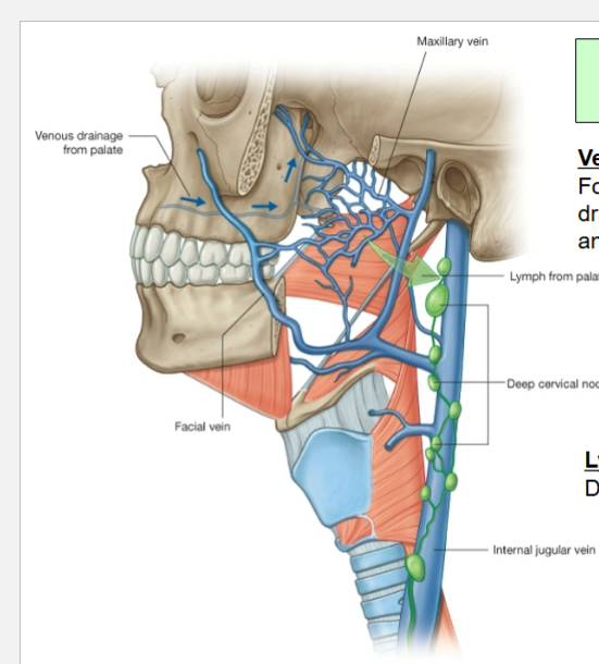
Image occlusion: name the hidden labels
152
Image occlusion: name the hidden labels
153
Image occlusion: name the hidden labels
154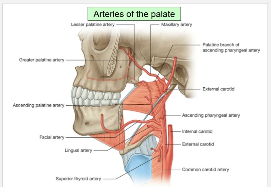
Image occlusion: name the hidden labels
155
Image occlusion: name the hidden labels
156
Image occlusion: name the hidden labels
157
Image occlusion: name the hidden labels
158
Image occlusion: name the hidden labels
159
Image occlusion: name the hidden labels
160
Image occlusion: name the hidden labels
161
Image occlusion: name the hidden labels
162
the veins of the palate drain the same as the arteries and ultimately drain into the ___________- and _________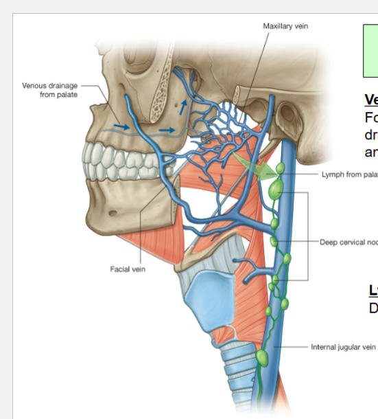
pterygoid plexus and pharnyngeal plexus
163
where does lymph from the palate go?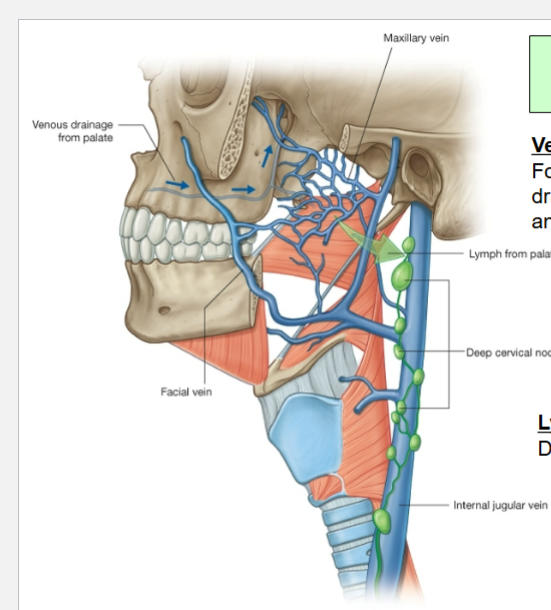
deeo cervical lymph nodes
164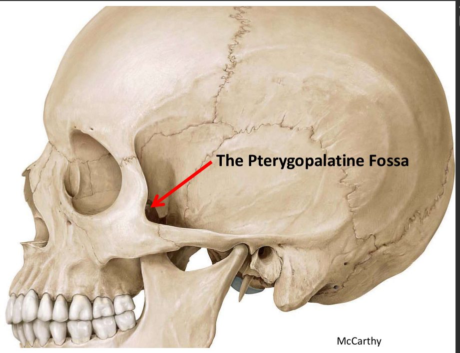
Image occlusion: name the hidden labels
165
boundaries f the pterygopalatine fossa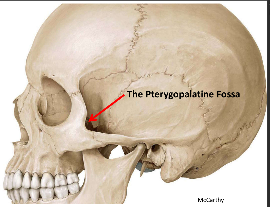
Posterior: Sphenoid
Anterior: Maxilla
Medial: Palatine bone
Lateral: Infratemporal fossa
(Pterygo(spheno)
maxillary fissure)
Anterior: Maxilla
Medial: Palatine bone
Lateral: Infratemporal fossa
(Pterygo(spheno)
maxillary fissure)
166
what communicates through the pp fossa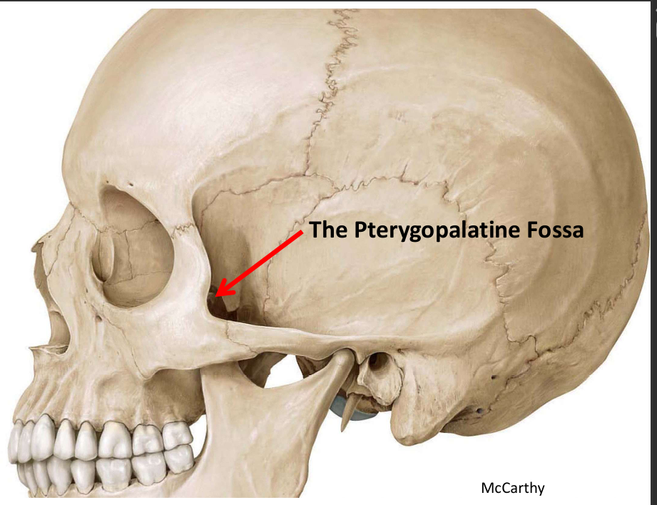
1. Middle Cranial Fossa
2. Nasal cavity
3. Orbit
4. Nasopharynx
5. Oral cavity
6. Infratemporal fossa
2. Nasal cavity
3. Orbit
4. Nasopharynx
5. Oral cavity
6. Infratemporal fossa
167
how does the nasal cavty communicate via the pp fossa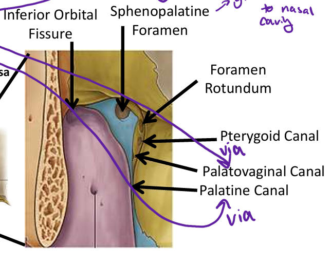
via the sphenopalatine foramen
168
how does the orbit communicate through the pp fossa`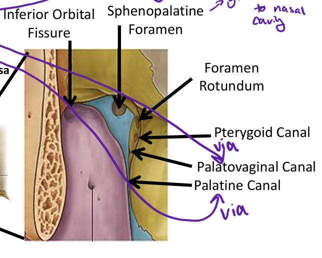
inferior orbital fissure
169
how does the nasopharynx communicate through the pp fossa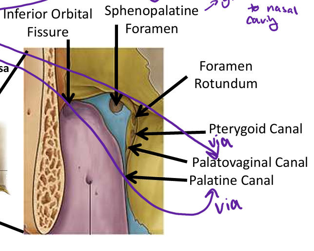
via the palatovaginal canal
170
how does the oral cavity communicate through the pp fossa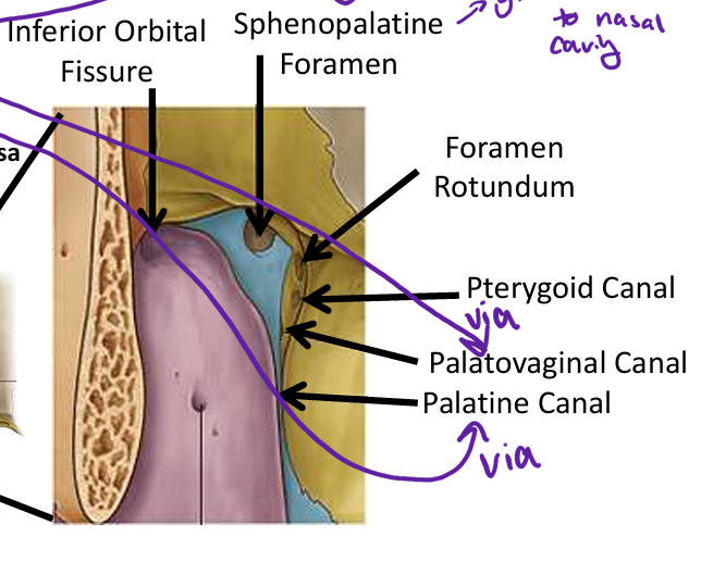
via the palatine canal
171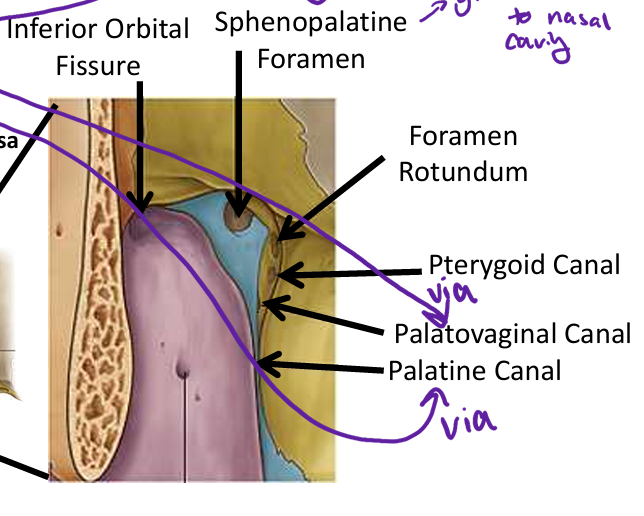
Image occlusion: name the hidden labels
172
Image occlusion: name the hidden labels
173
Image occlusion: name the hidden labels
174
Image occlusion: name the hidden labels
175
Image occlusion: name the hidden labels
176
Image occlusion: name the hidden labels
177
where is the ppg fossa?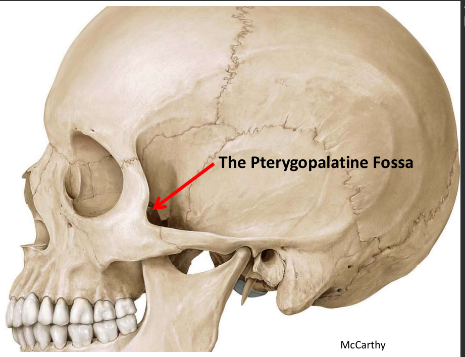
posterior to the maxillary bone
178
what CN is the pterygopalatine ganglion associated with
CN VII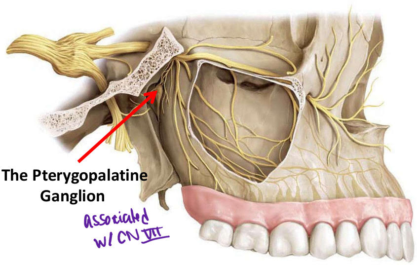
179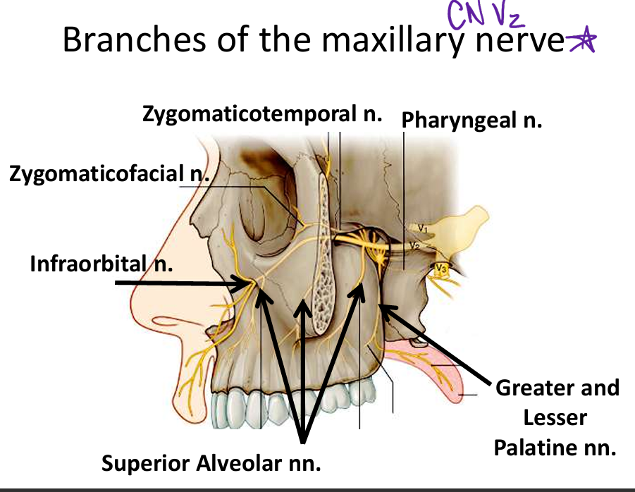
Image occlusion: name the hidden labels
180
Image occlusion: name the hidden labels
181
Image occlusion: name the hidden labels
182
Image occlusion: name the hidden labels
183
Image occlusion: name the hidden labels
184
Image occlusion: name the hidden labels
185
this n carries postganglionic GVE fibers to the lacrimal gland
Zygomaticotemporal n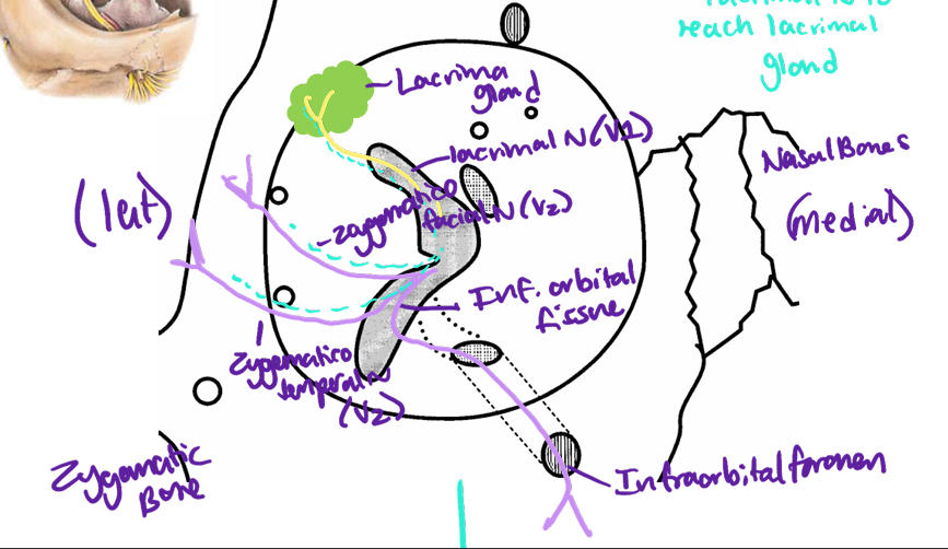
186
GVE and GVA hitchike on this n to reach the lacrimal gland
lacrimal n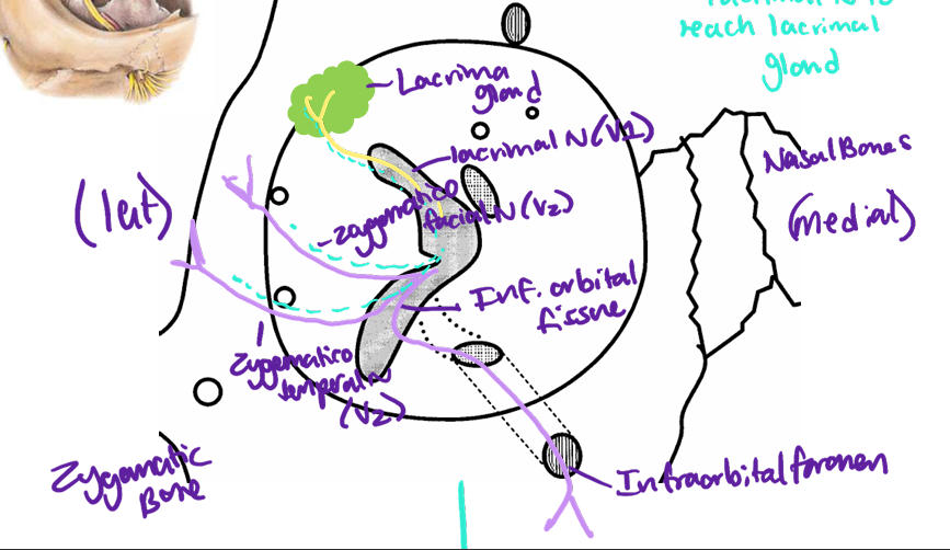
187
these n supply the medial and lateral walls of the nasal cavity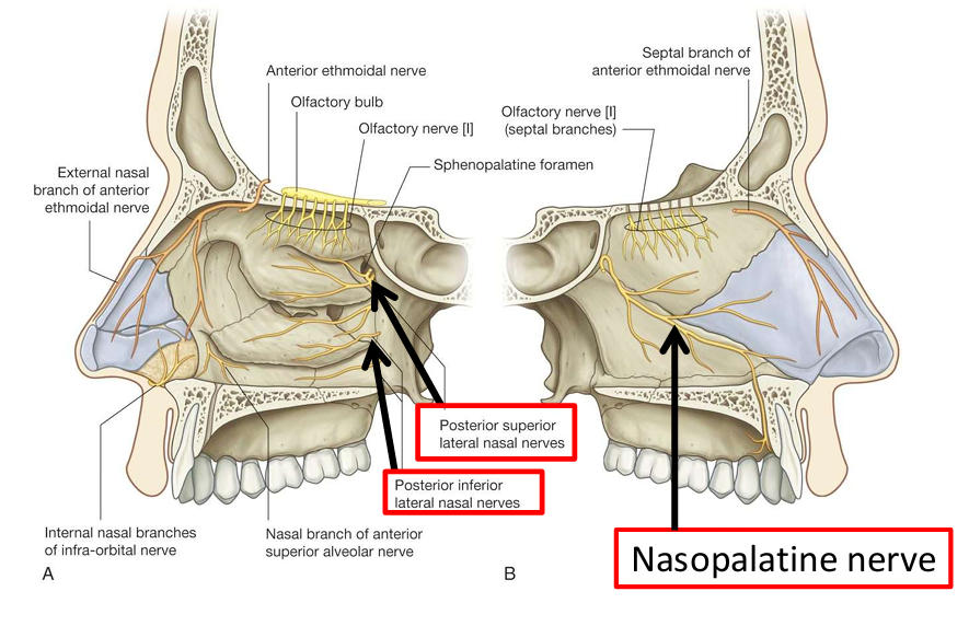
nasal nerves
188
this n innervates the nasal cavity than passes into the oral cavity via the incisive canal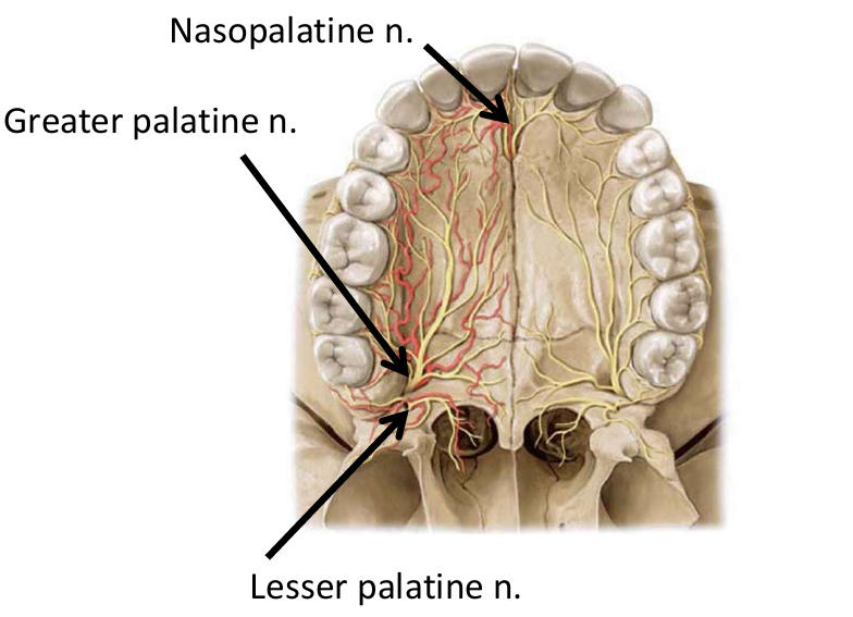
nasopalatine n
189
these nerves form the superior dental plexus, a network of nerves located in the upper jawbone (maxilla) that innervates the teeth and surrounding structures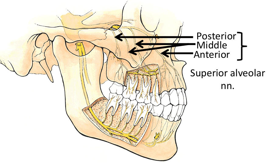
superior alveolar nerves
190
Image occlusion: name the hidden labels
191
Image occlusion: name the hidden labels
192
Image occlusion: name the hidden labels
193
Image occlusion: name the hidden labels
194
Image occlusion: name the hidden labels
195
Image occlusion: name the hidden labels
196
innervation to the cheek mucosa
buccal n (CN V3)
197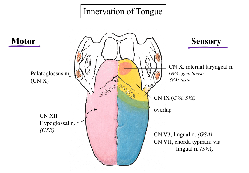
Image occlusion: name the hidden labels
198
Image occlusion: name the hidden labels
199
Image occlusion: name the hidden labels
200
Image occlusion: name the hidden labels
201
Image occlusion: name the hidden labels
202
fx genioglossus m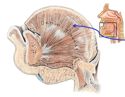
protrude tongue
203
innervation geniogkossus m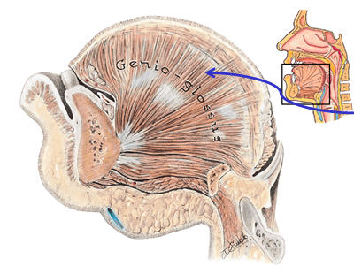
GN XII
204
hyoglossus action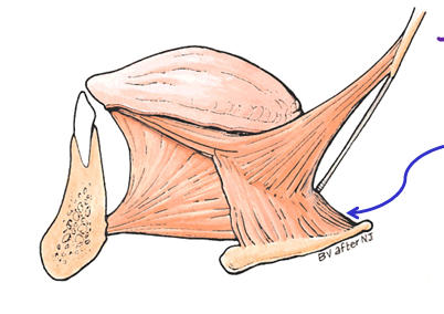
depress sides if tongue, retract tongue
205
innervationhyoglossus m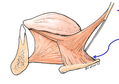
CN XII
206
action styloglossus m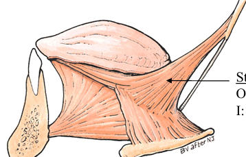
elevate and retract tongu
207
innervation styloglossus m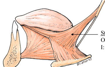
CNXII
208
extrinsic tongue muscles
styloglossus, palatoglossus, hyoglossus, genioglossus
209
fx intrinsic m of tongue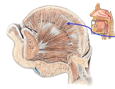
shorten/narrow/flatten tongue
210
when there is a cranial n injury, the tonmgue deviates to the (same side/op[posite side) of the injury
same side
211
in terms of a CN injury, the uvula deviates to the (same side/opposure side) of the injury
opposite side
212
Gag reflex ns
CN IX (afferent)
CN X and CN IX (efferent)
CN X and CN IX (efferent)
213
supplies post tongue, tonsils, epiglottois, soft palate
dorsal lingual a
214
supplies the tongue tip, R/L anastasomes and is contimuatiom of lingual a
deep lingual a
215
this a is under the mucosa and supplies the sublingual gland and mylohyoid m
sublingual a
216
Accompany lingual a. (vena comitantes)
Receive dorsal lingual and sublingual vv.
Drain to IJV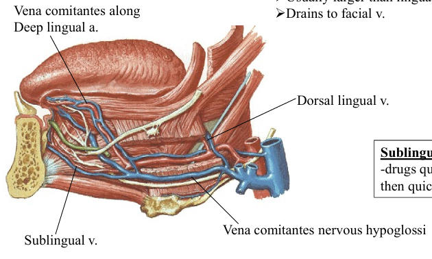
Receive dorsal lingual and sublingual vv.
Drain to IJV
lingual v
217
Accompany CN XII
Usually larger than lingual v.
Drains to facial v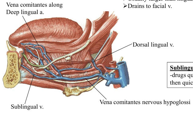
Usually larger than lingual v.
Drains to facial v
vena comitantes nervous hypiglossi
218
how are drugs absorbed sublingually
-drugs quickly enter sublingual vv. and
then quickly into IJV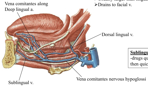
then quickly into IJV
219
Image occlusion: name the hidden labels
220
Image occlusion: name the hidden labels
221
Image occlusion: name the hidden labels
222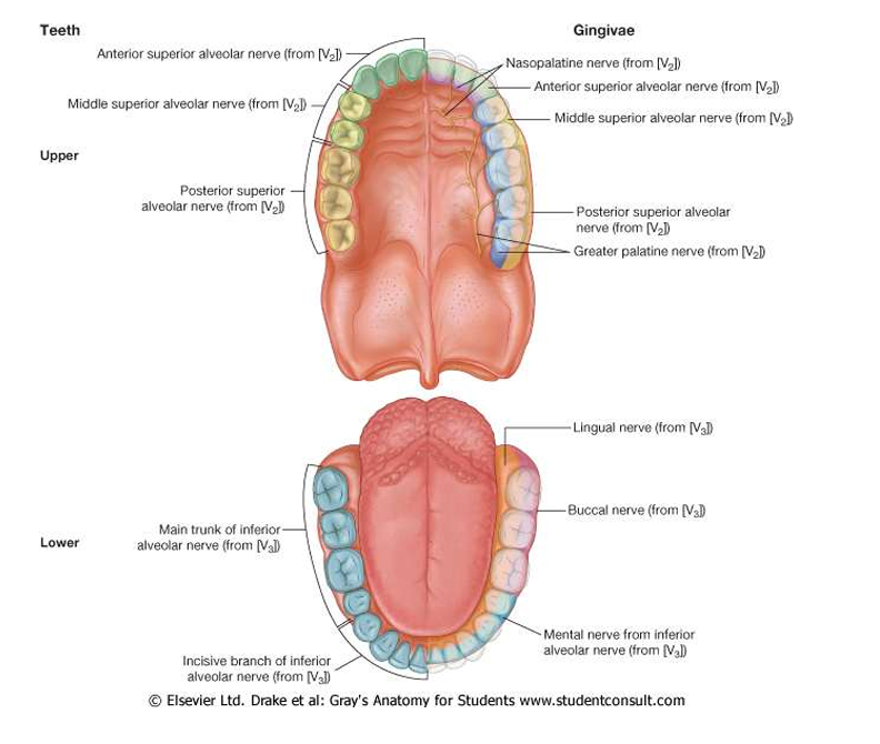
Image occlusion: name the hidden labels
223
Image occlusion: name the hidden labels
224
Image occlusion: name the hidden labels
225
Image occlusion: name the hidden labels
226
Image occlusion: name the hidden labels
227
Image occlusion: name the hidden labels
228
Image occlusion: name the hidden labels
229
Image occlusion: name the hidden labels
230
Image occlusion: name the hidden labels
231
Image occlusion: name the hidden labels
232
Image occlusion: name the hidden labels
233
Image occlusion: name the hidden labels
234
Image occlusion: name the hidden labels
235
innervations of submandibular and lingual glands
Parasympathetic – Facial N. (CN VII; chorda tympani)
Submandibular ganglion (site of Parasympathetic synapse)
Sympathetic – postganglionic fibers from superior cervical
ganglion give external carotid branches that form a
plexus on facial a. and accompany its branches to the
submandibular/sublingual glands
Submandibular ganglion (site of Parasympathetic synapse)
Sympathetic – postganglionic fibers from superior cervical
ganglion give external carotid branches that form a
plexus on facial a. and accompany its branches to the
submandibular/sublingual glands
236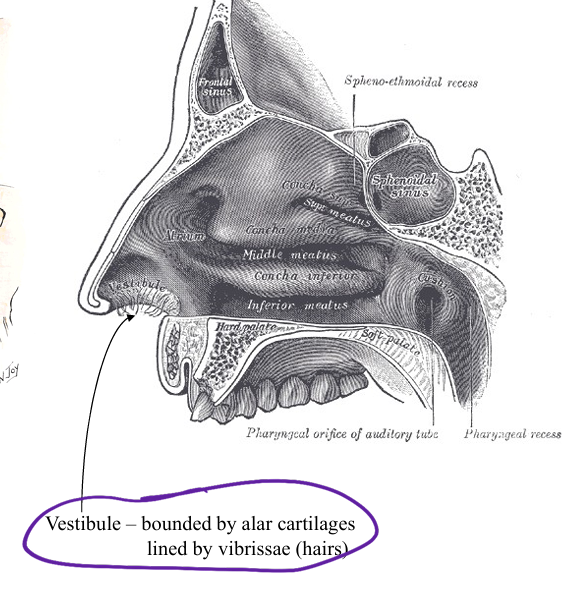
Image occlusion: name the hidden labels
237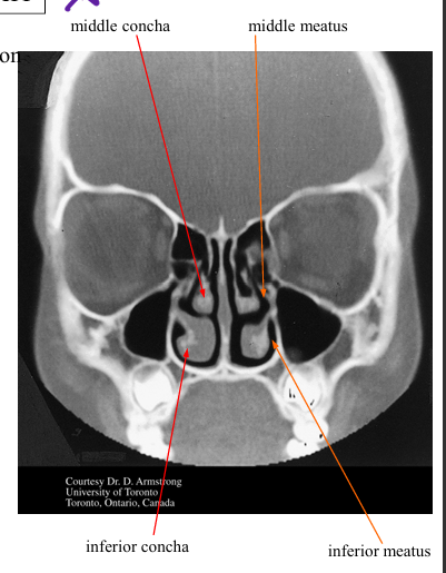
Image occlusion: name the hidden labels
238
Image occlusion: name the hidden labels
239
Image occlusion: name the hidden labels
240
Image occlusion: name the hidden labels
241
how does the frontal sinus drain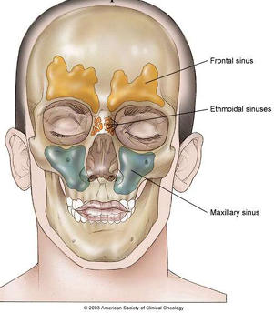
drains via frontonasal duct into anterior middle meatus and hiatus semilunaris
242
how do the ethmoidal air cells drain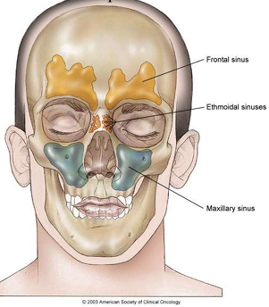
anterior: middle meatus at hiatus semilunaris--middle: on ethmoidal bulla in middle meatus--posterior: superior meatu
243
what drains the sphenoidal sinus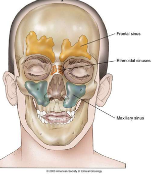
sphenoethmoidal recess drainage
244
what drains the maxillary sinus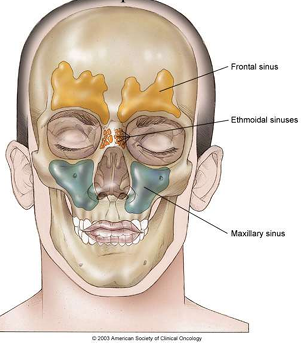
-posterior/inferior hiatus semilunaris drainage
245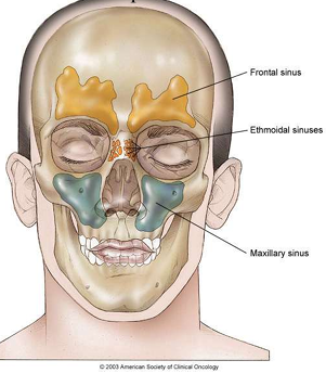
Image occlusion: name the hidden labels
246
Image occlusion: name the hidden labels
247
Image occlusion: name the hidden labels
248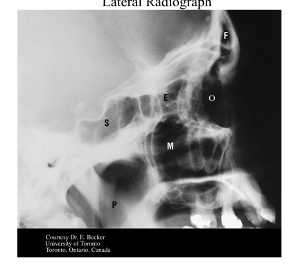
Image occlusion: name the hidden labels
249
Image occlusion: name the hidden labels
250
Image occlusion: name the hidden labels
251
Image occlusion: name the hidden labels
252
Image occlusion: name the hidden labels
253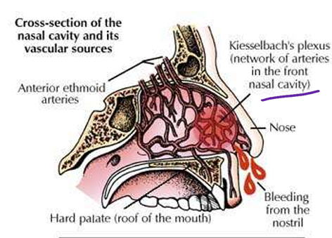
Image occlusion: name the hidden labels
254
anterior cavity lymph drainage nasal
facial/submandibular nodes
255
posterior cavity lymoh drainage
superior deep cervicak nodes
256
smell
SVA fibers via olfactorny n
257
general sense nasal cavity and paranasal sinuses
GSA via CN V1 and 2
258
parasympathetics to secretomotor to nasal mucosal glands
greater petrosal n via CN VII hitciking on CN V2; sympathetics via deep petrosal n hitchuke CN V2 (regulate blood flow)
259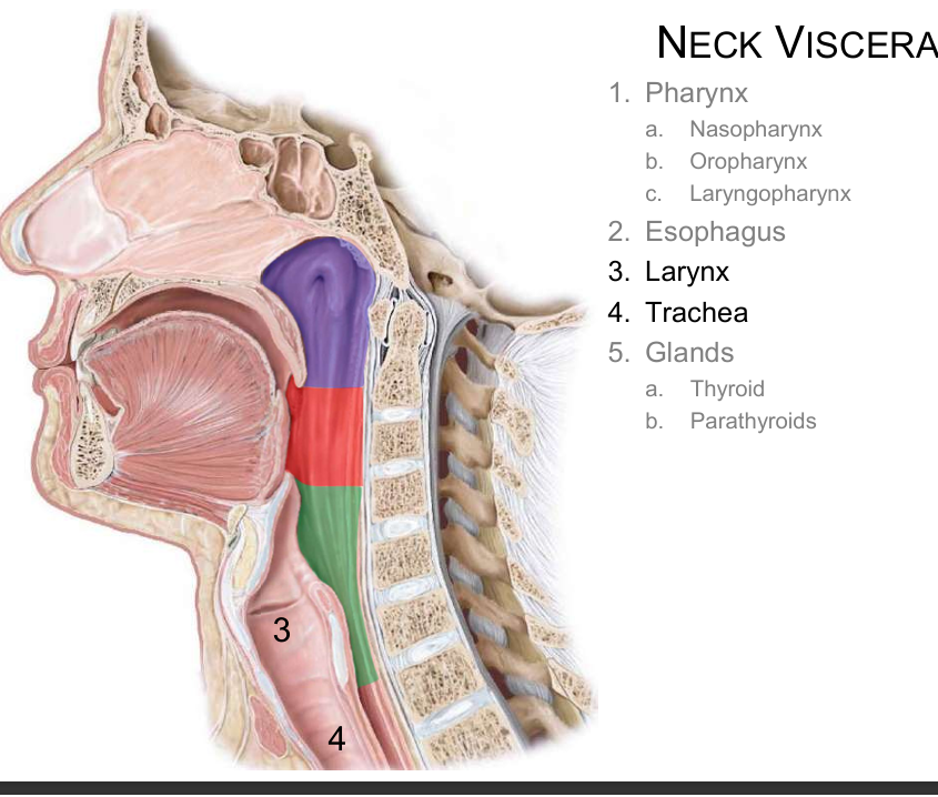
Image occlusion: name the hidden labels
260
Image occlusion: name the hidden labels
261
what is the movement at the cricothyroid jint?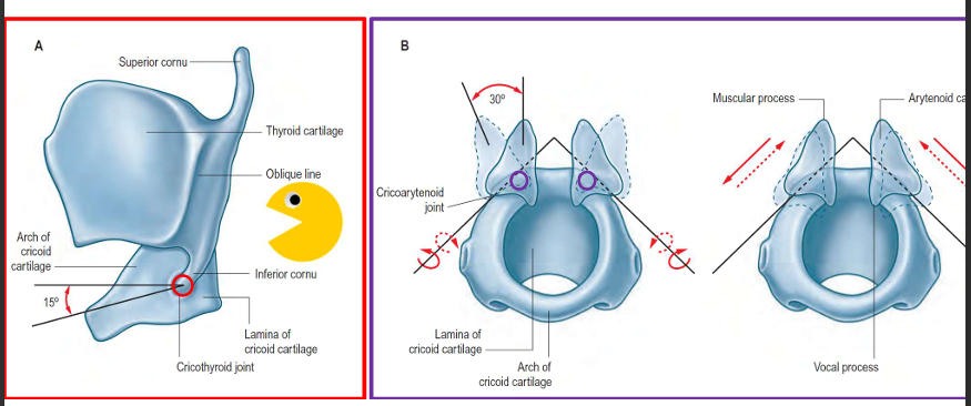
thyroid cartilage swings up and down
262
what is the movement at the cricoarytenoid articulation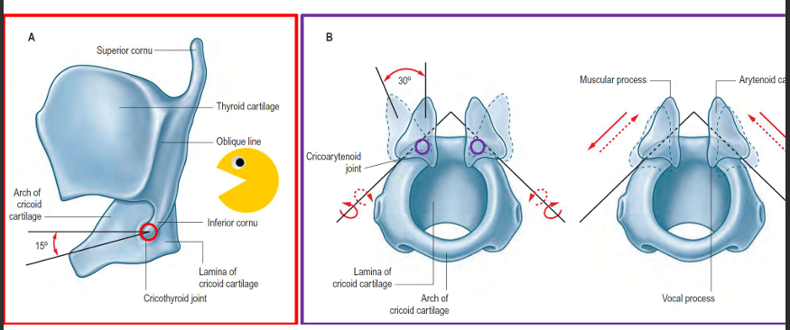
arytenoid cartilages swivel and slide on the cricoid
263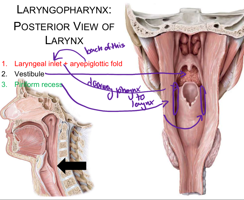
Image occlusion: name the hidden labels
264
Image occlusion: name the hidden labels
265
Image occlusion: name the hidden labels
266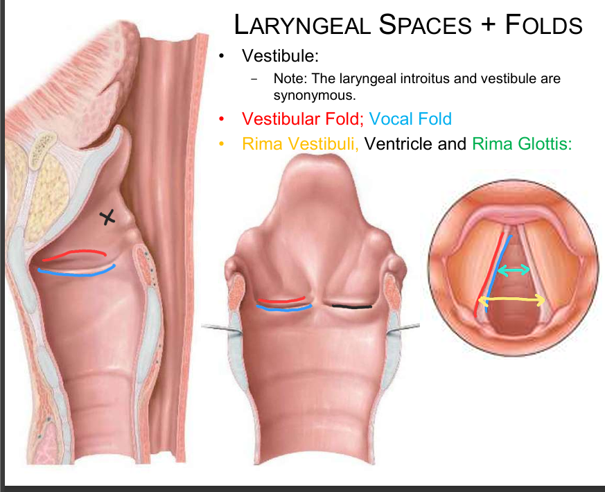
Image occlusion: name the hidden labels
267
Image occlusion: name the hidden labels
268
Image occlusion: name the hidden labels
269
Image occlusion: name the hidden labels
270
Image occlusion: name the hidden labels
271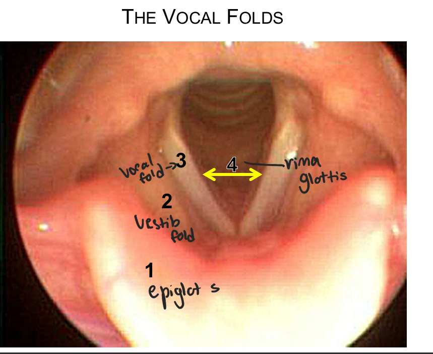
Image occlusion: name the hidden labels
272
Image occlusion: name the hidden labels
273
Image occlusion: name the hidden labels
274
Image occlusion: name the hidden labels
275
ddx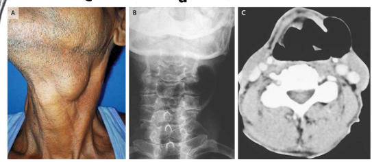
laryngeocoele
276
fx of thyroarytenoid m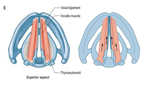
decrease tension of vocal cords; lower pitch
277
innervation thyroartenoid m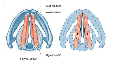
recurrent laryngeal n
278
innervation cricothyroid m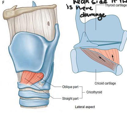
external laryngeal n
279
fx cricothyroid m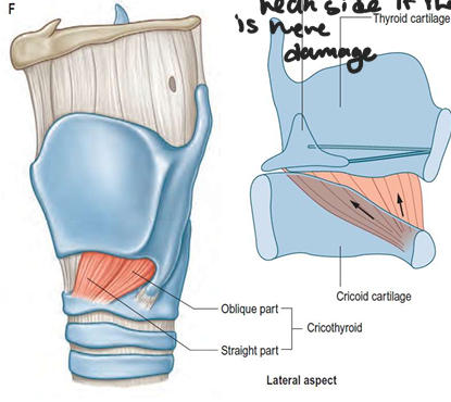
increase tension on vocl cords and increase pitch
280
if one of the cricothyroid m is weak, it will deviate to the side...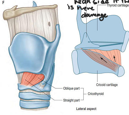
that is weak
281
fx posterior cricoarytenoid m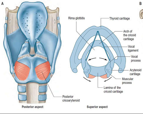
open rima glottis
282
fx lateral cricoarytenoid m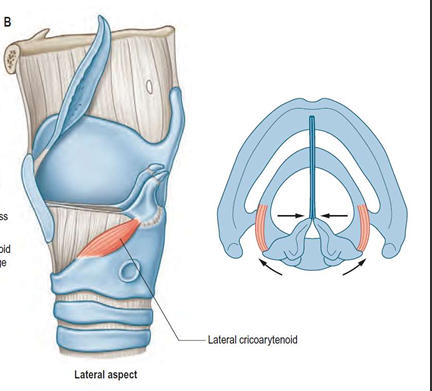
close rima glottis
283
fx aryepiglottcus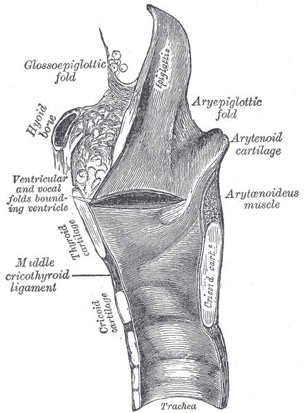
pulls epiglottis down and prevemts aspiring food
284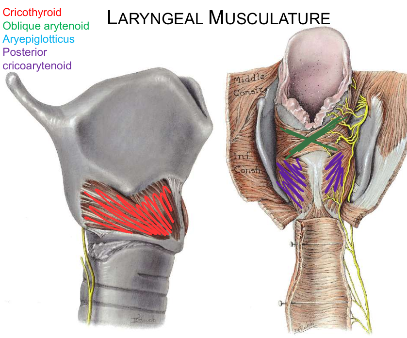
Image occlusion: name the hidden labels
285
Image occlusion: name the hidden labels
286
Image occlusion: name the hidden labels
287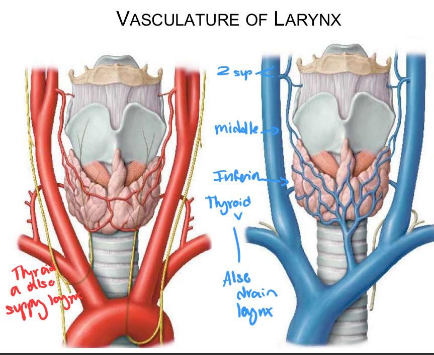
Image occlusion: name the hidden labels
288
Image occlusion: name the hidden labels
289
Image occlusion: name the hidden labels
290
what drains the larynx supraglottic
level II
(superior deep cervical)
(superior deep cervical)
291
what drains the infraglottic larynx
level IV
(inferior deep cervical) >
prelaryngeal, pretracheal
(inferior deep cervical) >
prelaryngeal, pretracheal
292
what structures are at risk in a cricothyrotomy
parasternal lobe ogf thyroid; thyroglossal duct
293
what structures are at risk when performing a treacheostomy
infrahyoid v; thyroidema
294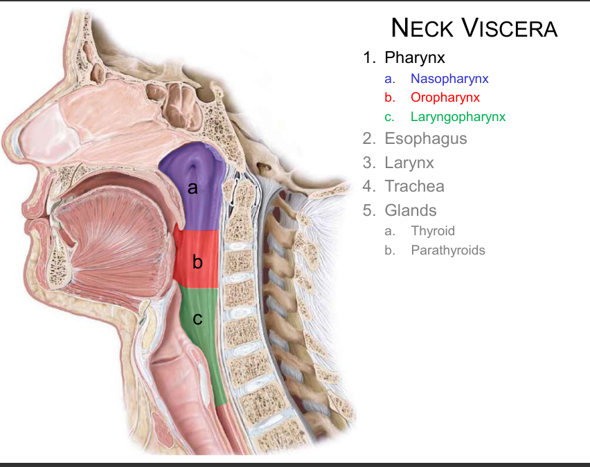
Image occlusion: name the hidden labels
295
Image occlusion: name the hidden labels
296
Image occlusion: name the hidden labels
297
boundaries of nasopharynx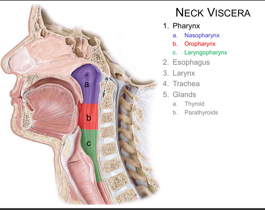
skull base to soft palate
298
boundaries of oropharynx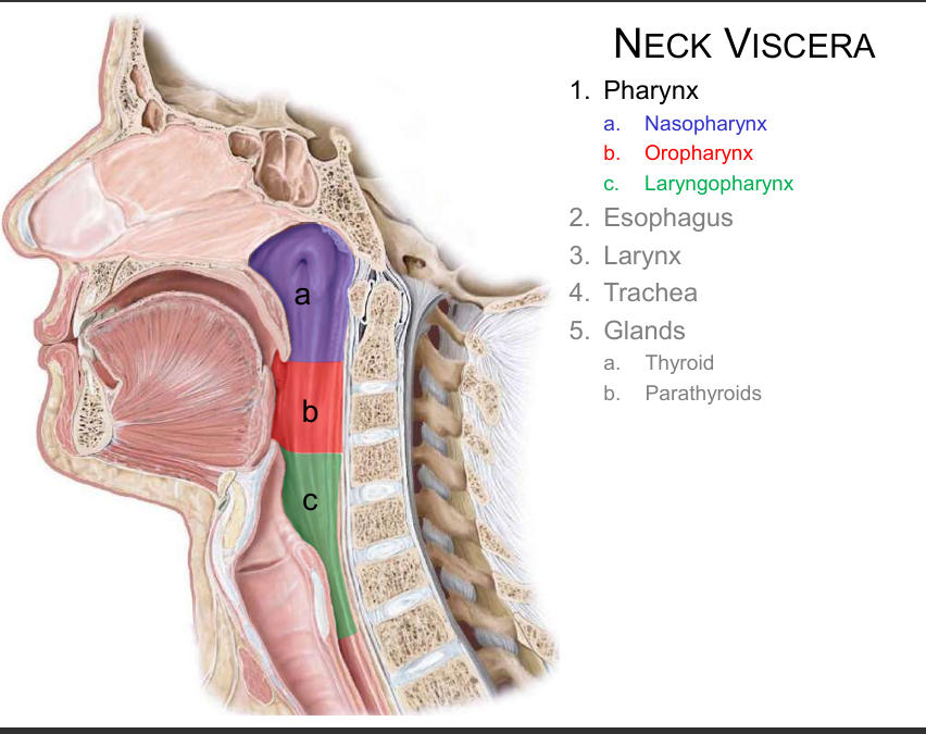
soft oalkate to epiglottis
299
boundaries of laryngopharynx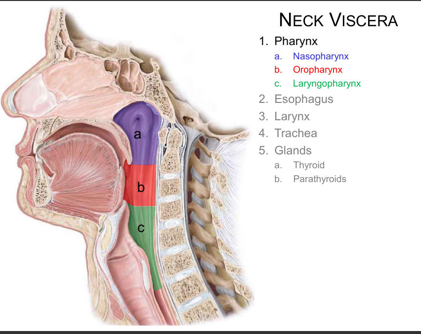
epiglottis to bottom of cricoud cartilage
300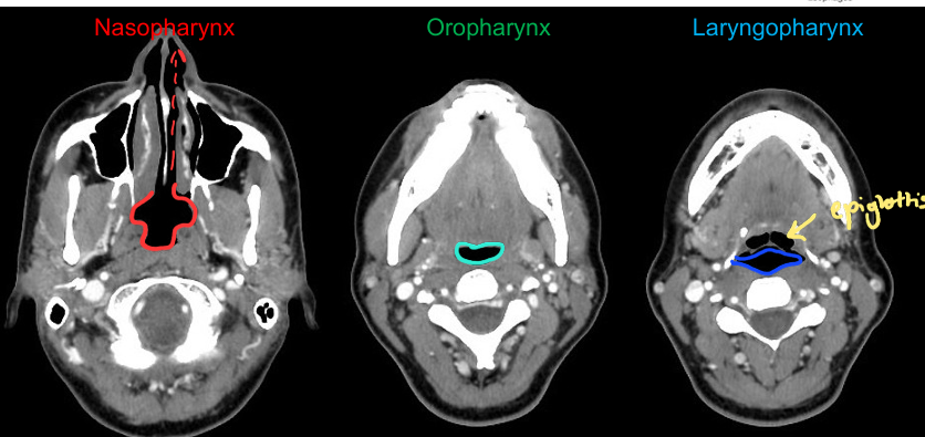
Image occlusion: name the hidden labels
179 more cards in OutLearn. Open the full deck