Anatomy S1 U2
Study these flashcardsFree · spaced repetition · iPhone, Android, Mac & web
All cards
1
parietalPleura related to the ribs and intercostal spaces is termed
costal part 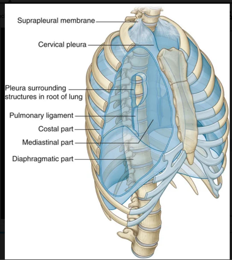
2
parietal Pleura covering the diaphragm is
diaphragmic part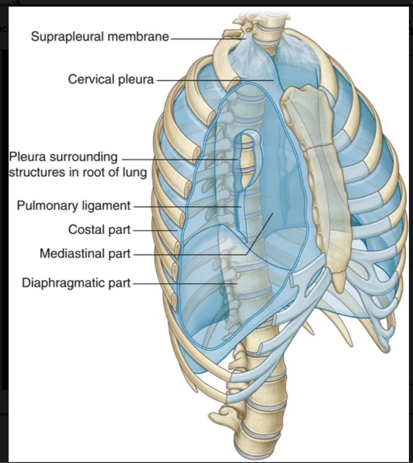
3
parietal Pleura covering the mediastinum is
mediastinal part
4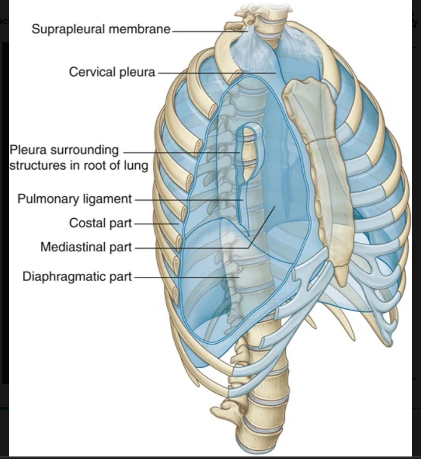
Image occlusion: name the hidden labels
5
Image occlusion: name the hidden labels
6
Image occlusion: name the hidden labels
7
Image occlusion: name the hidden labels
8
Image occlusion: name the hidden labels
9
Covering the superior surface of the cervical pleura is a distinct dome-like layer of fascia, the ____________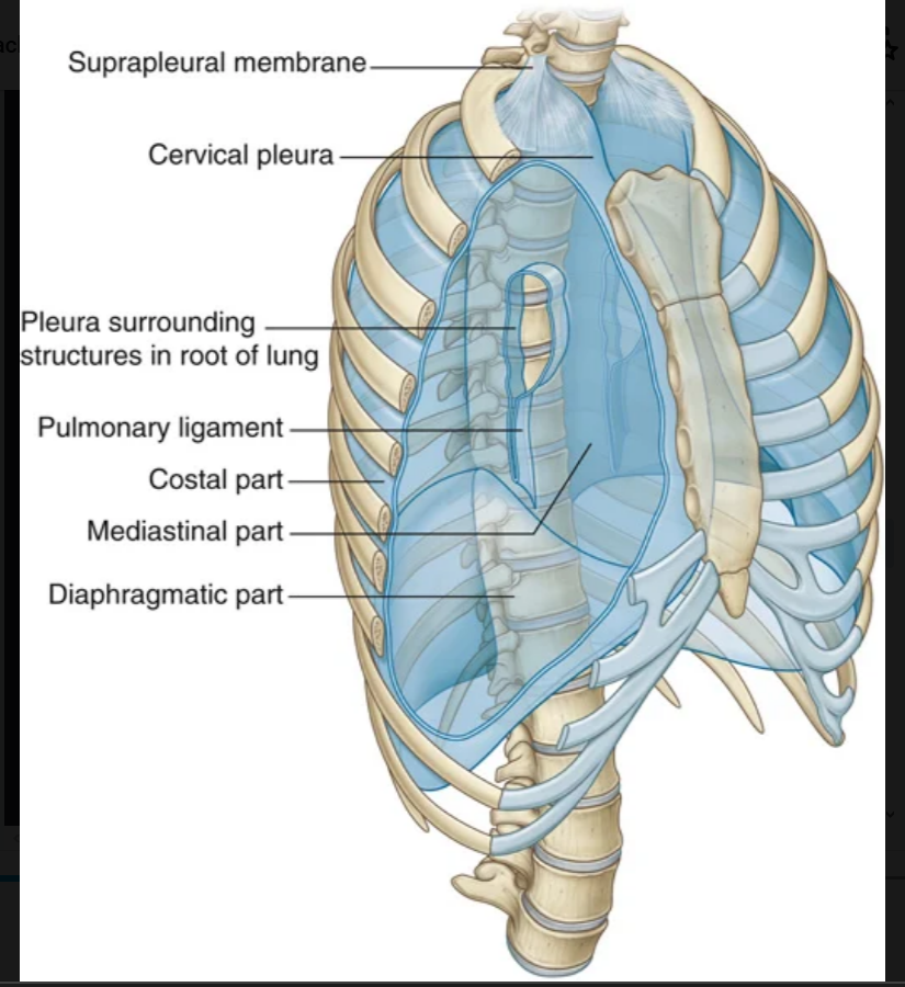
suprapleural membrane
10
the parietal pleura innervation
somatic afferent fibers
11
what is the costal pleura innervated by
intercostal neves
12
wjat is the diaphragmatic and mediastinal pleura innervated by?
phrenic n (C3-5)
13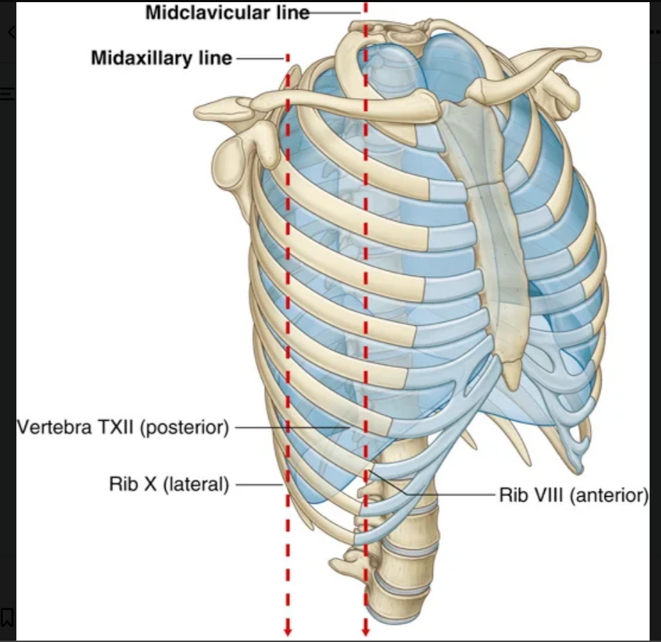
Image occlusion: name the hidden labels
14
Image occlusion: name the hidden labels
15
what innervates the visceral pleura
visceral efferent nerves
16
expansion of the lungs into the entire pleural cavity only occurs furing
forced inspiration
17
occurs when excess fluid accumulates within the pleural space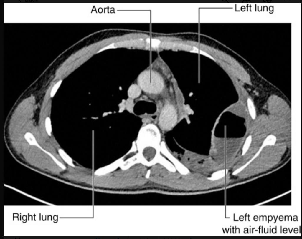
pleural effusin
18
pneumothorax: Occasionally, the gas within the pleural cavity may accumulate to such an extent that the mediastinum is “pushed” to the opposite side, compromising the other lung. This is termed a _______ pneumothorax and requires urgent treatment.
tension
19
A thin blade-like fold of pleura projects inferiorly from the root of the lung and extends from the hilum to the mediastinum. This structure is the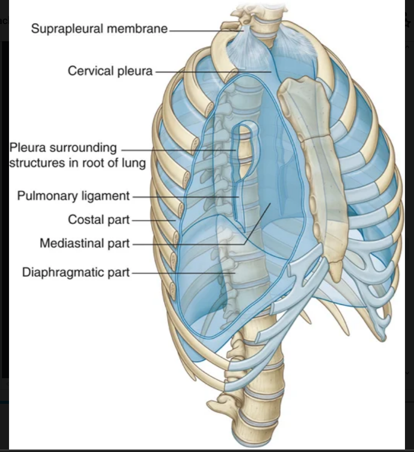
pulmonary ligament
20
what is located in the hilum of the lung? (x6)
pulmonary artery, 2 pulmonary veins, a main brnchus, broncjial vessels, nerves, lymohatics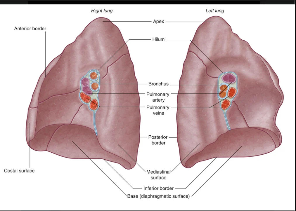
21
what is the medial srface f the RIGHT lung adjavent to
▪ heart,
▪ inferior vena cava,
▪ superior vena cava,
▪ azygos vein, and
▪ esophagus.
▪ inferior vena cava,
▪ superior vena cava,
▪ azygos vein, and
▪ esophagus.
22
the medial surface of the left lung lies adjacent to this
▪ heart,
▪ aortic arch,
▪ thoracic aorta, and
▪ esophagus.
▪ aortic arch,
▪ thoracic aorta, and
▪ esophagus.
23
the smallest functionally independent region of a lung and the smallest area of lung that can be isolated and removed without affecting adjacent regions.
bromchiopulmonary segment
24
what does the right bronchial a originate from
third posterior intercostal artery (but occasionally, it originates from the upper left bronchial artery).
25
what does the left bronchial a arise from
directly from the anterior surface of the thoracic aorta
26
what do the broncjial v drain into
▪ either the pulmonary veins or the left atrium, and
▪ into the azygos vein on the right or into the superior intercostal vein or hemiazygos vein on the left.
▪ into the azygos vein on the right or into the superior intercostal vein or hemiazygos vein on the left.
27
a tough connective tissue outer layer that defines the boundaries of the middle mediastinum.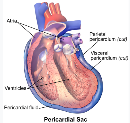
fibrous pericardium
28
two layers of the serous pericardium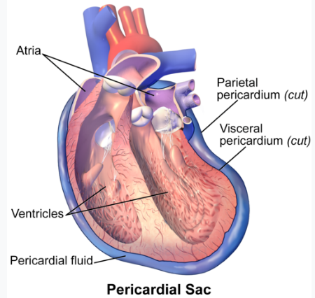
parietal layer and visceral layer
29
these attachments help retain the heart in its position in the thoracic cavity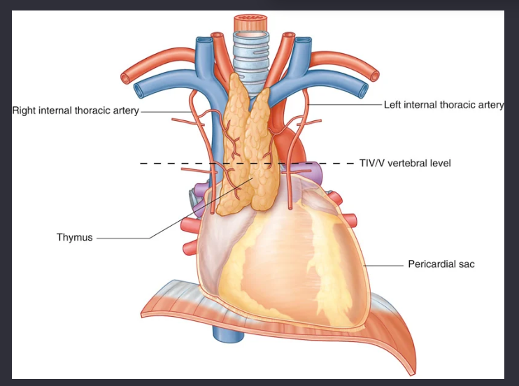
sternopericardial ligaments
30
an inflammatory condition of the pericardium. Common causes are viral and bacterial infections, systemic illnesses (e.g., chronic renal failure), and after myocardial infarction.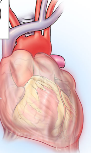
pericarditis
31
pericarditis symptomology
complaints of central chest pain that may radiate to one or oth arms, pain may be relieved y sitting forward
-shows diffuse ST elevation
-shows diffuse ST elevation
32
Abnormal thickening of the pericardial sac (___________-), which usually involves only the parietal pericardium, but can also less frequently involve the visceral layer, can compress the heart, impairing heart function and resulting in heart failure.
constrictive pericarditis
33
circles the heart, separating the atria from the ventricles (Fig. 3.70). As it circles the heart, it contains the right coronary artery, the small cardiac vein, the coronary sinus, and the circumflex branch of the left coronary artery.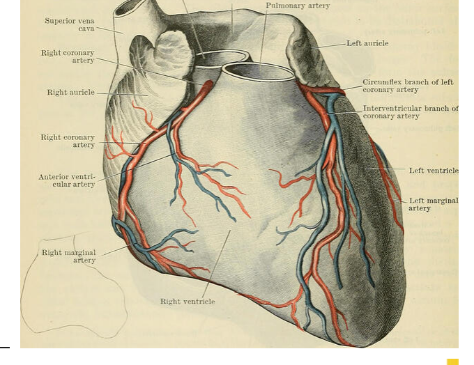
coronary sulcus
34
From the right atrium, blood passes into the right ventricle through the ______________. This opening faces forward and medially and is closed during ventricular contraction by the tricuspid valve.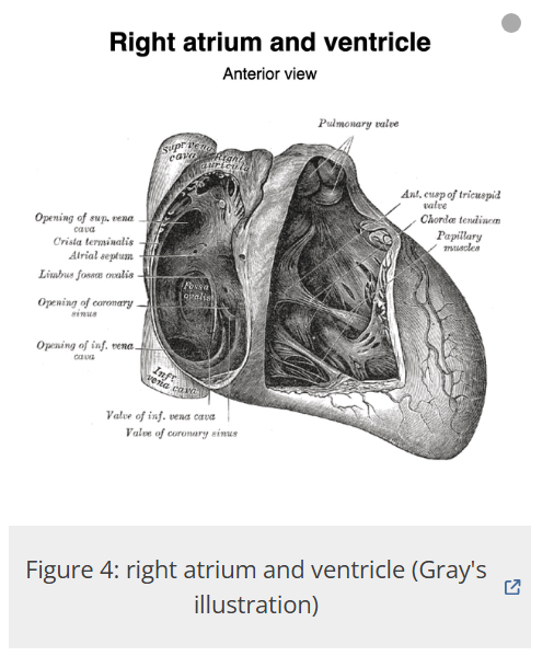
right atrioventricular orifice
35
right ventricle: the largest and most constant papillary muscle, and arises from the anterior wall of the ventricle.
anterior pappilary musle
36
right ventricle: may consist of one, two, or three structures, with some chordae tendineae arising directly from the ventricular wall.
posterior pappillary muscle
37
right ventricle: most inconsistent papillary muscle, being either small or absent, with chordae tendineae emerging directly from the septal wall.`
septal papillary muscle
38
orms a bridge between the lower portion of the interventricular septum and the base of the anterior papillary muscle.
-carries a portion of the cardiac conduction system, the right bundle of the atrioventricular bundle, to the anterior wall of the right ventricle.
-carries a portion of the cardiac conduction system, the right bundle of the atrioventricular bundle, to the anterior wall of the right ventricle.
septomarginal trabecula (moderator band)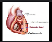
39
heart changes in mitral va;lve disease (x4)
▪ left ventricular hypertrophy (this is appreciably less marked in patients with mitral stenosis);
▪ increased pulmonary venous pressure;
▪ pulmonary edema; and
▪ enlargement (dilation) and hypertrophy of the left atrium.
▪ increased pulmonary venous pressure;
▪ pulmonary edema; and
▪ enlargement (dilation) and hypertrophy of the left atrium.
40
what causes aortic valve stenosis
atherosclerosis causing calcification of the valve leaflets
41
what does the right coronary artery supply
upplies the right atrium and right ventricle, the sinu-atrial and atrioventricular nodes, the interatrial septum, a portion of the left atrium, the posteroinferior one-third of the interventricular septum, and a portion of the posterior part of the left ventricle.
42
what does it mean to be a dominant coronaru artery
the posterior interventricular branch arises from that artery (right or left)
43
when does a heart attack occur?
when the perfusion to the myocardium is insufficient to meet the metabolic needs of the tissue, leading to irreversible tissue damage.
44
This is a technique in which a long fine tube (a catheter) is inserted into the femoral artery in the thigh and passed through the external and common iliac arteries and into the abdominal aorta.
- A fine wire is then passed into the coronary artery and is used to cross the stenosis. A fine balloon is then passed over the wire and may be inflated at the level of the obstruction, thus widening it; this is termed angioplasty.
- A fine wire is then passed into the coronary artery and is used to cross the stenosis. A fine balloon is then passed over the wire and may be inflated at the level of the obstruction, thus widening it; this is termed angioplasty.
percutaneous coronary intervention
45
The great saphenous vein, in the lower limb, is harvested and used as a graft. It is divided into several pieces, each of which is used to bypass blocked sections of the coronary arteries. The internal thoracic and radial arteries can also be used.
coronary artery bypass graft
46
allows oxygenated blood to flow from the left atrium (higher pressure) across the ASD into the right atrium
ASD
47
When this occurs, the oxygenated blood in the aortic arch (higher pressure) passes into the left branch of the pulmonary artery (lower pressure) and produces pulmonary hypertension and left atrial and ventricular enlargement
PDA
48
women heart attack symptoms
abdominal pain, achiness in the jaw or back, nausea, shortness of breath, or simply fatigue
49
order of the cardiac conducting system
sinus atrial node
atrioventricular node
atroventricular budle wight and left bunfdle branches
purkinje fibers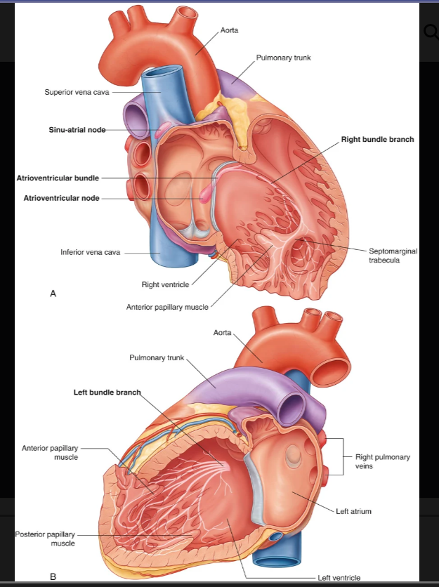
atrioventricular node
atroventricular budle wight and left bunfdle branches
purkinje fibers
50
where is the sinu atrial node located
superior end of the crista terminalis at the junction of the superior vena cava and the right atrium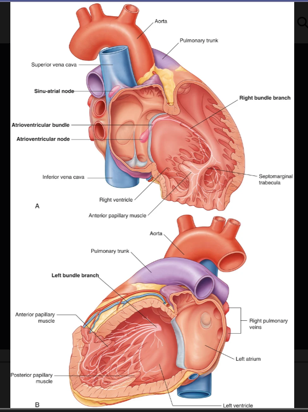
51
where is the AV node located
near the opening of the coronary sinus, close to the attachment of the septal cusp of the tricuspid valve, and within the atrioventricular septum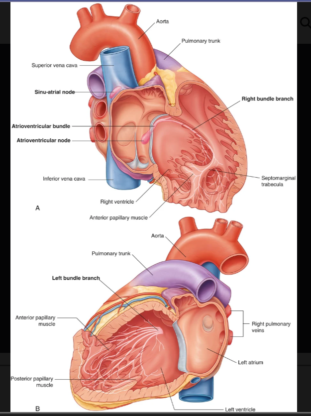
52
what is the autonomic division of the NS respnsible for regiulating in the hjeart
increases hjeart rate, increases force of contraction, cardiac output
53
what is the parasympathetic innervation of the NS responsible for regulating in the heart
decrease heart rate, reduce force of contraction, constricts the coronary arteries
54
what is the sympathetic innervation of the NS responsible for in regulating the heart
increasing heart rate and increasing force of contraction