Histo Exam 1
Study these flashcardsFree · spaced repetition · iPhone, Android, Mac & web
All cards
1
these allow proteins in the membrane to interact with other cellsroups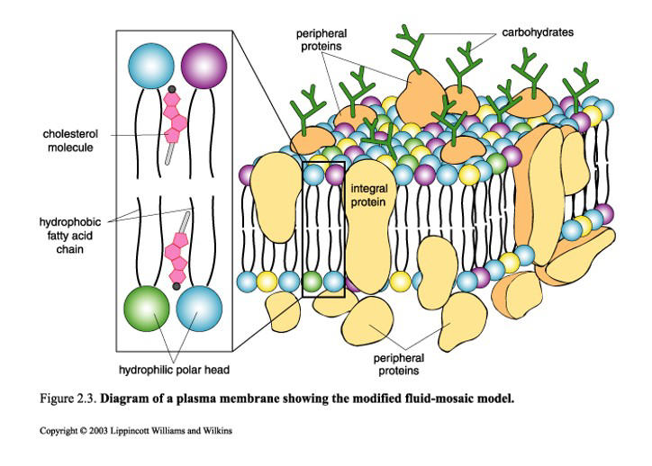
carbohydrates
2
these allow proteins in the membrane to have more motility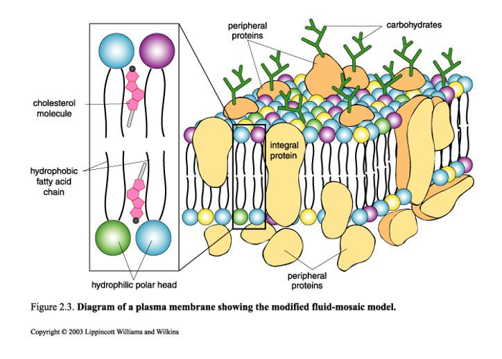
cholesterol
3
what are the different types of integral membrane proteins?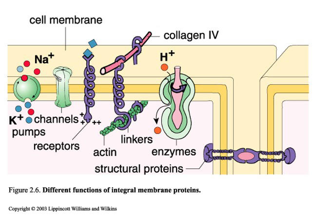
pumps, channels, receptors, linkers, enzymes, structural
4
move substances against the concentration gradient, integral membrane protein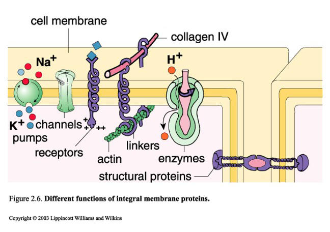
pumps
5
favorable membrane transport, energy npt required for things to move across this thing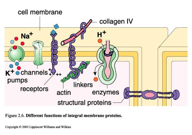
channels
6
rigid scafolding so cells dont move freely along the medium, integral membrane proteins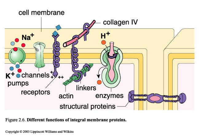
linkers
7
diffusion with no mediation needed
EX; uncharged small nonpolar molecules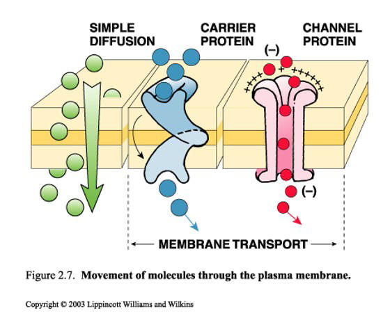
EX; uncharged small nonpolar molecules
simple diffusion
8
used in active (pumps) or passive transport, highly selective for what they are moving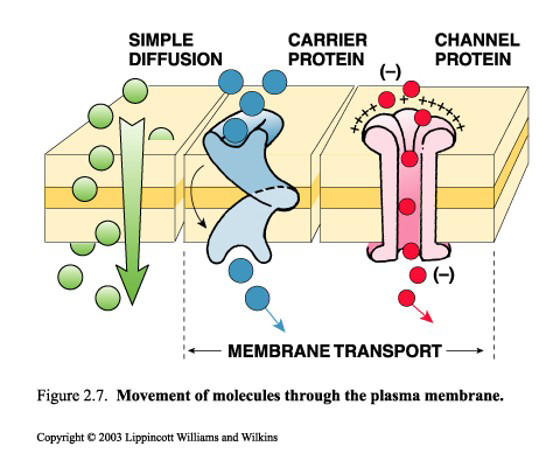
carrier proteins
9
ligand gated or voltage gated, regulated by membrane potentials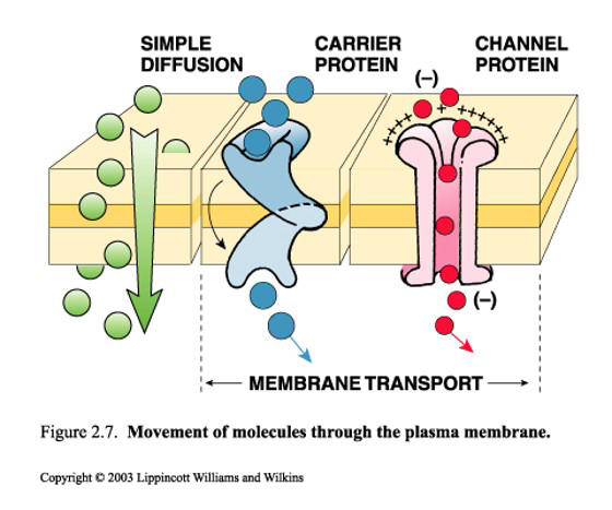
channel proteins
10
phrase for favorable (conc)
down conc. gradient
11
phrase for nonfavorable (conc)
up conc gradient
12
types of endocytosis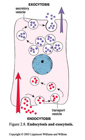
pinocytosis, phangocytosis, receptor mediated endocytosis
13
types of exocytosis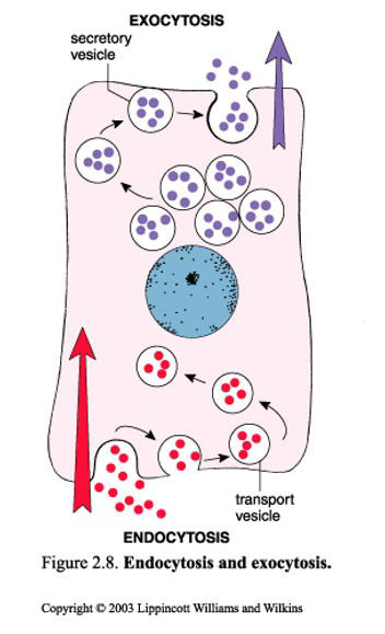
constitutive: constant
regulated: needs signaling to occur
regulated: needs signaling to occur
14
cell drinking, nonspecific, small proteins and fluids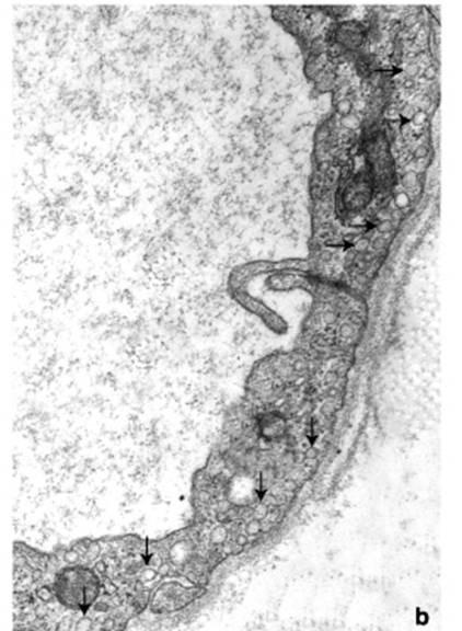
pinocytosis
15
Engulf cell debris and bacteria
Requires rearrangement of the actin cytoskeleton
Form phagosomes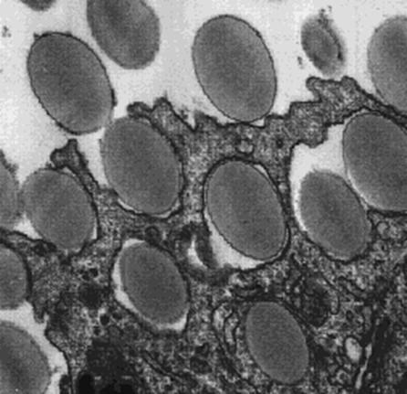
Requires rearrangement of the actin cytoskeleton
Form phagosomes
phangocytosis
16
steps to receptor mediated endocytosis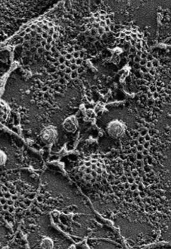
1) cargo protein binds to cargo receptor as adaptin also binds to intracellular cargo receptor
2) clarthrin binds to adaptin, coated pin forming
3) coated pit formed and vessel invaginates
4) vessel completely invaginates and moves into the cell
5) vessel begins uncoating as adaptin/clarthrin bindings break off
6) uncoated vesicle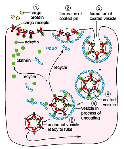
2) clarthrin binds to adaptin, coated pin forming
3) coated pit formed and vessel invaginates
4) vessel completely invaginates and moves into the cell
5) vessel begins uncoating as adaptin/clarthrin bindings break off
6) uncoated vesicle
17
what is the functio and pH of early endosomes
sory and recycle proteins; 6.2
18
what is the function and ph of late endosomes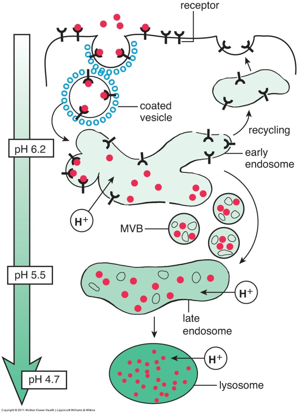
pre lysosomes, some sorting still occurs; 5.5
19
what is the structure and ph of lyososomes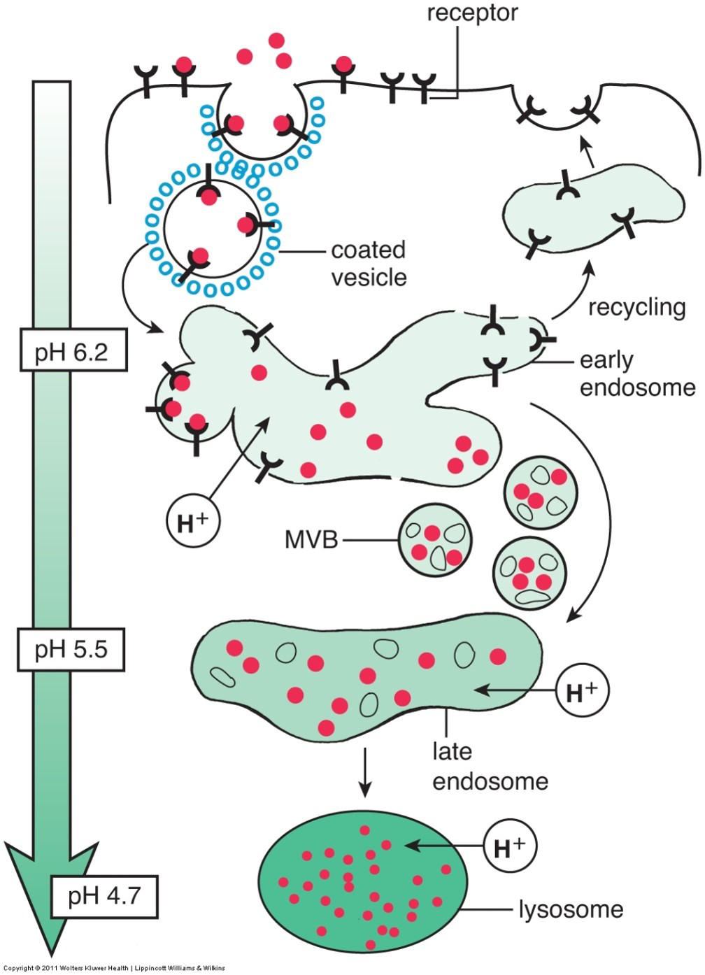
degradation, ph 4.7
20
what 4 pathways lead to intracellular digestion in lysosomes?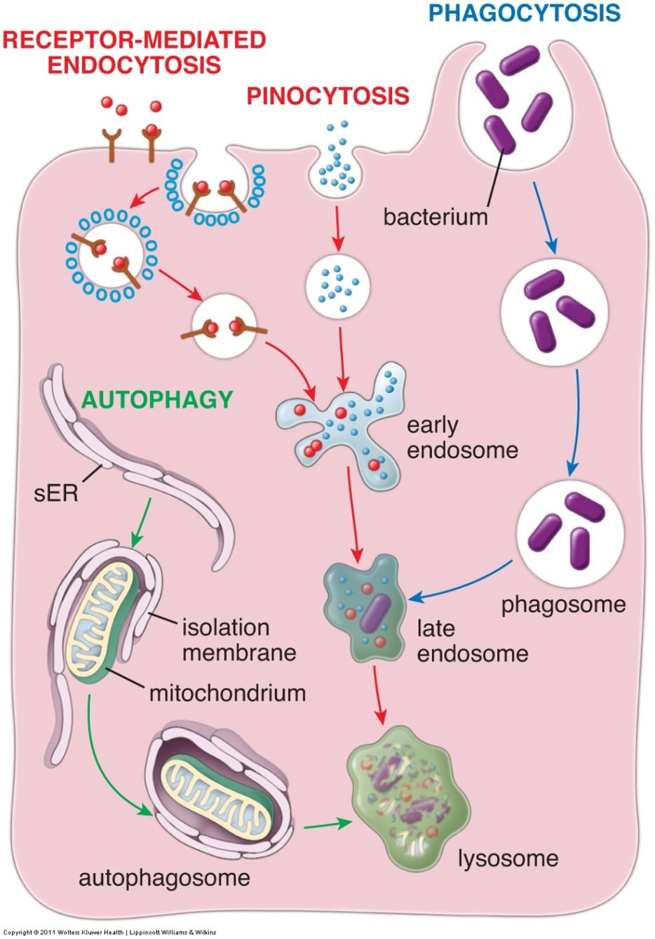
receptor mediated endocytosis, pinocytosis, phangocytosis, autophagy
21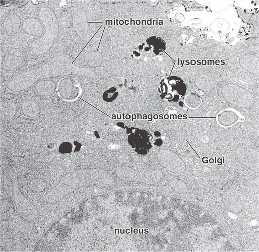
Image occlusion: name the hidden labels
22
Image occlusion: name the hidden labels
23
Image occlusion: name the hidden labels
24
Image occlusion: name the hidden labels
25
Image occlusion: name the hidden labels
26
Loss of vision & hearing, muscle atrophy from loss of nervous tissue, early death (< 5y)
Tay Sachs
27
what enzyme is deficient in tay sachs
hexosaminidase
28
what is the function of hexosaminidase (tay sachs)
Gm2 ganglioside breakdown, builds up in lysosomes and neuronal cells when deficient
29
Skeletal abnormalities, hepatomegaly, intellectual disability, early death (< 8y)
I-cell disease
30
what enzyme is deficient in I cell disease?
N-acetylglucosaminyl-1-phosphotransferase
31
what is the function of N-acetylglucosaminyl-1-phosphotransferase?
lysosomal hydrolases are not phosphorylated and get secreted instead leading to multiple substances accumulating in the lysosomes
32
Hepatosplenomegaly, neurodegeneration, progressive wasting due to sphingomyelin accumulation; early death (< 3y)
Niemann-Pick (type A)
33
Sphingomuelinase
what enzyme is deficient in niemann pick type a
34
what happens with sphingomuelinase deficiency? (niemann pick type a)
sphingomyelin accumulates
35
Affects the spleen, liver, lungs, bone marrow
Gaucher
36
what enzyme is effected in gaucher?
B glycocerebrosidase
37
what does a deficiency in B glucocerebrosidase cause?
glucosyleramide accumulaes in macrophage (monocyte system)
38
contains enzymes that break down H2O2
peroxisomes
39
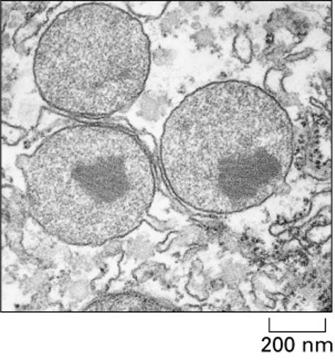
functions of peroxisomes
β-oxidation of fatty acids
Detoxification of ethanol
Synthesis of plasmalogens
Detoxification of ethanol
Synthesis of plasmalogens
40
what is the deficiency in zellweger syndrome?
Mutations in at least 12 genes ID’d, code for peroxins
41
what does the mutation in zellweger syndrome cause?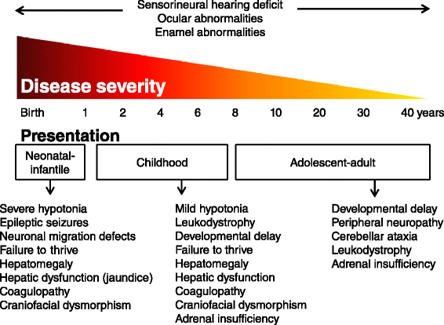
reduce myelin synthesis and loss of white matter
42
Rough ER function?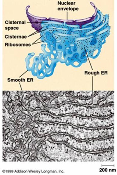
synthesis of secreted and membrane proteins
43
smooth ER function
lipid metabolism
detoxification
steroid synthesis
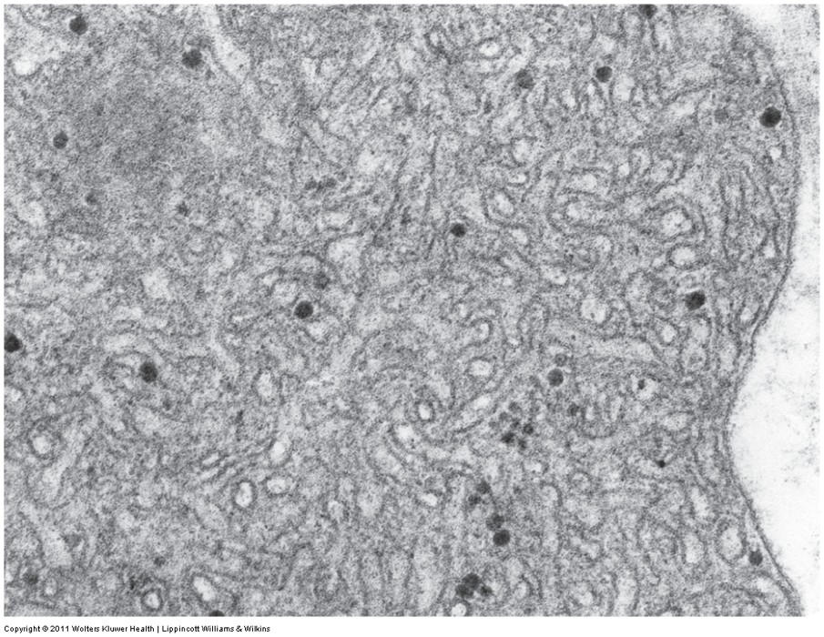
detoxification
steroid synthesis
44
Golgi function?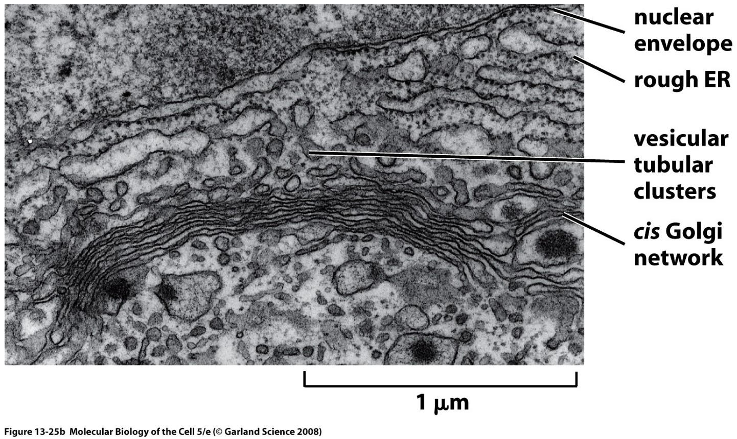
post translational modification, sorting, pavkaging of proteins
45
what are the types ofnonliving inclusions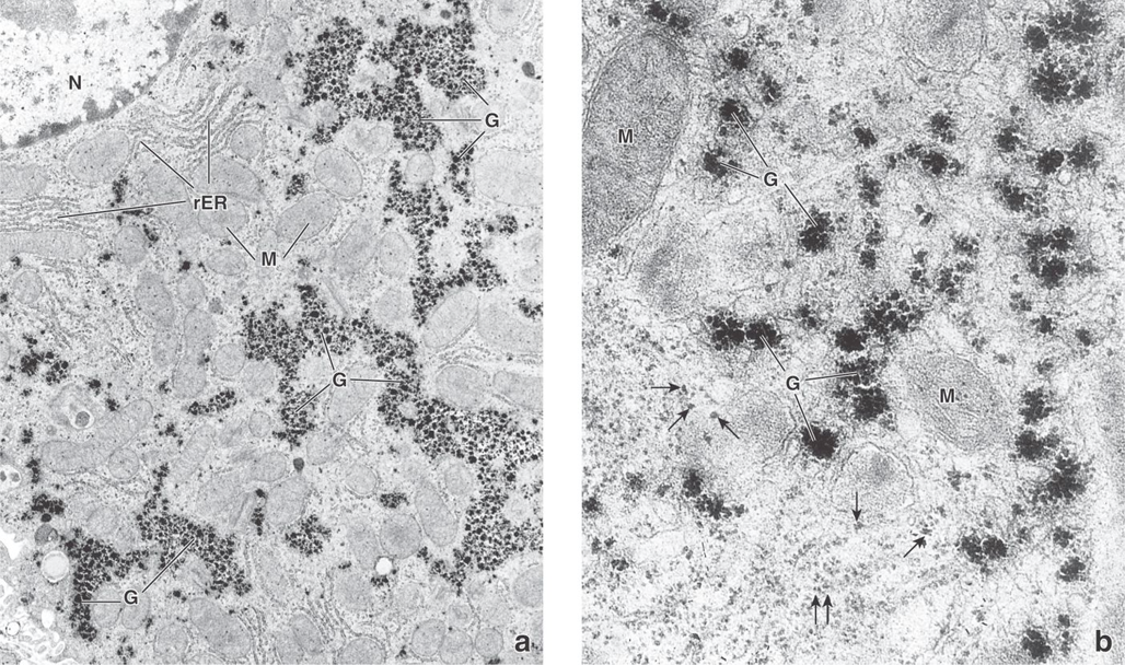
Secretory granules
Stored energy (e.g., glycogen and fat)
Pigments (e.g., hemoglobin and melanin)
Stored energy (e.g., glycogen and fat)
Pigments (e.g., hemoglobin and melanin)
46
Cytoskeleton parts
microtubles, actin, intermediate filaments
47
what happens if you have more GTP than GDP
polymerization
48
functions of microtbles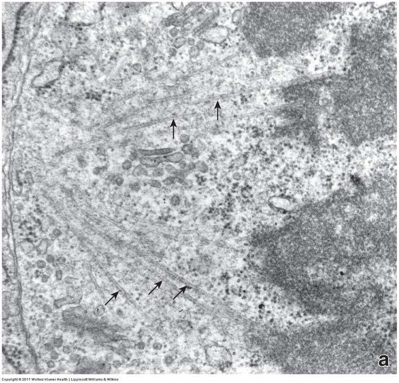
Vesicular transport
Movement of cilia and flagella
Alignment, separation of chromosomes during cell division
Movement of cilia and flagella
Alignment, separation of chromosomes during cell division
49
movement + to -
dyneins
50
movement - to +
kinesins
51
align mitotic spindle during cellular division
centrosomes
52
Central point of MTOC assembly
9 sets of microtubule triplets
centrosome is 2 of these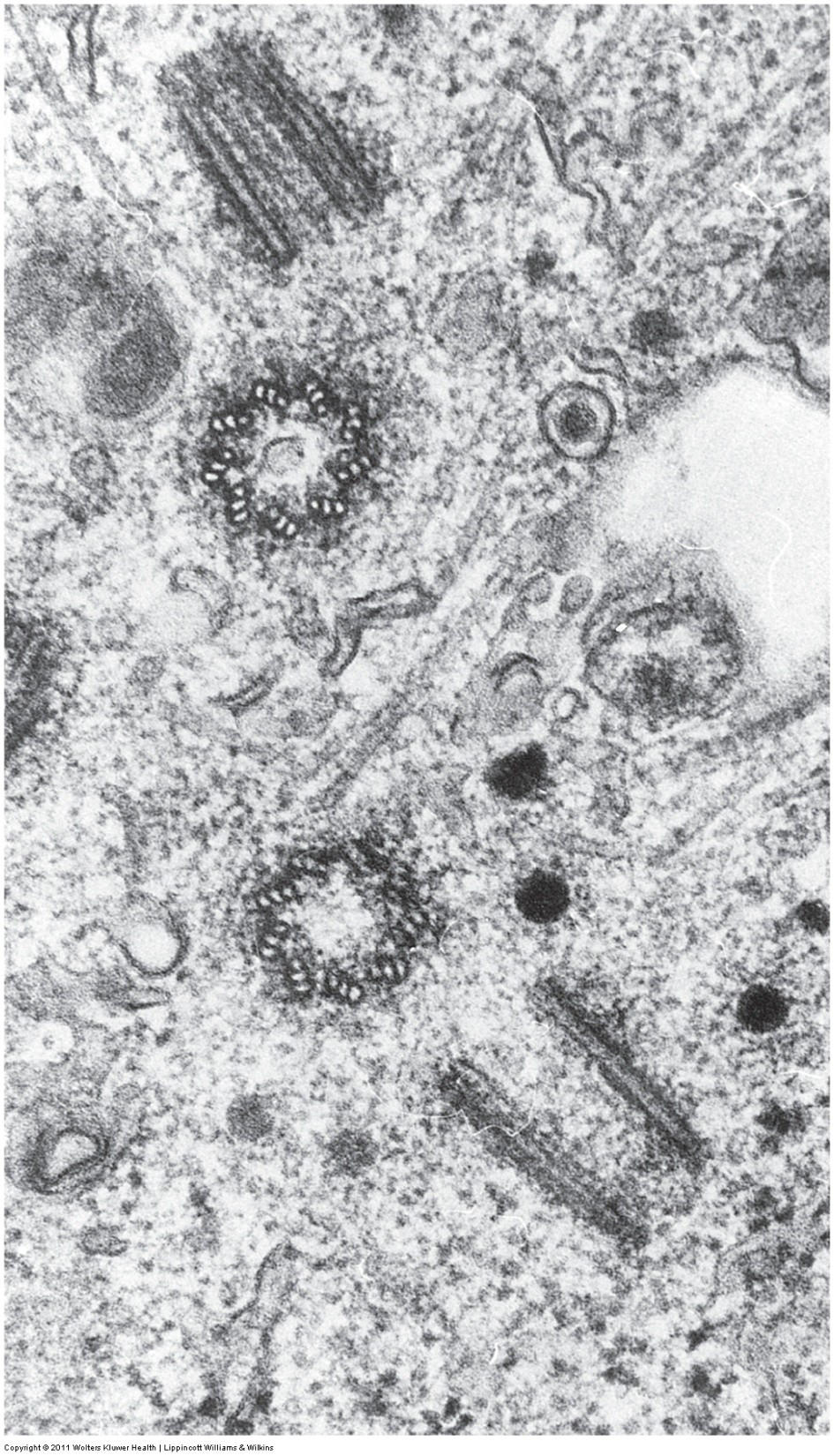
9 sets of microtubule triplets
centrosome is 2 of these
centrioles
53
microtuble disorder
kartagener syndrome
54
what happens in kartagener syndrome
immobility of cilla and infertility
55
what are the chemotherapy drugs? (microtuble drugs)
taxol (paclitaxel) and vinblastine/vincristine
56
prevents depolymerization microtubles
taxol (paclitaxel)
57
inhibits formation of mitotic spindke for cell division
vinblastine/vincristine
58
actin functions (x5)
Cell shape/structure
Microvilli
Anchorage and movement
Extension of cell processes
Locomotion
Microvilli
Anchorage and movement
Extension of cell processes
Locomotion
59
prevents depolymerization by binding to F actin
phalloidin
60
prevents polymerization of actin; this inhibits lymphocyte migration, phangocytosis, and cell division
cytochalasin B and D
61
intermediate fillaments function
structural role
62
what are the types of intermediate filaments
Keratins
All epithelial cells
Vimentins
Fibroblasts, neurons
Neurofilaments
Neurons
Lamins
Most differentiated cells, all nucleated cells
All epithelial cells
Vimentins
Fibroblasts, neurons
Neurofilaments
Neurons
Lamins
Most differentiated cells, all nucleated cells
63
intermediate filament disorders
alcohol liver cirrhosis: keratin accumulation as mallory bodies, alzheimers: neurofilaments
64
function of nucleolus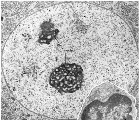
rRNA synthesis
65
scaffolding for the chromosomes and the nuclear pores
nuclear lamina
66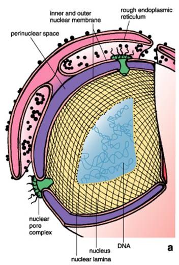
Image occlusion: name the hidden labels
67
Image occlusion: name the hidden labels
68
Image occlusion: name the hidden labels
69
impaired nuclear lamina disorders (x2)
progeria, emery-dreifuss muscular dystrophy
70
what gene is mutated in progeria
lamin A/C gene
71
what happens with a lamin A/C gene mutation (progeria)
splicing of lamina A mRNA, lamin A tight;y associated with nuclar memnrane si there are problems with chromatin scaffolding in nucleus
72
what is mutated/effected n emery dreifuss muscular dystrophy
multiple genes, affects the nuclear envelope; muscle weakness and stiff joints (onset age 10)
73
X chromosome is repressed and stays tightly condensed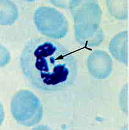
Barr body
74
what happens during the interphase phases?
G1: proteins neeed for DNA synthesis
S: DNA synthesis
G2: growth and reorganization
S: DNA synthesis
G2: growth and reorganization
75
what is the Go phase
cells are stable and nondividing
76
what is the Gtd phase
permanent nondividing cells phase
77
what regulates the cell cycle?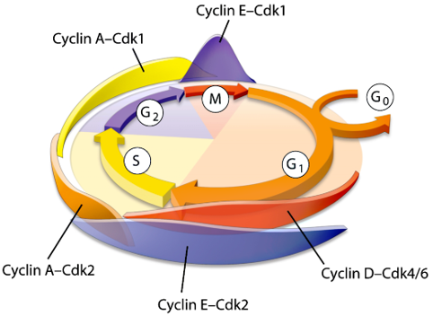
cyclins which activate cyclin dependent kinases at checkpoint regulators
78
accidental cell death where the cell lyses and swells
necrosis
79
basophilic dye
hematoxylin
80
what do basic dyes color
heterochromatin and nucleoli
cytoplasm
ECM
cytoplasm
ECM
81
acidc dye
eosin
82
what does acidophilic dye dye?
cytoplasmic filaments
Intracellular memnrane filamennts
extracelular fibers
Intracellular memnrane filamennts
extracelular fibers
83
where is epithelium not found
articular cartilage, anterior surface of iris, enamel of teeth
84
where is pseudostratified epithelium found?
airway and mail reproductive tract
85
where is transitional epithalium found
urinary system, bladder, urethra
86
what is the issue with transition zones like the gastro esophageal junction?
cancers (metaplasia)
87
Finger-like projections involved in absorption
“brush border”
Complex cytoskeleton anchored to terminal web; core of actin filaments – contractile properties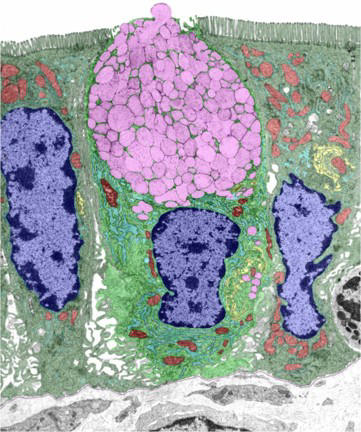
“brush border”
Complex cytoskeleton anchored to terminal web; core of actin filaments – contractile properties
microvilli
88
Unusually long, immotile microvilli (see internal structure)
Limited to epididymis, ductus deferens, cochlear & vestibular hair cells
Limited to epididymis, ductus deferens, cochlear & vestibular hair cells
stereocilia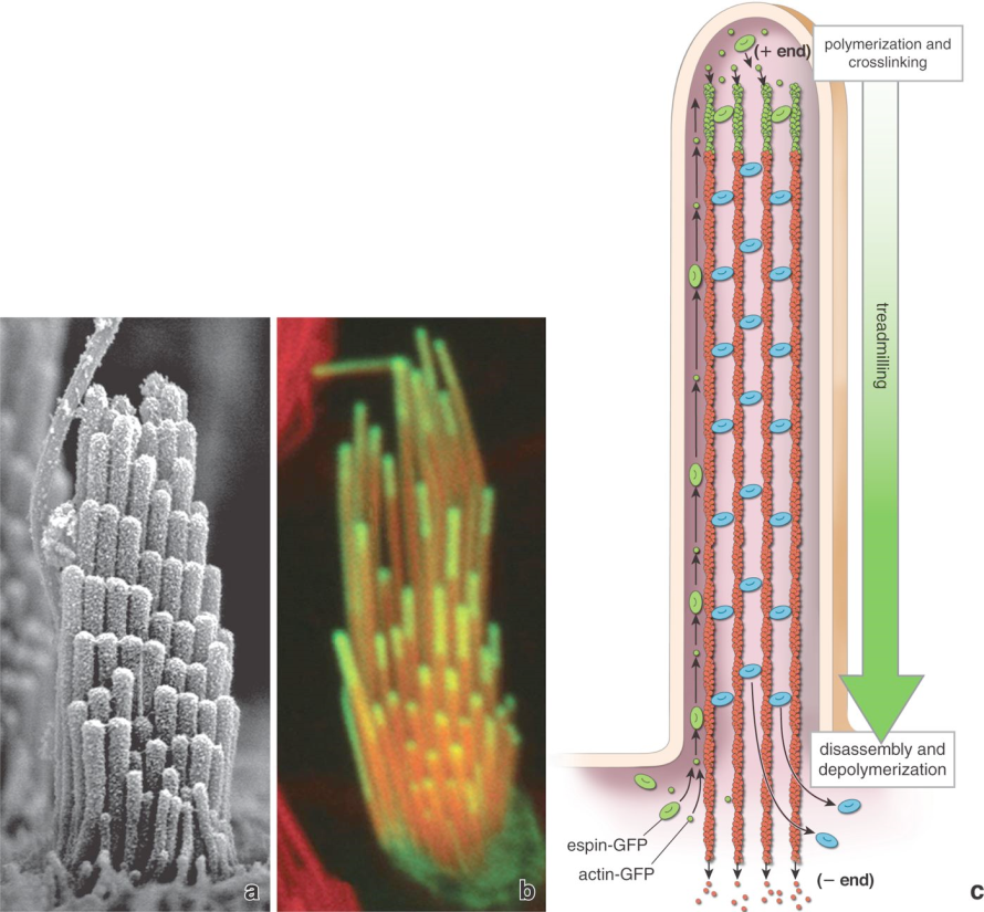
89
cilia types
motile and flagella (9+2): trachea, bronchi, oviducts
primary cilia
nodal cilia
primary cilia
nodal cilia
90
what is the lateral domain made up of (x2)
terminal bar, junctional complex, lateral folds
91
what is the terminal bar/junctional complex made up of?
occluding tight junctions (linked to actin) which seperate cellular tissue
anchoring junctions: zona adherins (actin), macula adherins (intermediate filaments)
communicating junctions (gap)
anchoring junctions: zona adherins (actin), macula adherins (intermediate filaments)
communicating junctions (gap)
92
what is the basal domain made up of?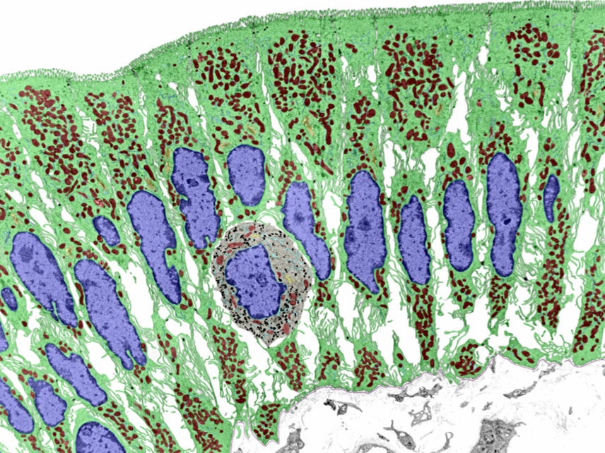
basement membrane
cell to ECM junctions: focal adhesions (actin), hemidesmosome (intermediate filaments)
basal cell membrane foldings
cell to ECM junctions: focal adhesions (actin), hemidesmosome (intermediate filaments)
basal cell membrane foldings
93
what are the two types of endocrine glands
cord and clump, follicle
94
what gland?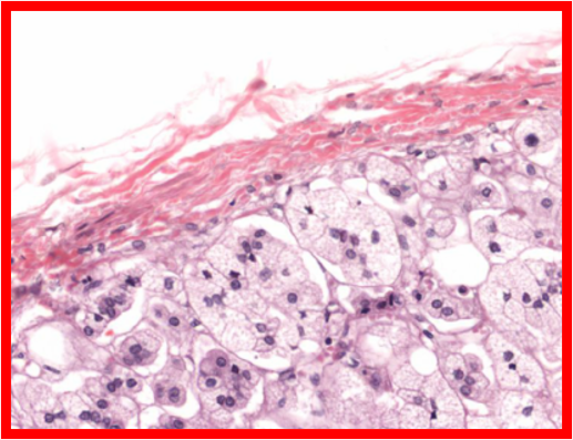
cord and clump
95
what gland?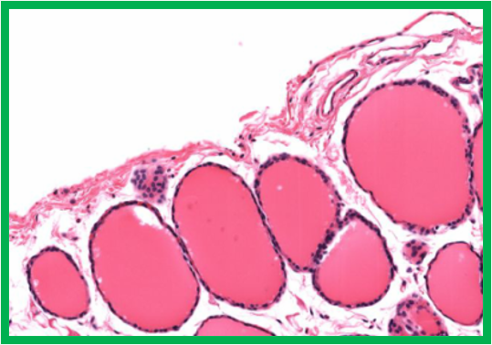
follicle
96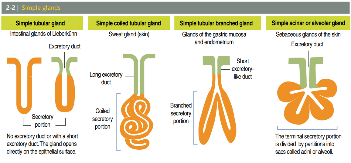
Image occlusion: name the hidden labels
97
Image occlusion: name the hidden labels
98
Image occlusion: name the hidden labels
99
Image occlusion: name the hidden labels
100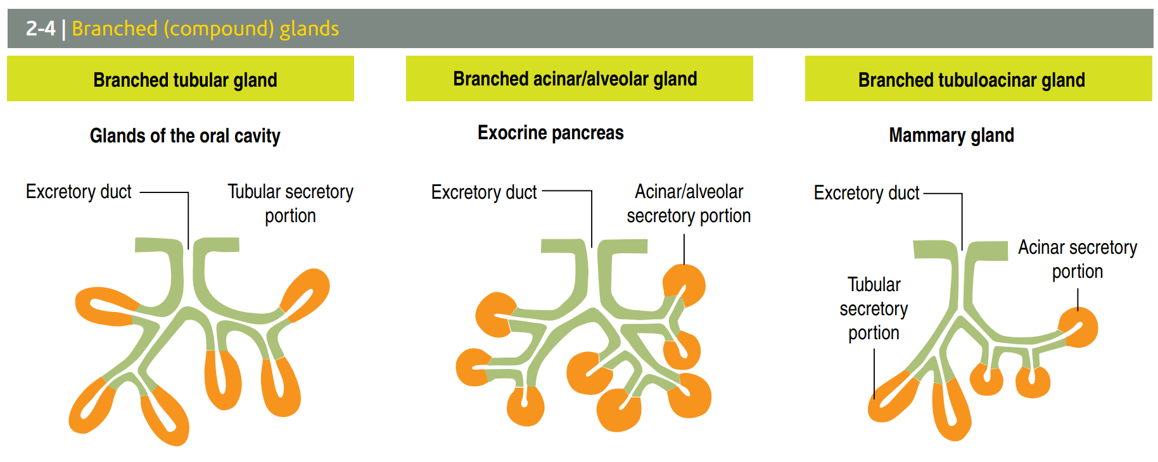
Image occlusion: name the hidden labels
101
Image occlusion: name the hidden labels
102
Image occlusion: name the hidden labels
103
purple in image (exocrine gland)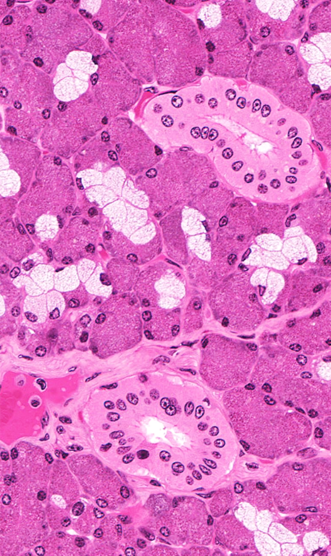
nuclei
104
light pink in photo (exocrine )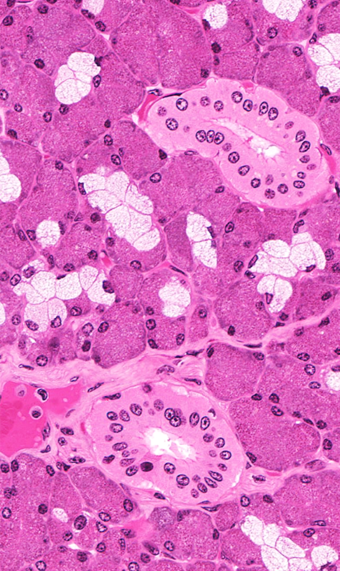
mucous secreting exocrine glands
105
dark pink in photo (exocrine)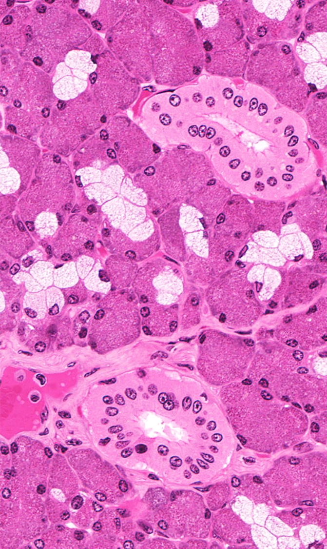
serous secreting exocrine glands
106
what type of exocrine gland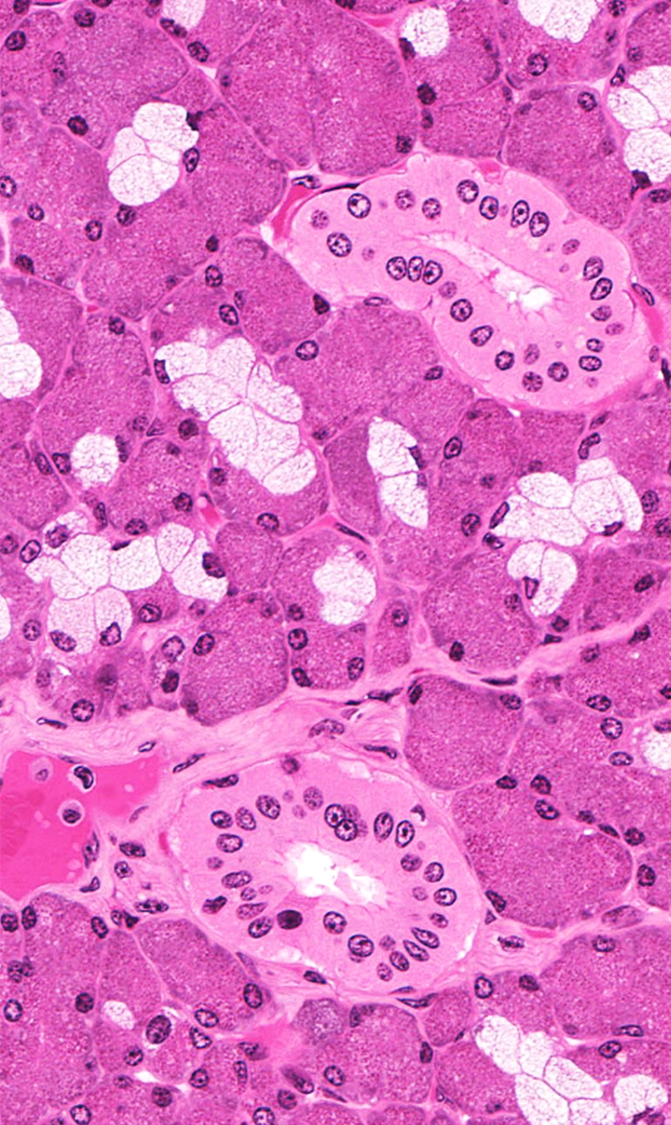
mixed, has both serous and mucous secreting glands
107
Thin filaments muscles?
actin
108
thick filaments muscle
myosin II
109
three typoes of striated muscle
skeletal, visceral striated, cardiac
110
may have attachments to bone but is associated with the gut tube, same histo as skeletal but different embryology
visceral striated
111
where is smooth muscle found
viscera: blood/lymph vessels, organs, glands
112
muscle fibers arise from aggregations of ________________ these cells fuse to form a ____________________ muscle fiber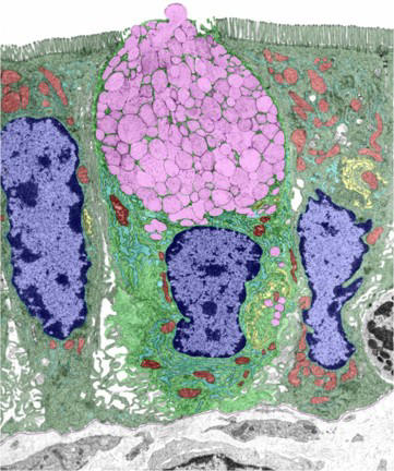
myoblasts; multinucleated
113
means many nuclei, this is what muscle fibers are
syncythium
114
strength of muscle depends on this
number of fibers
115
attach muscle to bone
tendons
116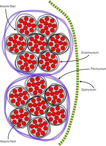
Image occlusion: name the hidden labels
117
Image occlusion: name the hidden labels
118
Image occlusion: name the hidden labels
119
Image occlusion: name the hidden labels
120
Image occlusion: name the hidden labels
121
black arrow?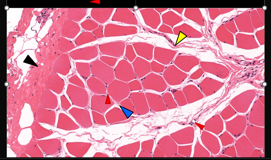
epimysium
122
yellow arrow?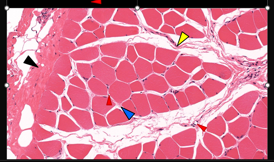
perimysium
123
blue arrow?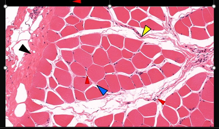
endomysium
124
different fiber types/colors are attributed to (x3)
Succinic dehydrogenase
NADH-TR
Myoglobin
NADH-TR
Myoglobin
125
what are the characteristics of white/fast glycolitic/type IIb fibers
low succinate dehydrogenase, atpase
less extensive blood supply
fewer mitochondria
adapted for rapid and powerful cotraction
less extensive blood supply
fewer mitochondria
adapted for rapid and powerful cotraction
126
what are the characteristics of red/slow twitch/aerobic/type I fibers
smaller, extensive blood supply, many mitochondria and myoglobin
for prolonged and continued actvity
for prolonged and continued actvity
127
invaginatuons of the plasma membrane (sarcolemma) used to have SR closer to membrane to speed up muscle contractions 9easier ease of access for Ca2+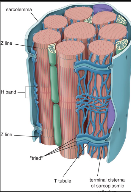
transverse tubles
128
contractile unit of muscle fbers
myofibrils
129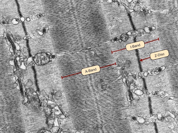
Image occlusion: name the hidden labels
130
Image occlusion: name the hidden labels
131
Image occlusion: name the hidden labels
132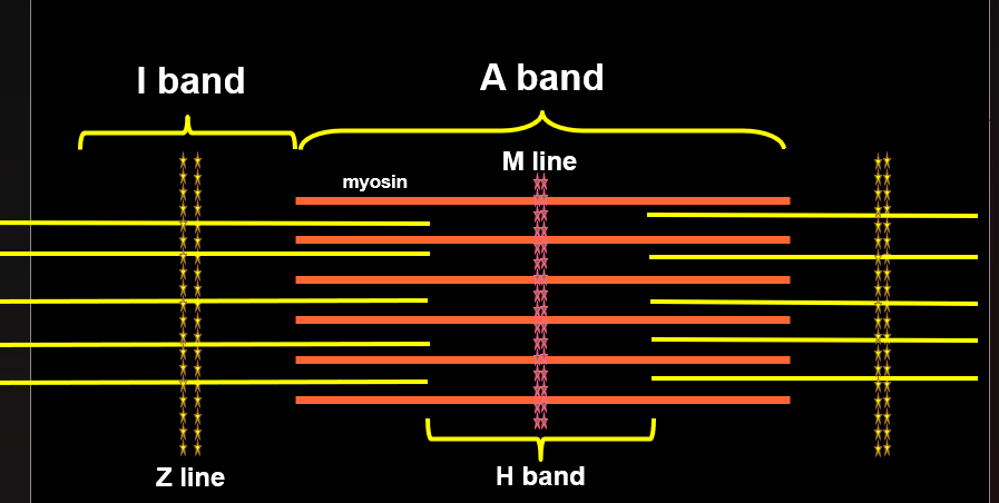
Image occlusion: name the hidden labels
133
Image occlusion: name the hidden labels
134
Image occlusion: name the hidden labels
135
Image occlusion: name the hidden labels
136
modified smooth ER, is arranged around myofibers; serves as a reservoir for Ca++
sarcoplasmic reticulum
137
tissue type? whats happening here?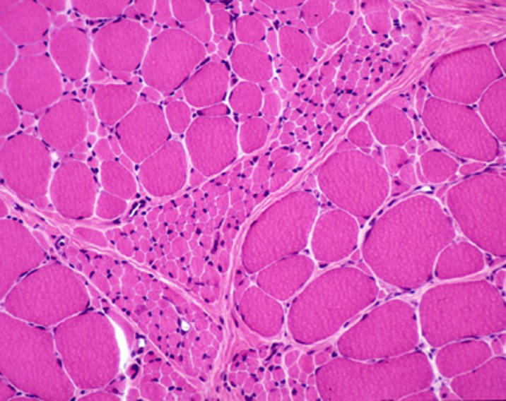
skeletal; muscle atrophy
138
intrafusal fiber types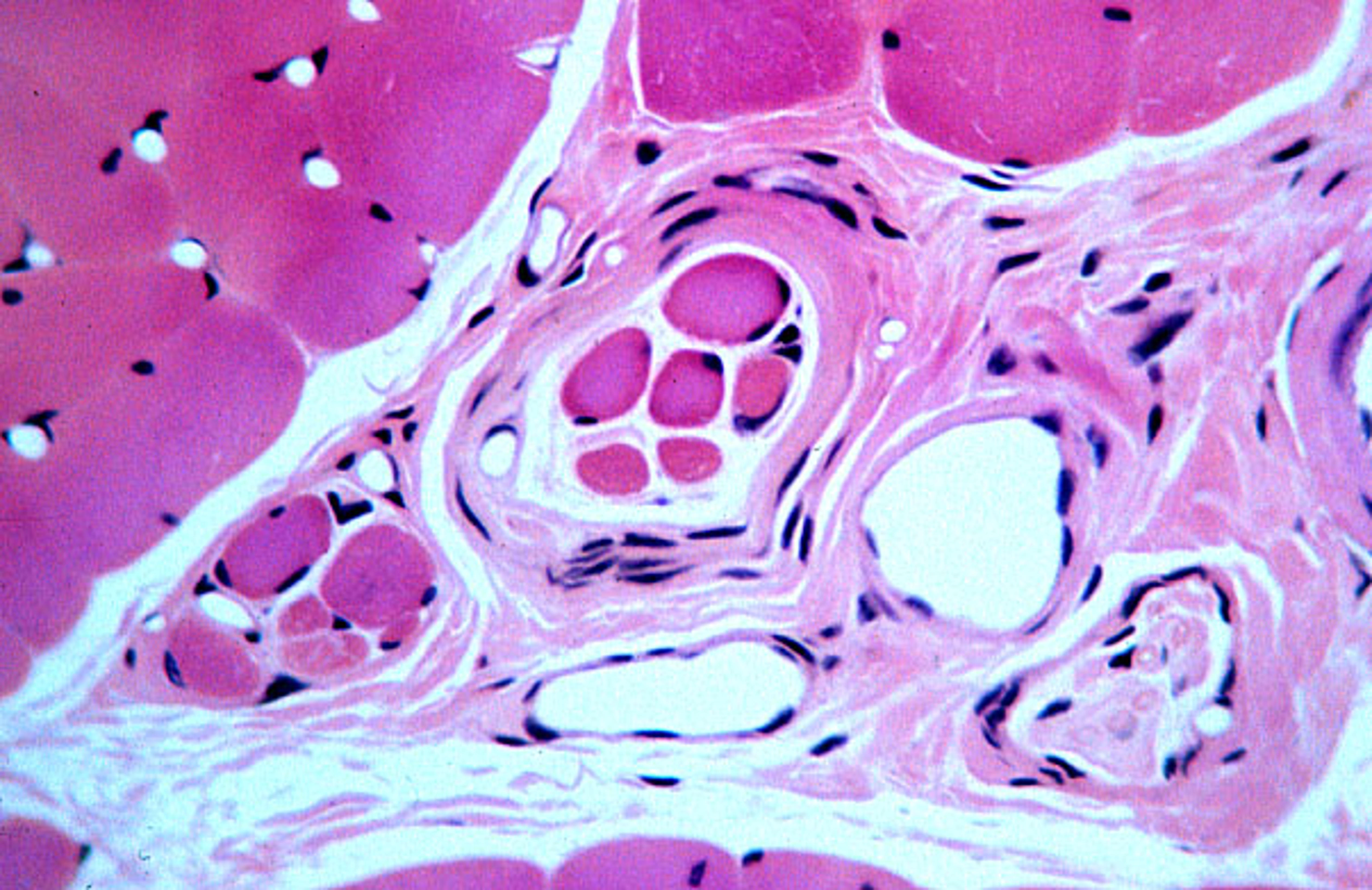
muscle spindles, golgi tendon
139
what is this?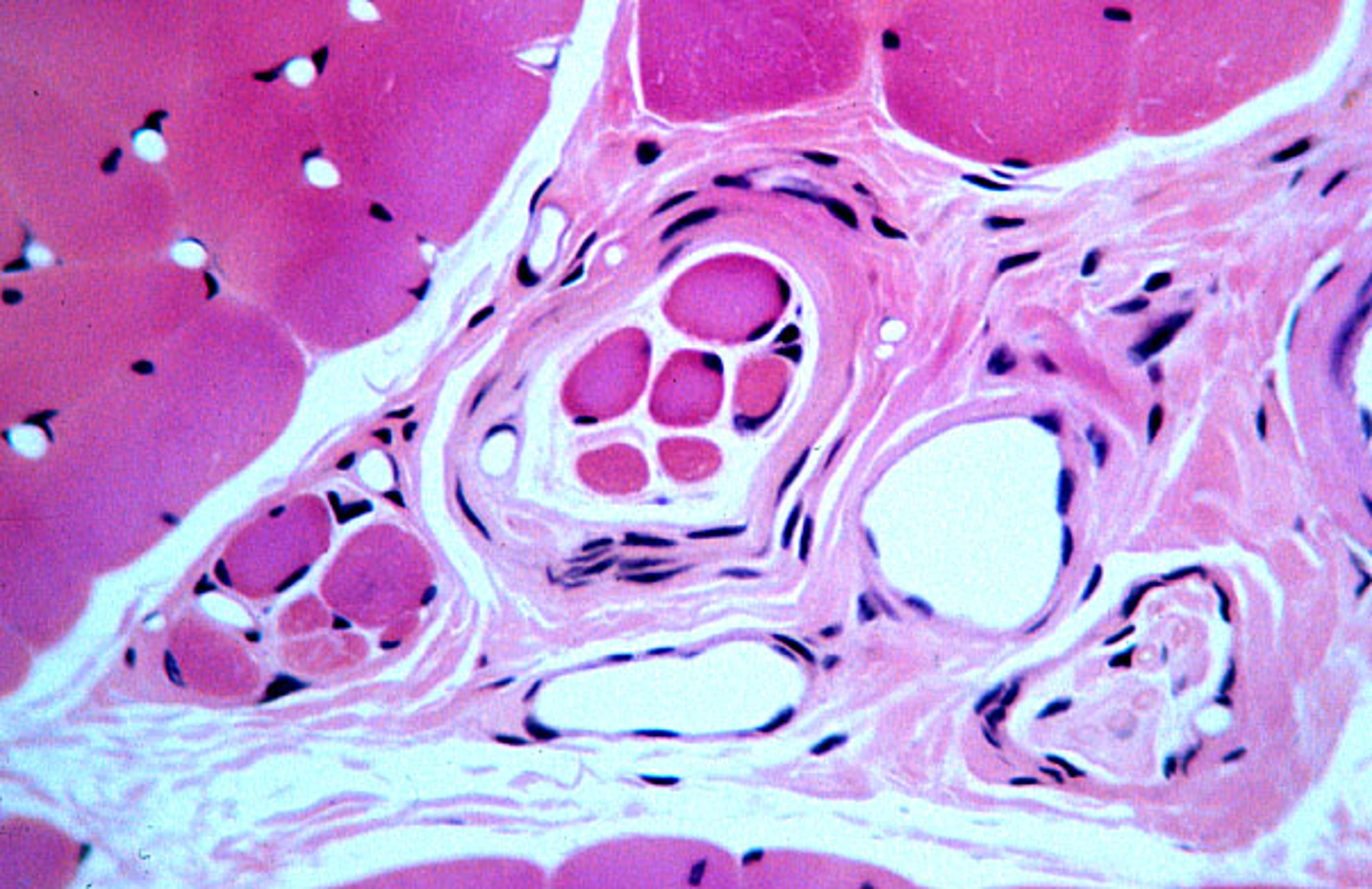
muscle spindle
140
these can regenerate skeletal muscle fibers to a minimum capacity
satellite cells
141
what is this muscle tissue? specifics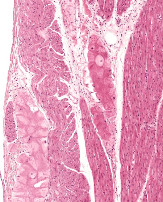
cardiac muscle; coducting fibers
142
how many T tubles per SR in cardiac? in skeletal?
1; 2
143
what do cardiac cells do in repair?
scar tissue; limited repair
144
smooth muscle cells regeneration
can regenerate; pericytes
145
dense bodies are a characteristic of this muscle type
smooth
146
what fibers make up ECM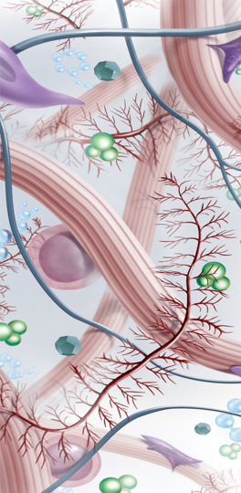
collagen
reticular
elastic
reticular
elastic
147
what ground substances make up ECM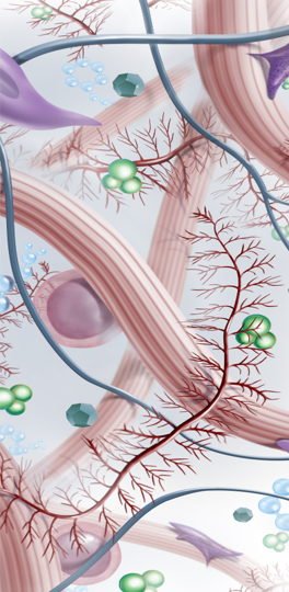
glycoproteins (fibronectn and laminin), glycosaminoglycans (sulfate), proteoglycans
148
what is made up of type 1 collagen?
bone, skin, tendons, ligaments
149
what is made of type 2 collagen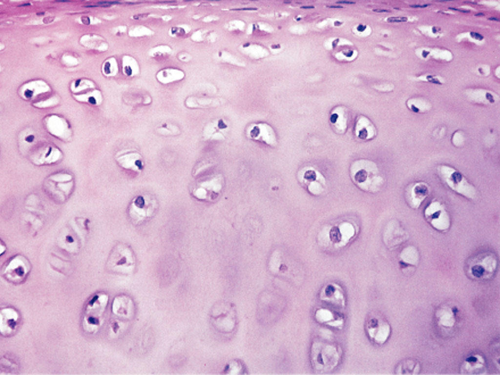
hyaline cart
150
what is made of type 3 collagen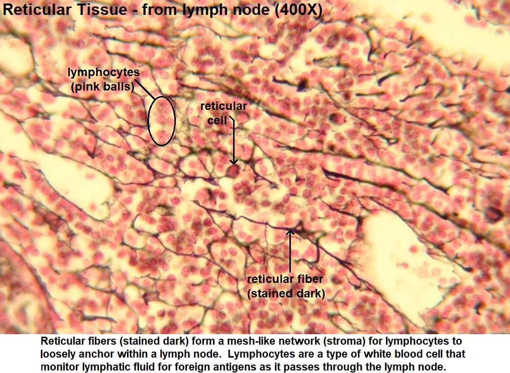
reticular fibers
151
what is made og type 4 collagen
basement mambrane
152
what forms collagen I fibers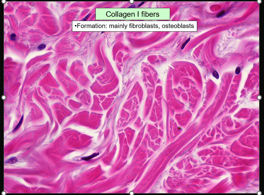
fibroblasts and osteoblasts
153
where can you find reticular fibers?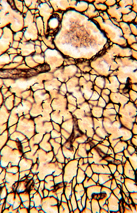
adipocytes, muscle cells, small blood vessels hemopoietic and lymphatic tissues
154
what type of stain?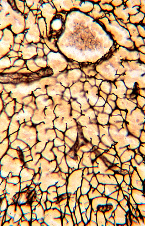
silver salts
155
what forms reticular fibers?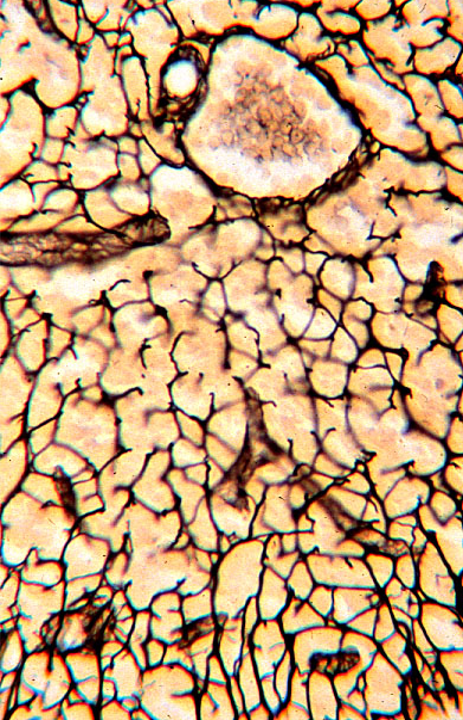
fibroblasts, smooth muscle cells, schwann cells
156
where are elastic fibers found?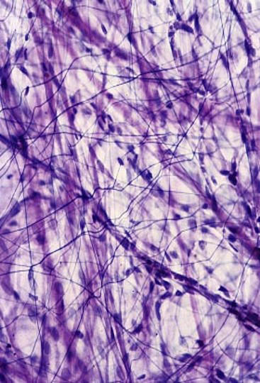
elastic cartilage, elastc arteries, elastic ligaments
157
what stain?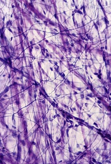
resorcin fuchsin
158
what makes elastoc fibers?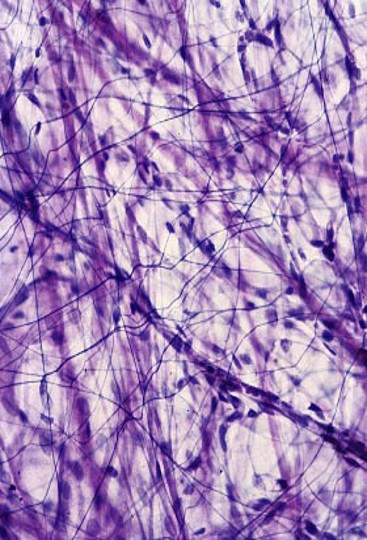
fibroblasts and smooth muscle cells
159
what makes up elastic fibers?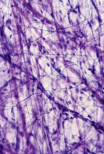
Elastin (central core)
Fibrillin microfibrils around the core embedded into elastin
Fibrillin microfibrils around the core embedded into elastin
160
what is the deficiency in marfan syndrome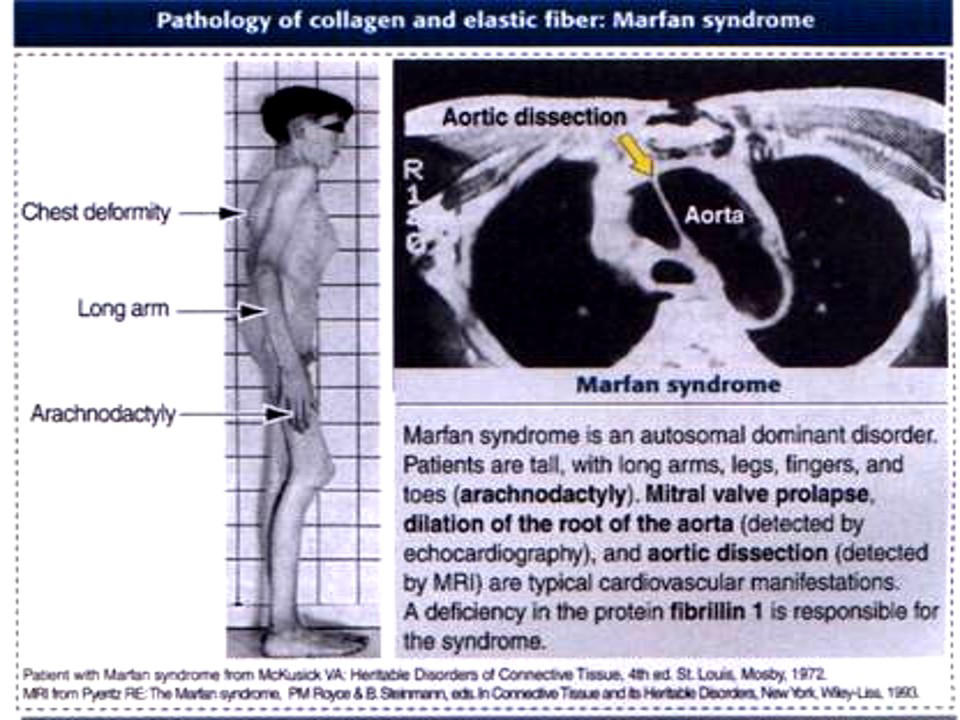
mutation in fibrilin 1 gene, fibil imp for maintaining elastic fibers
161
what is the deficiency/issue in ehlers danlos syndrome?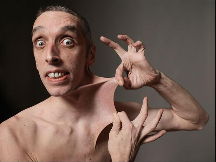
mutation in genes affecting collahen polypeptide chains
162
what is the issue/deficiency in osteogenesis imperfecta
type 1 collagen mutation: less severe
type II collagen mutation: small and fragile rib cage
type II collagen mutation: small and fragile rib cage
163
Star or spindle shaped cells
Pluripotential – capable to form various cell types
In adult, they are located usually around the vessels
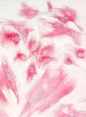
Pluripotential – capable to form various cell types
In adult, they are located usually around the vessels
mesenchymal cell
164
where are mesenchymal cells located? 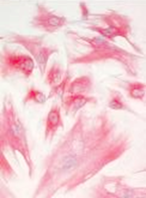
around the vessels
165
what can mesenchyme cells form (x5)
chondroblasts, lipoblasts, fibroblasts, osteoblasts, myoblasts
166
what are the funct of fibroblasts
synthesize fibers and ground substances, protein synthesis
167
what are the functions of myofibroblasts
regulating the shape and empotying f glands and wound contraction and closure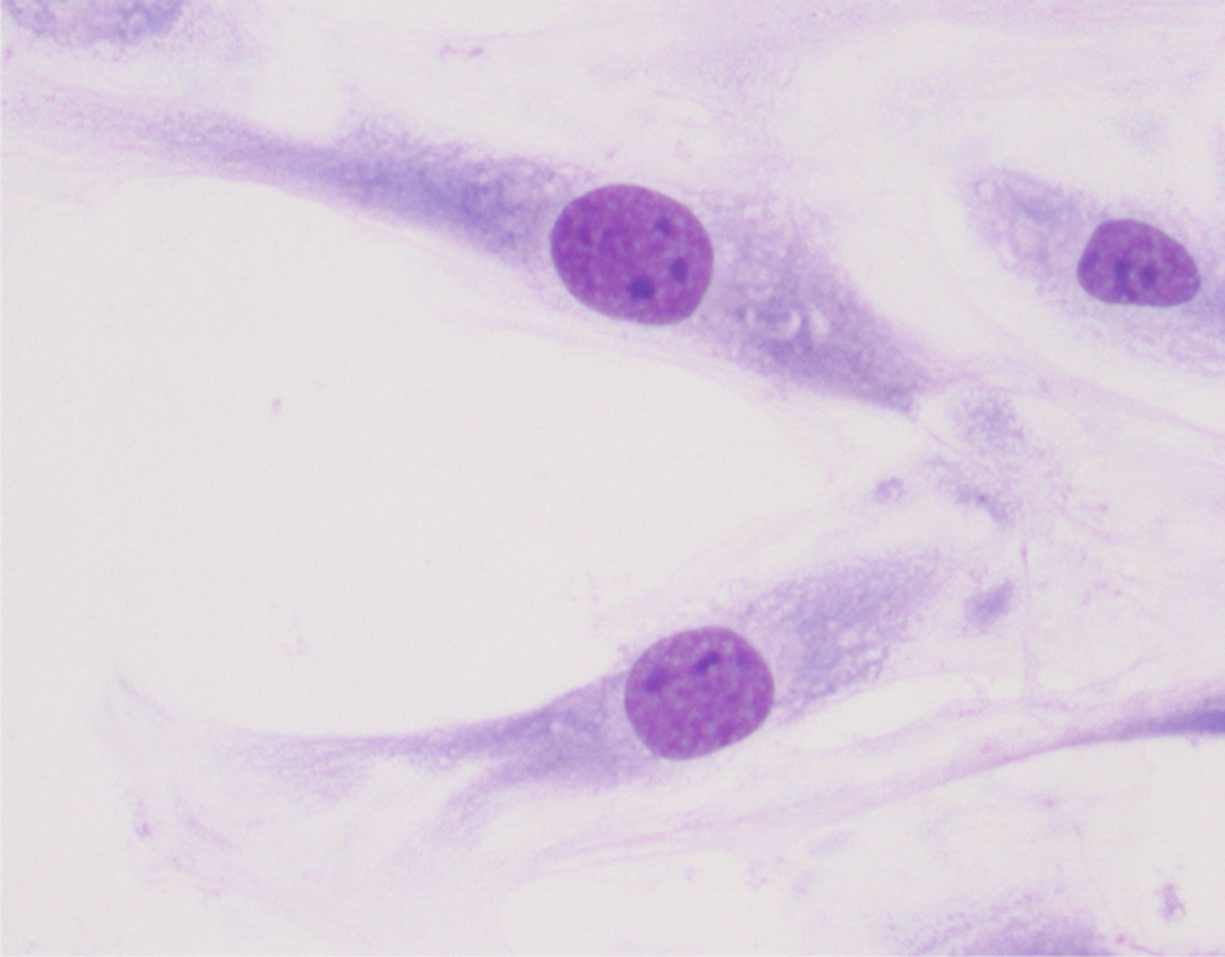
168
where are myofibroblasts located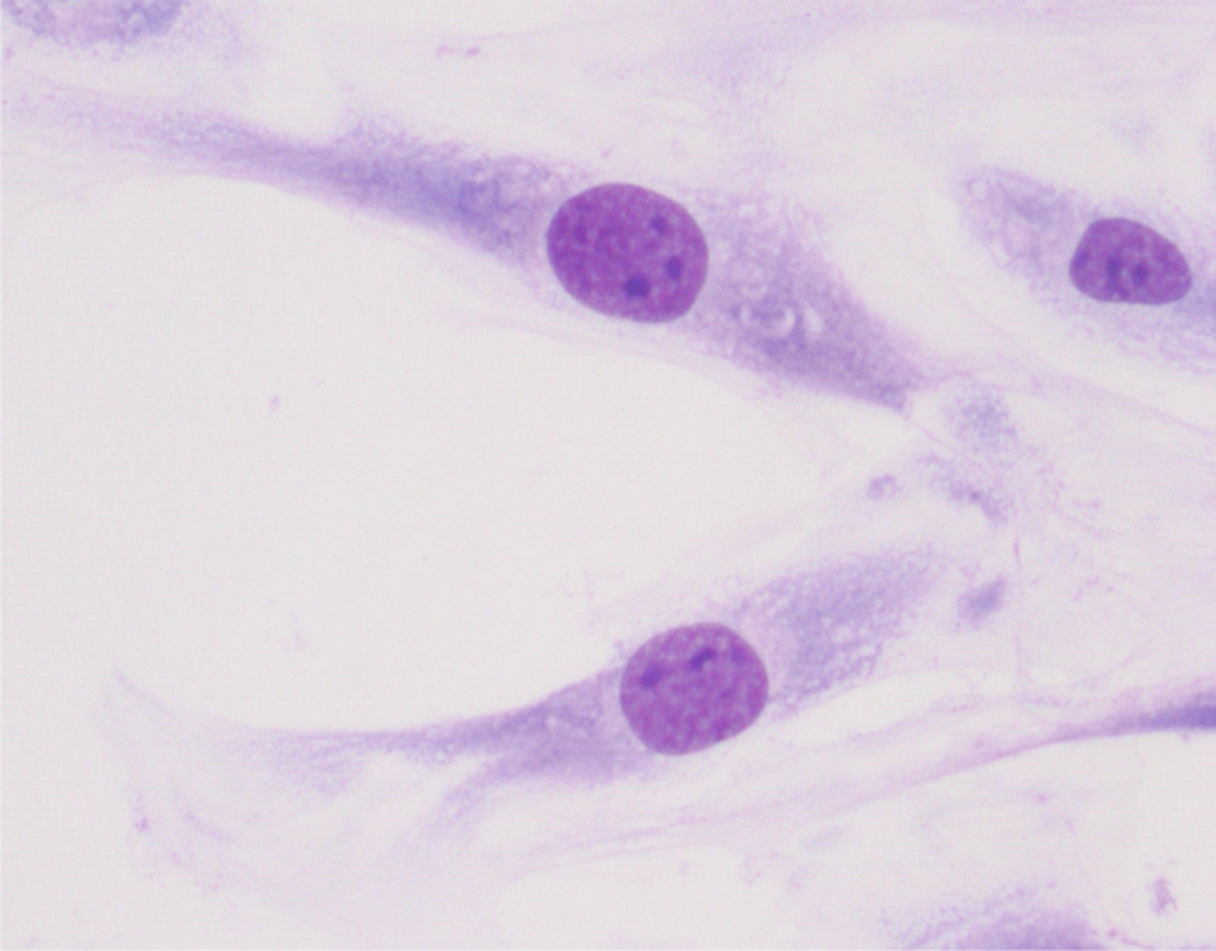
loose connective tissue
169
Numerous mitochondria
Characteristic for newborns – rapid energy release
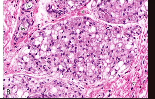
Characteristic for newborns – rapid energy release
brown adipose
170
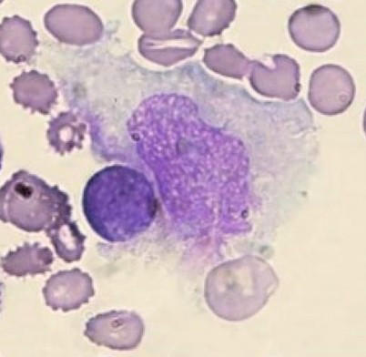
macrophages function
Phagocytic cells derived from blood monocytes - garbage collectors of the tissue
171
where are mast cells located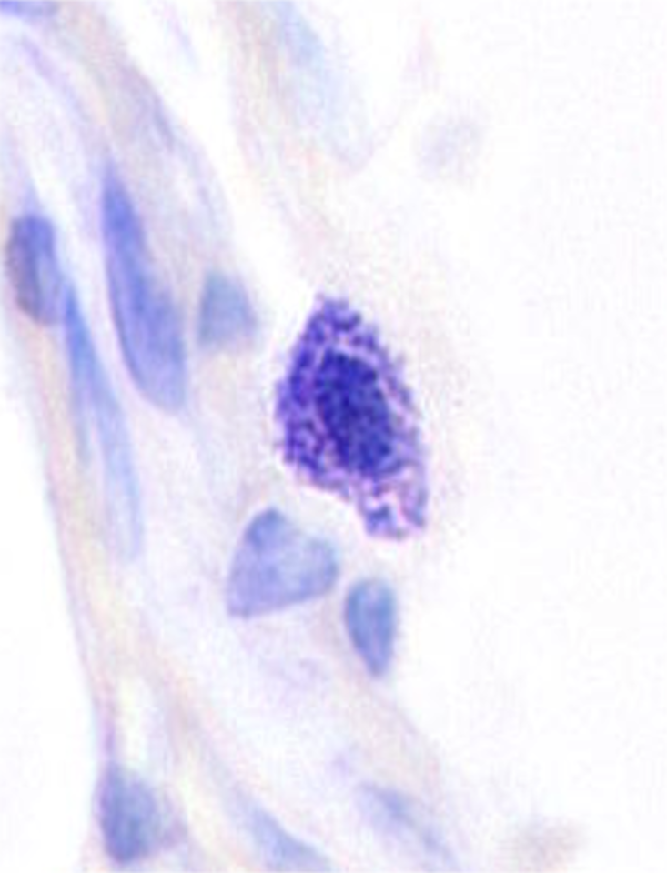
small vessels in connective tissue, capsules of organs
172
fx mast cells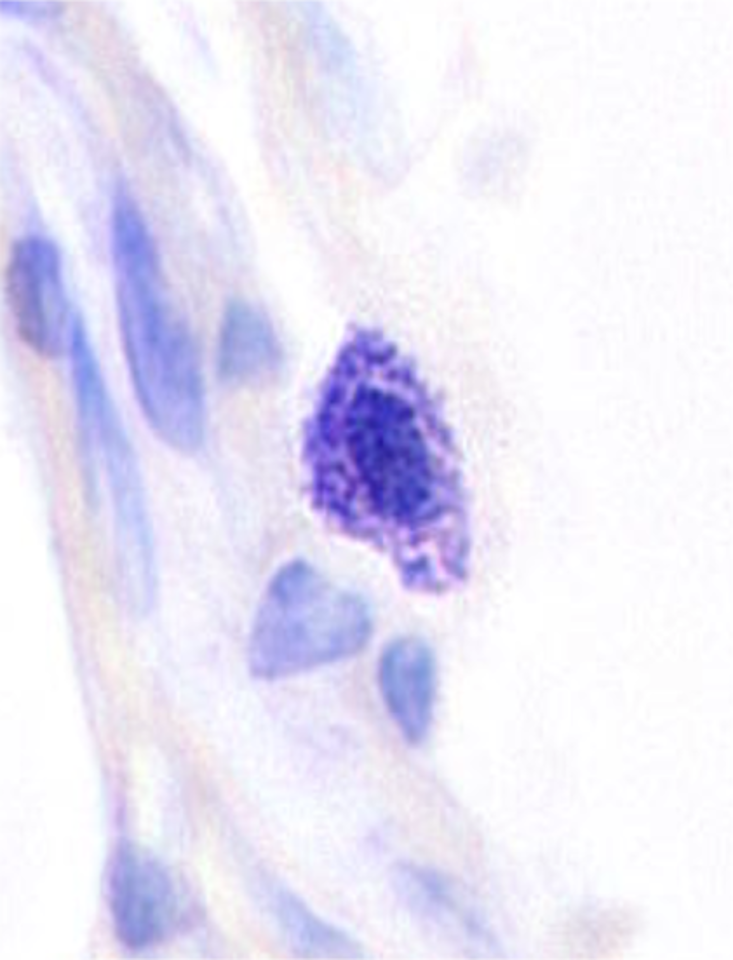
anaphylactoc oversensitive reactions
173
what do heparan sulphate and heparin do in mast cells
block coagulkation, resp for basophilic apprearance
174
what are histamine and SRS-A resp for in mast cells
vasodilation, edema
175
what are ECF and NCF resp for in mast cells
celll attraction
176
what are the different types of lymphocytes and where do they differentiate?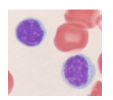
T lymphocytes (differentiating in the thymus) – 60-80 %
B lymphocytes (differentiating in the bone marrow) – 20-30 %
NK (natural killer) cells – 5-10% (bone marrow)
B lymphocytes (differentiating in the bone marrow) – 20-30 %
NK (natural killer) cells – 5-10% (bone marrow)
177
what are plasma cells derived frpom?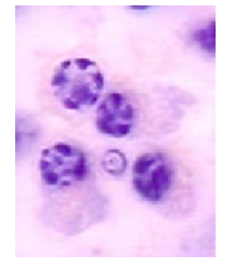
B cells
178
what are plasma cells fxn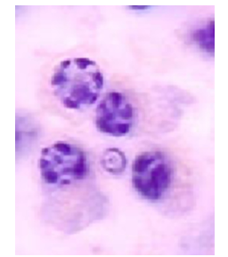
antibody producing
179
Capable of migration
Granules –small, purple, barely visible in the light microscope
Nucleus multilobed (thus also called polymorphs)
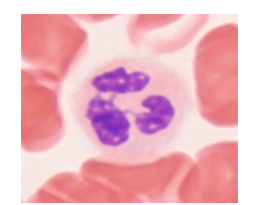
Granules –small, purple, barely visible in the light microscope
Nucleus multilobed (thus also called polymorphs)
neurotrophils
180
Named after the large eosinophilic granules in the cytoplasm
Typically bilobed nucleus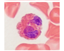
Typically bilobed nucleus
eosinophils
181
Large basophilic granules in the cytoplasm
Nucleus obscured by the granules
Closely related to mast cells of the connective tissue
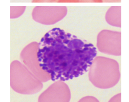
Nucleus obscured by the granules
Closely related to mast cells of the connective tissue
basophils
182
what are the types of embryonic connective tissue (x2)
mesenchyme, mucous connective tissue
183
where can you find mucous connective tissue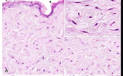
umbilical cord
184
what cells are present in mucous connective tissue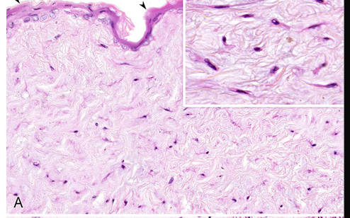
fibroblasts and mesenchymal cells
185
Primitive pluripotent connective tissue – capable of turning into other tissue types
Cells: pluripotential mesenchymal cells
Cells are more closely packed than in Wharton’s Jelly
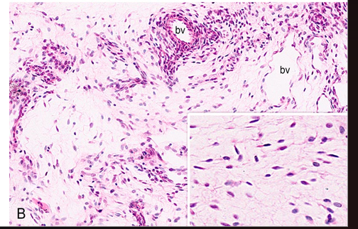
Cells: pluripotential mesenchymal cells
Cells are more closely packed than in Wharton’s Jelly
mesenchyme
186
Few fibroblast
Little ground substance
Large amount of collagen fibers between them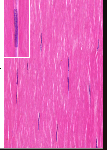
Little ground substance
Large amount of collagen fibers between them
Dense connective tissue
187
Fibers are arranged in parallel array
Present in tendons, aponeuroses and ligaments
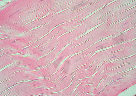
Present in tendons, aponeuroses and ligaments
dense regular connective tissue
188
Fibers are arranged in bundles running in every direction
Present in the submucosa of the hollow organs – connecting the mucous membrane to the underlying muscle layer
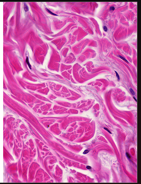
Present in the submucosa of the hollow organs – connecting the mucous membrane to the underlying muscle layer
dense irregular connective tissue
189
Loosely arranged fibers and cells
Found mostly beneath the epithelia (layer is called lamina propria mucosae)
Contains blood cells (migrated from local blood vessels)
In inflammation it can undergo significant swelling
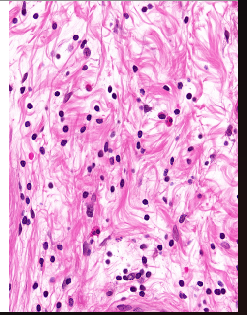
Found mostly beneath the epithelia (layer is called lamina propria mucosae)
Contains blood cells (migrated from local blood vessels)
In inflammation it can undergo significant swelling
loose connective tissue