Path S1 U1
Study these flashcardsFree · spaced repetition · iPhone, Android, Mac & web
All cards
1
what are the 5 major classes of functional non protein coding sequences in the human genome?
promoters and enhancers
binding sites for factors that organize and maintain higher order chromatin structyres
noncoding regulatory RNAs
special stuctural regions DNA (teomeres and centromeres)
transposons
binding sites for factors that organize and maintain higher order chromatin structyres
noncoding regulatory RNAs
special stuctural regions DNA (teomeres and centromeres)
transposons
2
variants at single nucleotide positions and are almost always biallelic (only two choices exist at a given site within the population, such as A or T)
Single nucleotide polymorphisms
3
orm of genetic variation consisting of different numbers of large contiguous stretches of DNA; these can range from 1000 base pairs to millions of base pairs. can be biallelic and simply duplicated or, alternatively, deleted in some individualscautosomal hematopoeticskeletal nend
CNVs
4
composition of a nucleosome
DNA segments 147 bp long that are wrapped around a central core structure of highly conserved low-molecular-weight proteins called histones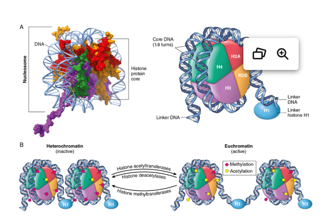
5
In histone methylation, both ______ abd ____- xa ve nmethylated and can lead to transcriptional activation or repression
lysines and arginines
6
In histone acetylation, ________-- residues are acytylated by histone actyltransferase (HATs) and undone by histone deacetylases (HDACs)
lysine)
7
In histone phosphorylation, ____- residues can be modified by phosphorylation, can be made actove or repressed for transcription
serine
8
high levels of DNA methy;lation tend to result in ____- of genes
silencing
9
what is the fx of chromatin organizing factors
believed to bind to noncoding regions and contrl long range looping of DNA
10
do not encode proteins; they modulate translation of target messenger RNAs (mRNAs)
miRNAs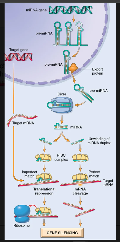
11
short RNA sequences that can be introduced experimentally into cells where they serve as substrates for Dicer and interact with RISC, thereby reproducing endogenous miRNAs function
siRNAs
12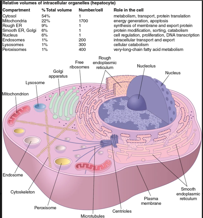
Image occlusion: name the hidden labels
13
Image occlusion: name the hidden labels
14
Image occlusion: name the hidden labels
15
Image occlusion: name the hidden labels
16
Image occlusion: name the hidden labels
17
Image occlusion: name the hidden labels
18
Image occlusion: name the hidden labels
19
Image occlusion: name the hidden labels
20
disposal” complexes that degrade denatured or otherwise “tagged” cytosolic proteins.
proteosomes
21
intracellular organelles containing degradative enzymes that permit digestion of a wide range of macromolecules, including proteins, polysaccharides, lipids, and nucleic acids. They are the site of intracellular organelle breakdown (a process called autophagy) and where phagocytosed microbes are killed and catabolized.
lysosomes
22
ontain catalase, peroxidase, and other oxidative enzymes; they play a specialized role in the breakdown of very long-chain fatty acids, generating hydrogen peroxide in the process
peroxisomes
23
what are the fx of phosphatidylinositol
an be phosphorylated, serving as an electrostatic scaffold for intracellular proteins; alternatively, polyphosphoinositides can be hydrolyzed by phospholipase C
24
fx of phosphatidylserine
electrostatic protein interactions;
25
n general, proteins associate with the lipid bilayer in one of four ways.
As integral or transmembrane proteins, having one or more relatively hydrophobic α-helical segments that traverse the lipid bilayer.
• Through posttranslational addition of prenyl groups (e.g., farnesyl, related to cholesterol) or fatty acids (e.g., palmitic or myristic acid) that insert into the cytosolic side of the plasma membrane.
• Through glycosylphosphatidylinositol (GPI) tails that are added posttranslationally, a linkage used to associate extracellular proteins with the membrane.
• Indirectly, through noncovalent interactions with true transmembrane proteins.
• Through posttranslational addition of prenyl groups (e.g., farnesyl, related to cholesterol) or fatty acids (e.g., palmitic or myristic acid) that insert into the cytosolic side of the plasma membrane.
• Through glycosylphosphatidylinositol (GPI) tails that are added posttranslationally, a linkage used to associate extracellular proteins with the membrane.
• Indirectly, through noncovalent interactions with true transmembrane proteins.
26
he movement of endocytosed vesicles between the apical and basolateral compartments of cells.
transcytosis
27
eal adjacent epithelial cells together to create a continuous barrier that restricts the paracellular (between cells) movement of ions and other molecules.
occluding junctions
28
mechanically attach cells—and their cytoskeletons—to other cells or the ECM. Adherens junctions are often closely associated with and beneath tight junctions.
anchoring junctions
29
permit the diffusion of chemical or electrical signals from one cell to another. The junction consists of a dense planar array of 1.5- to 2-nm pores (called connexons) formed by a pair of hexamers (one on each cell) of transmembrane connexin proteins.
communicating junctions
30
the site of synthesis of all transmembrane proteins and lipids found in the plasma membrane and cellular organelles, including the ER itself. It is also the initial site of synthesis of secreted proteins.
ER
31
excess accumulation of misfolded proteins—exceeding the capacity of the ER to edit and degrade them—leads to the ER stress response (also called
unfolded protein response
32
lysosomal enzymes are initially synthesized in the ER lumen and then tagged with ______________________________________ within the Golgi apparatus.
manose 6 phosphate (M6P)
33
the electron transport chain need not necessarily be coupled to ATP generation. An inner membrane protein enriched in brown fat called ____________________________________________ (also called uncoupling protein-1 [UCP-1]) is a hydrogen ion transporter that can dissipate the proton gradient, uncoupling it from oxidative phosphorylation
thermogenin
34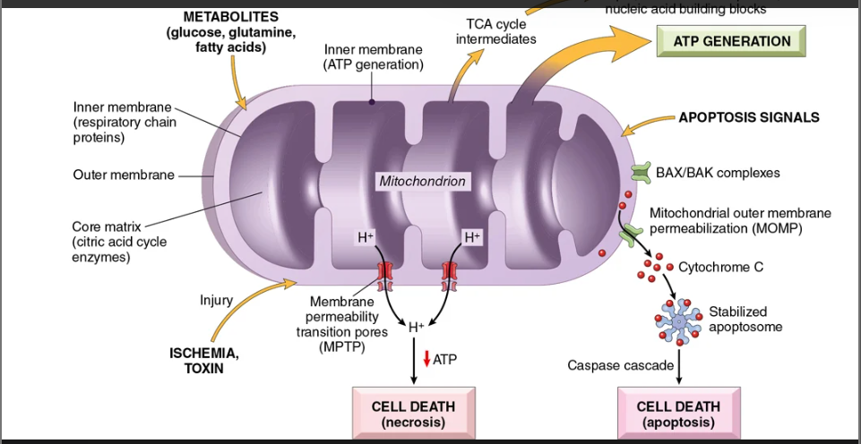
Image occlusion: name the hidden labels
35
Image occlusion: name the hidden labels
36
are integral membrane proteins; ligand-induced cross-linking activates intrinsic tyrosine kinase domains located in their cytoplasmic tails.
receptor tyrosine kinases
37
interacts with receptors after ligand binding and phosphorylates specific motifs on the receptor or other proteins.
nonreceptor tyrosine kinases
38
ligand binding to ___________________ receptors leads to proteolytic cleavage of the receptor and subsequent nuclear translocation of its cytoplasmic domain (intracellular _____________) to form a transcription complex.
Notch
39
Wnt protein ligands can also influence cell development through a canonical pathway involving transmembrane Frizzled family receptors, a distinct set of GPCRs that regulate intracellular levels of ___________________.
B catenin
40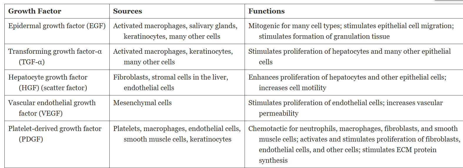
Image occlusion: name the hidden labels
41
Image occlusion: name the hidden labels
42
Image occlusion: name the hidden labels
43
Image occlusion: name the hidden labels
44
Image occlusion: name the hidden labels
45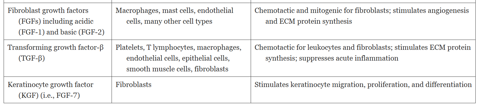
Image occlusion: name the hidden labels
46
Image occlusion: name the hidden labels
47
Image occlusion: name the hidden labels
48
Ito forms of ECM: occupies the spaces between stromal cells within connective tissue and between parenchymal epithelium and the underlying supportive vascular and smooth muscle structures in some organs. Interstitial matrix is synthesized by mesenchymal cells (e.g., fibroblasts)
interstitial matrix
49
two forms of ECM: interstitial matrix within connective tissues becomes highly organized around epithelial cells, endothelial cells, and smooth muscle cells, where it forms _______________________________________, specialized surfaces for cell growth.
basement membrane
50
components of he ECM (x3)
• Fibrous structural proteins such as collagens and elastin that confer tensile strength and recoil
• Water-hydrated gels such as proteoglycans and hyaluronan that permit compressive resistance and lubrication
• Adhesive glycoproteins that connect ECM elements to one another and to cell
• Water-hydrated gels such as proteoglycans and hyaluronan that permit compressive resistance and lubrication
• Adhesive glycoproteins that connect ECM elements to one another and to cell
51
Some collagen types (e.g., types I, II, III, and V) form linear fibrils stabilized by interchain hydrogen bonding; such fibrillar collagens comprise a major proportion of the connective tissue in bone, tendon, cartilage, blood vessels, and skin, as well as in healing wounds and scars
fibrillar collagens
52
refers to cell replication in which one daughter cell enters a differentiation pathway and gives rise to mature cells, while the other remains undifferentiated and retains its self-renewal capacity.
asymmetric division
53
occurs when both daughter cells retain self-renewal capacity. Such replication occurs early in embryogenesis—when stem cell populations are expanding—and under stress conditions, as in bone marrow repopulation after ablative chemotherapy.
symmetric divison
54
are the most undifferentiated. They are present in the inner cell mass of the blastocyst, have virtually limitless cell renewal capacity, and can give rise to every cell in the body
embryonic stem cells
55
are found in intimate association with the differentiated cells of a given tissue. They are normally protected within specialized tissue microenvironments called stem cell niches
tissue stem cells
56
related to mutations in single genes with large effects. These mutations typically critically affect gene function and directly cause or predispose to the disease. Such mutations result in highly penetrant phenotypes
-follow mendelian inheritance
-follow mendelian inheritance
monogenic disorders
57
human genetic disorders are grouped into three categories...
monogenic disorders related to mutations in a single gene with large effects, chromosomal disorders, complex multogenic disorders
58
These can be further considered as structural or numerical alterations in the autosomes and sex chromosomes
-an altered copy number of a region that may encompass many genes. While some chromosomal disorders are heritable, many spontaneously arise in the germline or early in development.
-an altered copy number of a region that may encompass many genes. While some chromosomal disorders are heritable, many spontaneously arise in the germline or early in development.
chromosomal disorders
59
They are most often caused by interactions between multiple genetic variants and environmental factors. risk conferred by any single haplotype is low, and susceptibility is attributed to the additive effects of several such polymorphisms—hence the term multigenic or polygenic
complex multigenic disorders
60
what does this show?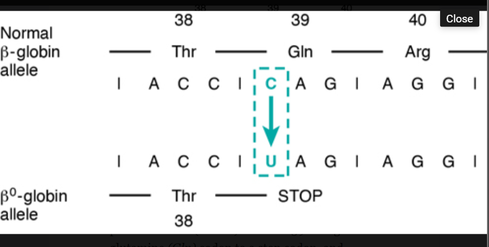
Nonsense mutation leading to premature chain termination.
61
what does this show?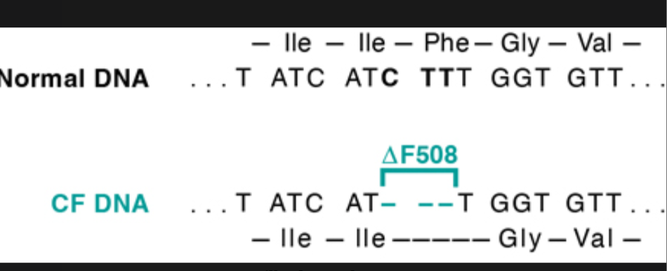
Three-base pair deletion in the common cystic fibrosis (CF) allele results in synthesis of a protein that lacks amino acid 508 (phenylalanine). Because the deletion is a multiple of three, this is not a frameshift mutation.
62
what does this show?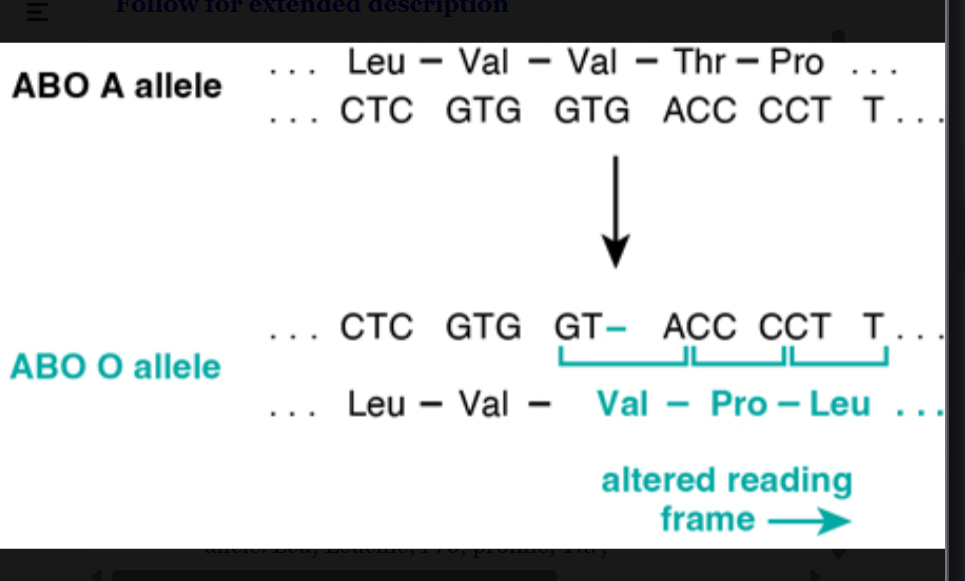
Single-base pair deletion at the ABO (glycosyltransferase) locus, leading to a frameshift mutation responsible for the O allele. Leu, Leucine; Pro, proline; Thr, threonine; Val, valine
63
what does this show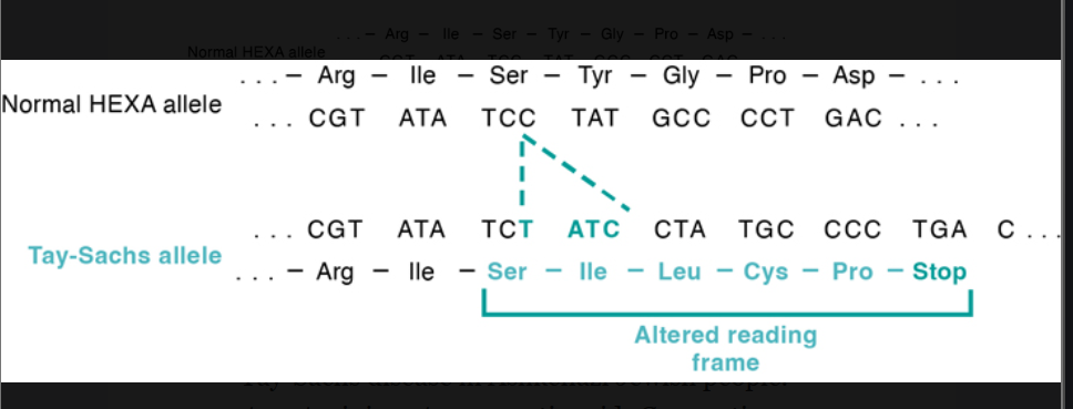
Four-base pair insertion in the hexosaminidase A gene, leading to a frameshift mutation. This mutation is the major cause of Tay-Sachs disease in Ashkenazi Jewish people.
64
result from mutations that have large functional effects and manifest in families with Mendelian inheritance patterns. Many mutations in autosomal genes produce no phenotype or a muted phenotype in individuals heterozygous for the mutation while affecting homozygotes more severely
single geneor monogenic disorder
65
Disorders caused by mutations involving single genes typically follow one of three patterns of inheritance:
autosomal dominant, autosomal recessive, or X-linked.
66
autosomal dominant nervous system disorders (x4)
Huntington disease
Neurofibromatosis
Myotonic dystrophy
Tuberous sclerosis
Neurofibromatosis
Myotonic dystrophy
Tuberous sclerosis
67
autosomal dominant urinary disorders
polycystic kidney doisease
68
autosomal dominant gastrointestinal disease
familial polyposis coli
69
hematopoetic autosomal dominant disorders (x2)
hereditary spherocytosis
von willevrand disease
von willevrand disease
70
skeletal autosomal dominant disorders
Marfan syndromea
Ehlers-Danlos syndrome (some variants)a
Osteogenesis imperfecta
Achondroplasia
Ehlers-Danlos syndrome (some variants)a
Osteogenesis imperfecta
Achondroplasia
71
metabolic autosomal dominant disorders
Familial hypercholesterolemiaa
Acute intermittent porphyria
Acute intermittent porphyria
72
metabolic autosomal recessive disorders (9)
Cystic fibrosis
Phenylketonuria
Galactosemia
Homocystinuria
Lysosomal storage diseasesa
α1-Antitrypsin deficiency
Wilson disease
Hemochromatosis
Glycogen storage diseasesa
Phenylketonuria
Galactosemia
Homocystinuria
Lysosomal storage diseasesa
α1-Antitrypsin deficiency
Wilson disease
Hemochromatosis
Glycogen storage diseasesa
73
hematopoetic autsomal recessive disorders
Sickle cell anemia
Thalassemias
Thalassemias
74
endocrine autosomal recessive disorders
congenital adrenal hyperplasia
75
Ehlers-Danlos syndrome (some variants)a
Alkaptonuria
skeletal autosomal recessive disorders
Ehlers-Danlos syndrome (some variants)a
Alkaptonuria
76
nervous autosmal recesisve disorders
Neurogenic muscular atrophies
Friedreich ataxia
Spinal muscular atrophy
Friedreich ataxia
Spinal muscular atrophy
77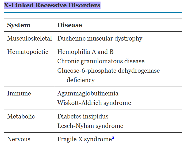
Image occlusion: name the hidden labels
78
Image occlusion: name the hidden labels
79
Image occlusion: name the hidden labels
80
Image occlusion: name the hidden labels
81
Image occlusion: name the hidden labels
82
Image occlusion: name the hidden labels
83
Image occlusion: name the hidden labels
84
Image occlusion: name the hidden labels
85
Image occlusion: name the hidden labels
86
Image occlusion: name the hidden labels
87
Image occlusion: name the hidden labels
88
Image occlusion: name the hidden labels
89
Image occlusion: name the hidden labels
90
Image occlusion: name the hidden labels
91
Image occlusion: name the hidden labels
92
Image occlusion: name the hidden labels
93
Image occlusion: name the hidden labels
94
Image occlusion: name the hidden labels
95
Image occlusion: name the hidden labels
96
Image occlusion: name the hidden labels
97
Image occlusion: name the hidden labels
98
Image occlusion: name the hidden labels
99
Image occlusion: name the hidden labels
100
Image occlusion: name the hidden labels
101
Image occlusion: name the hidden labels
102
Image occlusion: name the hidden labels
103
Image occlusion: name the hidden labels
104
Image occlusion: name the hidden labels
105
Image occlusion: name the hidden labels
106
Image occlusion: name the hidden labels
107
Image occlusion: name the hidden labels
108
Image occlusion: name the hidden labels
109
Image occlusion: name the hidden labels
110
Image occlusion: name the hidden labels
111
disorder of connective tissues, manifested principally in the skeleton, eyes, and cardiovascular system.
marfan syndrome
112
Marfan syndrome is caused by a mutation in the ______________ encoding fibrillin, which is required for structural integrity of connective tissues and regulation of TGF-β signaling.TGF B upreulated
FBN1 gene
113
FH is an autosomal dominant disorder caused by mutations in the genes encoding the (1) _______________________ (85% of cases), (2) _________________ (5% to 10% of cases), or (3) ________________ (1% to 2% of cases).
LDLR; ApoB protein; PCSK9
114
what does this show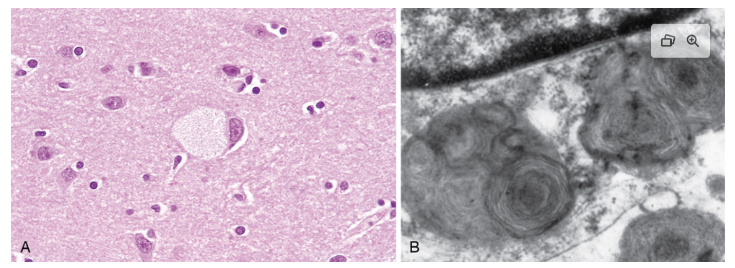
Ganglion cells in Tay-Sachs disease. (A) Under the light microscope, a large neuron has obvious lipid vacuolation. (B) A portion of a neuron under the electron microscope shows prominent lysosomes with whorled configurations. Part of the nucleus is shown above
115
what does this show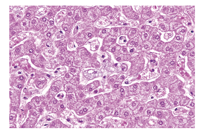
Niemann-Pick disease in liver. The hepatocytes and Kupffer cells have a foamy, vacuolated appearance due to deposition of lipids.
116
________________________ accumulate in massive amounts within phagocytic cells throughout the body in all forms of Gaucher disease. The distended phagocytic cells, known as Gaucher cells, are found in the spleen, liver, bone marrow, lymph nodes, tonsils, thymus, and Peyer patches.
glucocerebrosidases
117
a group of closely related syndromes that result from genetically determined deficiencies of enzymes involved in the degradation of mucopolysaccharides (glycosaminoglycans).
Mucopolysaccharidoses (MPSs)
118
caused by an inability to metabolize GM2 gangliosides due to lack of the α-subunit of lysosomal hexosaminidase. GM2 gangliosides accumulate in the central nervous system and cause severe intellectual disability, blindness, motor weakness, and death by 2 to 3 years of age.
tay sachs disease
119
are caused by a deficiency of sphingomyelinase. In the more severe type A variant, accumulation of sphingomyelin in the nervous system results in neuronal damage. Lipid also is stored in phagocytes within the liver, spleen, bone marrow, and lymph nodes, causing their enlargement. In type B, neuronal damage is not present.
niemann pick type a and b
120
esult from hereditary deficiency of one of the enzymes involved in the synthesis or sequential degradation of glycogen, the storage form of glucose.
glycigen storage diseases
121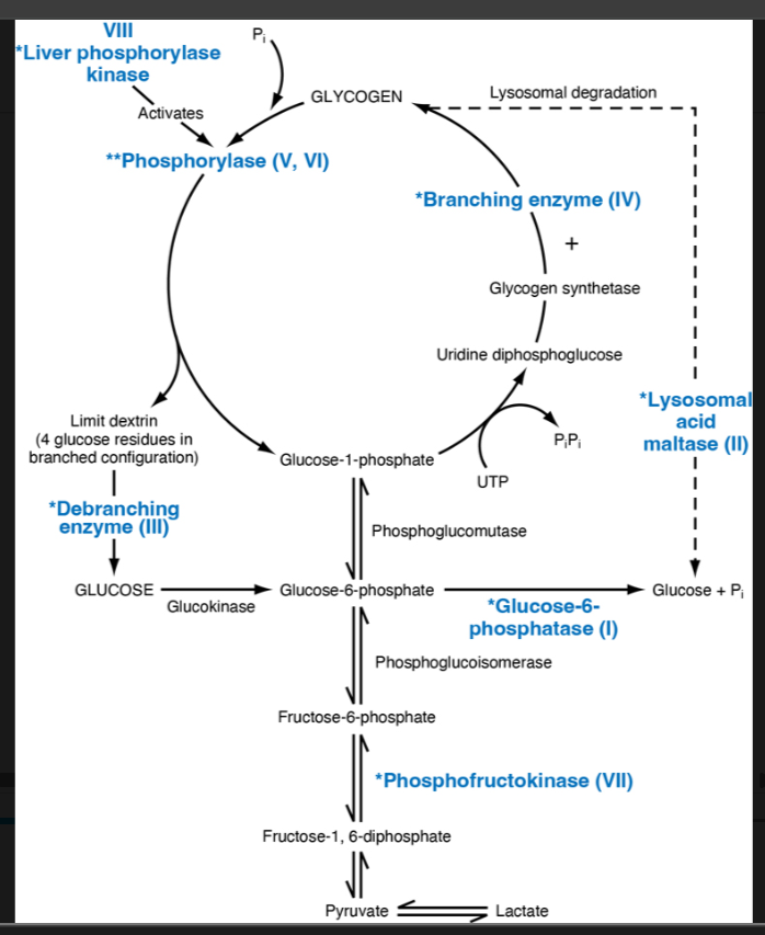
Image occlusion: name the hidden labels
122
Image occlusion: name the hidden labels
123
Image occlusion: name the hidden labels
124
Image occlusion: name the hidden labels
125
Image occlusion: name the hidden labels
126
Image occlusion: name the hidden labels
127
what does this show?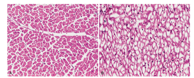
Pompe disease (glycogen storage disease type II). (A) Normal myocardium with abundant eosinophilic cytoplasm. (B) Patient with Pompe disease (same magnification) showing the myocardial fibers full of glycogen, seen as clear spaces.
128
midline malformations that involve some combination of neural tissue, meninges, and overlying bone or soft tissue; collectively, they are the most common CNS malformations.
neural tube defects
129
what are the 2 pathogenic mechanisms that contribute to neural tube defects
failure of neural tube closure, in which secondary mesenchymal tissue defects stem from aberrant skeletal modeling around the malformed tube (e.g., anencephaly and myelomeningocele); and (2) primary bony defects that are caused by abnormal axial mesoderm development and lead to secondary CNS abnormalities
130
a malformation of the anterior end of the neural tube that leads to absence of most of the brain and calvarium. Forebrain development is disrupted at approximately 28 days of gestatio
anencephaly
131
refers to extension of spinal cord tissue through a defect in the vertebral column; the term meningocele applies when there is only a meningeal extrusion.
myelomeningiocele
132
where to myelomeningoceles occur most commonly
lumbosacral region
133
refers to an extrusion of malformed brain tissue through a midline defect in the cranium. It most often occurs in the occiput, although nasofrontal variants involving the orbit, ethmoid, or cribriform plate (“nasal glial heterotopia,” sometimes misleadingly referred to as a “nasal glioma”) can be seen.
encephalocele
134
(the most common neural tube defect) ranges from an asymptomatic bony defect (spina bifida occulta) to a severe malformation with a flattened, disorganized segment of spinal cord, associated with an overlying meningeal outpouching.
spinal dysraphism or spinal bifida
135
Abnormalities in the generation and migration of neurons result in malformations of the _______
forebrain
136
how do neurons migrate?
The migration of neurons from the germinal matrix to the cerebral cortex follows two paths: radial migration, for progenitor cells destined to become excitatory neurons; and tangential migration, for those that will become inhibitory interneurons.
137
-brain abnirmally small
- accompanied by a small head circumference; it is associated with many different conditions, including chromosome abnormalities, fetal alcohol syndrome, and viral infection acquired in utero (e.g., with human immunodeficiency virus 1 [HIV-1] or Zika virus). It is postulated that the underlying anomaly is a reduction in the number of neurons that reach the neocortex, which leads to a simplification of gyral folding, a mechanism supported by experimental results in mouse models.
- accompanied by a small head circumference; it is associated with many different conditions, including chromosome abnormalities, fetal alcohol syndrome, and viral infection acquired in utero (e.g., with human immunodeficiency virus 1 [HIV-1] or Zika virus). It is postulated that the underlying anomaly is a reduction in the number of neurons that reach the neocortex, which leads to a simplification of gyral folding, a mechanism supported by experimental results in mouse models.
microcephally
138
a malformation characterized by reduction in the number of gyri, which in the extreme case may show no gyral pattern (agyria). Two patterns are observed, a classic smooth-surface form (type I), and a rough- or cobblestone-surface form (type II).
lissencephaly
139
forms are associated with mutations that disrupt mechanisms involved in cell migration, such as mutations in the cytoskeletal “motor” proteins that drive migration of neuroblasts.
type 1 lissencephaly
140
most commonly associated with genetic alterations that disrupt the “stop signal” for migration. This signal depends on a set of specifically glycosylated proteins, and mutations in the enzymes that place sugars onto these proteins are the most common causes of this form of lissencephaly.
type 2 lissencephaly
141
characterized by numerous small, irregularly formed cerebral convolutions with shallow sulci. The cerebral cortex is composed of four or fewer layers (instead of the normal six layers), with fusion of the molecular layers between gyri.
polymicrogyria
142
what causes polymicrogyria
can be induced by localized tissue injury toward the end of neuronal migration.
143
a group of neuron migration disorders defined by collections of subcortical neurons in inappropriate locations along the pathway of migration.
neuronal heterotopias
144
a spectrum of malformations characterized by incomplete separation of the cerebral hemispheres across the midline. Severe forms manifest midline facial abnormalities, including cyclopia; less severe variants (arrhinencephaly) show absence of the olfactory cranial nerves and related structures.
holoprosencephaly
145
a relatively common malformation, is the absence of the white matter bundles that carry cortical projections from one hemisphere to the other.
-Radiologic imaging studies show misshapen lateral ventricles (“batwing” deformity). Agenesis of the corpus callosum is sometimes associated with intellectual disability but may also be found in healthy individuals.
-Radiologic imaging studies show misshapen lateral ventricles (“batwing” deformity). Agenesis of the corpus callosum is sometimes associated with intellectual disability but may also be found in healthy individuals.
agenesis of the corpus callosum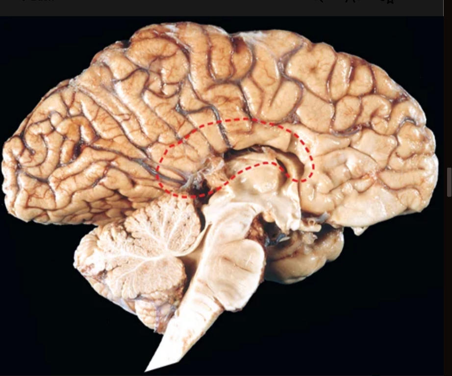
146
A distinct set of malformations primarily affects the brainstem and the cerebellum, which often show dramatic changes in size and shape.
posterior fossa anomalies
147
posterior fossa anomaly: consists of low-lying cerebellar tonsils that extend down into the vertebral canal. It may be a silent abnormality or may become symptomatic because of impaired CSF flow, medullary compression, or associated hydromyelia/syringomyelia (see later); if present, these symptoms can usually be corrected by neurosurgical intervention.
Chiari type 1 malformation
148
posterior fossa anomaly: more severe and produces a small posterior fossa, a misshapen midline cerebellum with downward extension of vermis through the foramen magnum (Fig. 28.8), and, almost invariably, hydrocephalus and a lumbar myelomeningocele. Other associated changes may include caudal displacement of the medulla, malformation of the tectum, aqueductal stenosis, cerebral heterotopias, and hydromyelia/syringomyelia.
chiari type II malformaton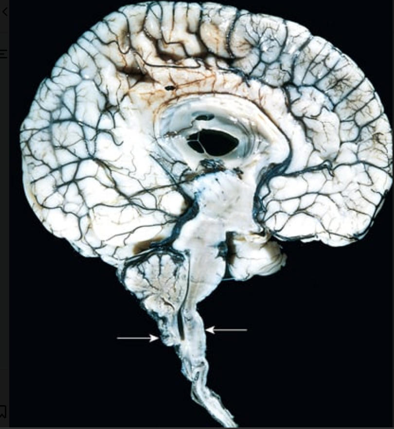
149
posterior fossa anomaly: characterized by an enlarged posterior fossa. The cerebellar vermis is absent or present only in rudimentary form in its anterior portion; in its place is a large midline cyst that is lined by ependyma and is contiguous with leptomeninges on its outer surface. This cyst represents the expanded, roofless fourth ventricle in the absence of a normally formed vermis. Dysplasias of brainstem nuclei are commonly found
dandy walker malformations
150
related disorders share hypoplasia of the cerebellar vermis with rotation and apparent elongation of the superior cerebellar peduncles and an altered brainstem shape; together these changes give rise to the “molar tooth sign” on imaging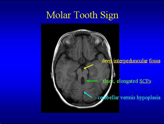
joubert syndrome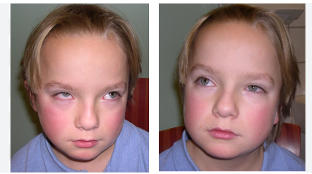
151
disorders are characterized by expansion of the ependyma-lined central canal of the cord (hydromyelia) or by the formation of a fluid-filled cleft-like cavity in the inner portion of the cord (syringomyelia, syrinx) that may extend into the brainstem (syringobulbia).
syringomyelia and hydromyelia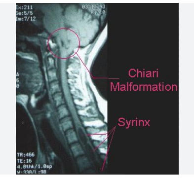
152
may be associated with a Chiari malformation; it may also occur in association with intraspinal tumors or following traumatic injury. In general, the histologic appearance is similar in all these conditions, with destruction of the adjacent gray and white matter, surrounded by dense reactive gliosis
-manifest 2nd or 3rd decade of life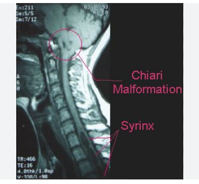
-manifest 2nd or 3rd decade of life
syringomuelia
153
injury to the brain because of altered blood flow—can be grouped into ischemic and hemorrhagic etiologies, with tissue infarction the ultimate consequence of both
cerebrovascular disease
154
what are the two etiologies of cerebrovascular disease
ischemic and hemorrhagic
155
what is the ultimate conclusion to both ischemic and hemorrhagic cerbrovascular disease
tissue infarction
156
what are the 2 major mechanisms of a stroke
• Hypoxia and/or ischemia resulting from impairment of oxygenation and blood supply of CNS tissue. This can be either a global or focal process, with the clinical manifestations determined by the region of brain affected. In the brain, embolism is a more common cause of vascular occlusion than thrombosis.
• Hemorrhage resulting from rupture of CNS vessels. Common etiologies include hypertension, vascular anomalies (aneurysms and malformations), and brain trauma (discussed later).
• Hemorrhage resulting from rupture of CNS vessels. Common etiologies include hypertension, vascular anomalies (aneurysms and malformations), and brain trauma (discussed later).
157
how may the brain be deprived of O2 (2x)
hypoxemia: low blood O2 content
ichemia: inadequate blood flow
ichemia: inadequate blood flow
158
ischemia in the CNS can result in inappropriate release of excitatory amino acid neurotransmitters such as glutamate, which can damage neurons by allowing excessive influx of ____________ through N-methyl-D-aspartate (NMDA)-type glutamate receptors; this phenomenon is termed ____________
calcium ions; excitotoxicity
159
region of the brain that is the transition between necrotic tissue and the normal brain. Can be rescued from cell death in many animal models with anti-apoptotic inervebtions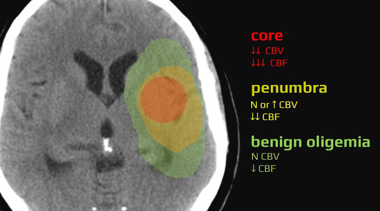
penumbra
160
follows reduction or cessation of blood flow to a localized area of the brain due to partial or complete arterial occlusion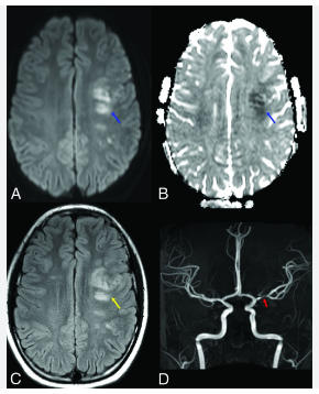
focal cerebral ischemia
161
what is the major source of collateral blood flow in the braon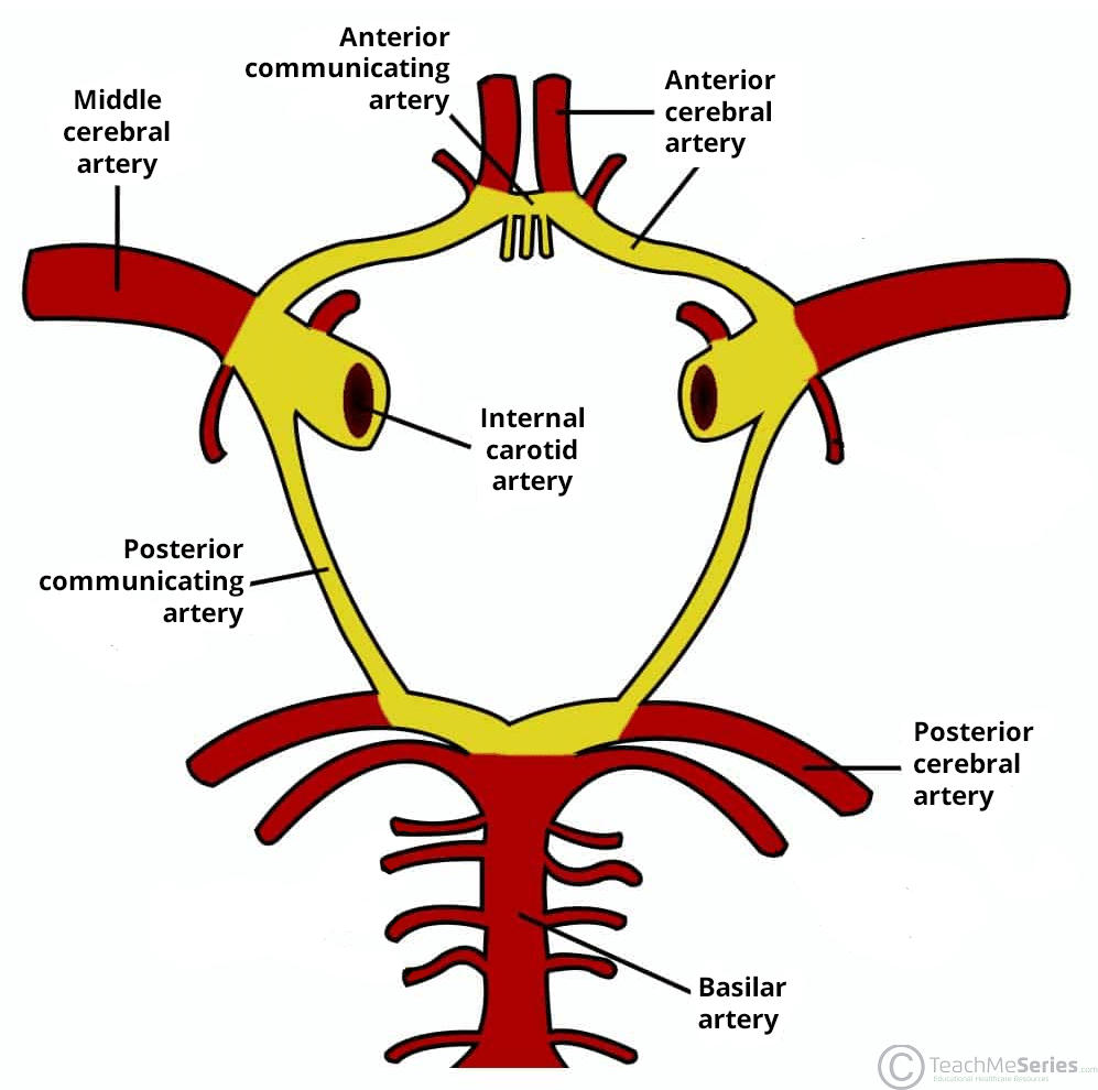
circle of willis
162
a medical condition where a foreign object, such as a blood clot, air bubble, or fat globule, travels through the bloodstream and blocks a blood vessel.
embolism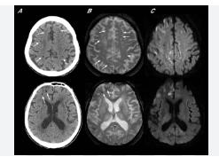
163
most common culprits of embolisms in te brain
Cardiac mural thrombi
164
what is the most affected by embolisms to the brain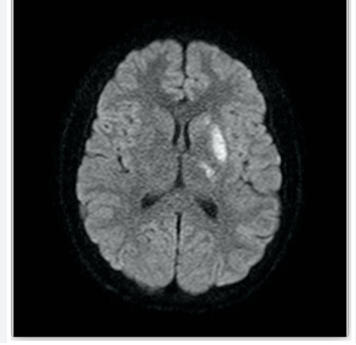
middle cerebral artery (either side) most ;ikely to lodge where blood vessels branch
165
what is the most common cause of thrombotic occlusions of the cerebral arteries
acute change of vulnerable atherosclerotic plaques, as in coronary artery disease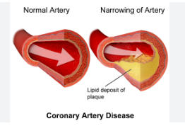
166
affects the deep penetrating arteries and arterioles that supply the basal ganglia and hemispheric white matter as well as the brainstem; these cerebral vessels develop arteriolosclerosis, also known as small vessel disease
hypertension
167
if atherosclerosis (small vessel disease) in the brain develops, what can happen?
disease process progresses to thrombosis and complete vessel occlusion, the end-result is development of small cavitary infarcts known as lacunes or lacunar infarcts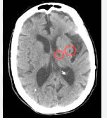
168
also known as ischemic strokes, occur when a blood vessel supplying the brain becomes blocked, typically by a clot, leading to an area of brain tissue death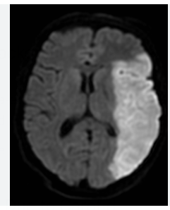
occlusive brain infacts
169
___________--- can occur from schemia-reperfusion injury following spontaneous or therapeutic dissolution or fragmentation of the intravascular occlusive material. (termed secondary hemorrhagic transformation)
secondary hemorrhage
170
what does A show?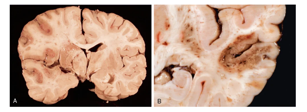
An ischemic infarct involves the territory of the middle cerebral artery, including the striatum, on the left side of this brain.
171
what does B show?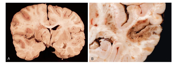
A hemorrhagic infarct with punctate hemorrhages, consistent with ischemia-reperfusion injury, is present in the temporal lobe.
172
first stage of hemorrhage: After the first 6 to 12 hours, neurons in the affected area show eosinophilic neuronal necrosis (increased eosinophilia of the cytoplasm followed by nuclear pyknosis and karyorrhexis (“dead red neurons”); both cytotoxic and vasogenic edema are present. There is loss of the usual tinctorial characteristics of white- and gray-matter structures. Endothelial and glial cells, mainly astrocytes, swell, and myelinated fibers begin to disintegrate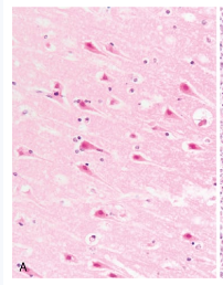
acute infact
173
hemorrhage: Phagocytic cells, derived from circulating monocytes and activated microglia, are evident at 48 to 72 hours and become the predominant cell type in the ensuing 2 to 3 weeks
-astrocytes at the edges of the lesion progressively enlarge, divide, and develop a prominent network of cytoplasmic extensions.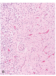
-astrocytes at the edges of the lesion progressively enlarge, divide, and develop a prominent network of cytoplasmic extensions.
subacute (evolving) infact
174
hemorrhage: After several months, the astrocytic response recedes, leaving behind a cystic cavity with dense meshwork of glial fibers admixed with new capillaries at the rim;
-he pia and arachnoid are not affected.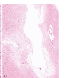
-he pia and arachnoid are not affected.
healed (remote) infarct
175
small occlusive infarcts that can be single or multiple and involve the putamen, globus pallidus, thalamus, internal capsule, deep white matter, caudate nucleus, and pons, in descending order of frequency.
-tissue loss surrounded by gliosis
-tissue loss surrounded by gliosis
lacunar infarcts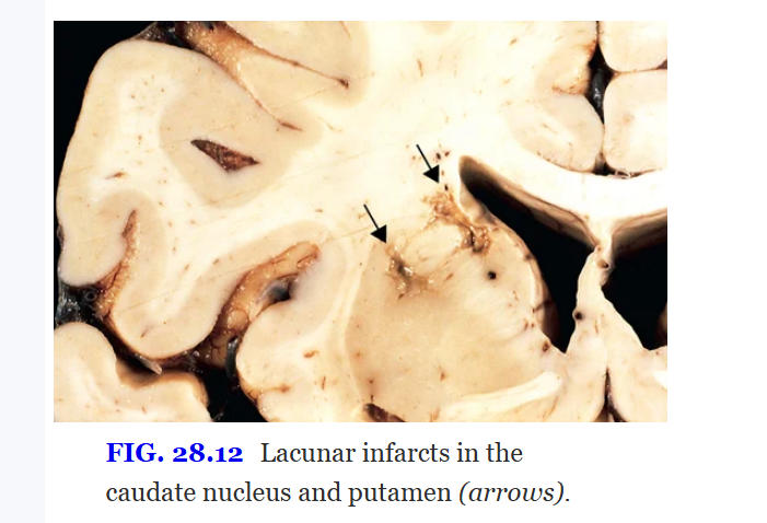
176
occurs when there is a generalized reduction of cerebral perfusion (as in cardiac arrest, shock, and severe hypotension) or decreased oxygen-carrying capacity of the blood (e.g., in carbon monoxide poisoning).
global cerebral hypoxia or ischemia
177
what is the higherarchy of cells sensitive to hypoxia/ischemia?
1) neurons
2) glial cells: specifically oligoidendrocytes and astrocytes
2) glial cells: specifically oligoidendrocytes and astrocytes
178
what neurons are the most sensitive in the brain to hypoxia or ischemia?
pyramidal neurons (hippocampus), cerebrall purkinje cells, pyramidal neurons (cerebral cortex layers III and V)
179
what can patients develop who survive severe global cerebral hypoxia/ischemia
-unresponsive wakefullness syndrome (persistent vegitative state)
-clinical criteria for brain death
-clinical criteria for brain death
180
when do border zone infacts develop?
after severe hypotensive episodes and are most commonly seen in patients resuscitated after cardiac arrest.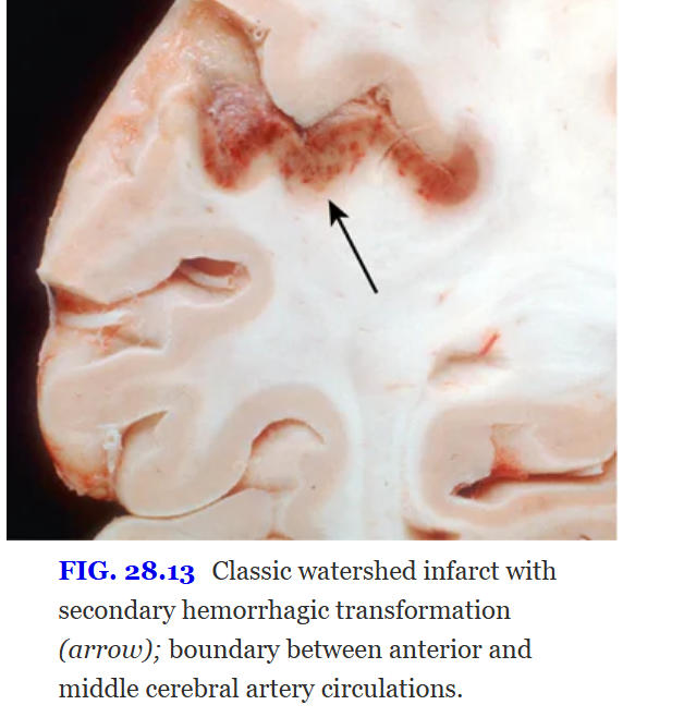
181
when the brain is undergoing global ischemia, the brain becomes edematous and swollen, producing widening of the _____ and narrowing of the ________
gyri; sulci
182
hemmorages within the brain
intraparenchymal hemorage
183
what is the common etiology of epidural space hemorrhages
trauma
184
what is the common etiology of subdural space hemorrhages
trauma
185
what is the common etiology of subarachnoid space hemorrhages
trauma or Vascular abnormality (arteriovenous malformation or aneurysm)
186
what is the common etiology of intraparenchymal space hemorrhages?
trauma (contusions) and Ischemia (hemorrhagic conversion of an ischemic infarct) and cerebral amyloid angiopathy and hypertension and tumors
187
Rupture of a _____________________ can result in a primary hemorrhage within the brain, often associated with sudden onset of neurologic symptoms (stroke)
small intraparenchymal vessels
188
Hemorrhages in the basal ganglia and thalamus are commonly designated __________________, whereas those that occur more superficially within the cerebral hemispheres are called___________________
ganglionic hemorrhages
lobar hemorrhages
lobar hemorrhages
189
this is the risk factor commonly associated with deep brain parenchumal hemorrhages
hypertension
190
what do these show?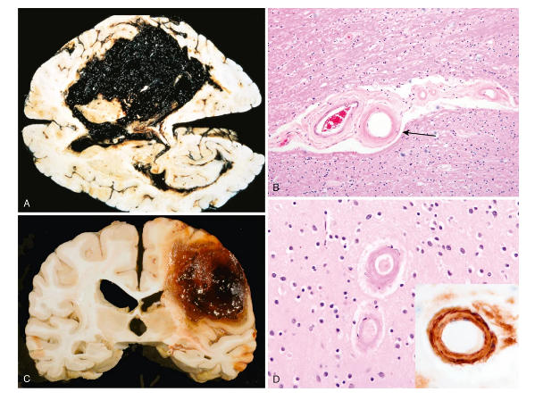
intraparenchymal cerebral hemorrhage: (A) Massive hypertensive ganglionic hemorrhage rupturing into a lateral ventricle. (B) Hyaline arteriolosclerosis (fibrosis and thickening of the arteriolar walls) in the basal ganglia and subcortical white matter of patients with long-standing hypertension, a risk factor for hypertensive hemorrhages and lacunar infarcts. (C) Large lobar hemorrhage due to cerebral amyloid angiopathy focally dissects into the subarachnoid space. (D) Amyloid deposition in cortical arterioles in cerebral amyloid angiopathyral hemorrhage
191
risk factor most often associated with lobar hemorrhages
cerebral amyloid angiopathy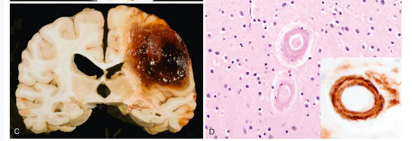
192
The most frequent cause of spontaneous subarachnoid hemorrhage is rupture of a ____________________in a cerebral artery.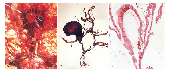
saccular berry aneurysm
193
aneurysm: Blood under arterial pressure is forced into the ______________, and affected individuals are stricken with a sudden, excruciating headache (“the worst headache I’ve ever had”) and rapidly lose consciousness. Between 25% and 50% of patients die with the first rupture, but patients who survive often improve and recover consciousness in minutes.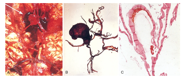
subarachnoid space
194
4 types of vascular malformations in the brain; which are associated with risk of hemorrhage and fevelopment of neurological symptoms?
risk: ateriovenous malformations, cavernous malformations
na: capillary telangiectasia, venous angiomas
na: capillary telangiectasia, venous angiomas
195
tangled networks of wormlike vascular channels with high blood flow due to prominent, pulsatile arteriovenous shunting that may involve vessels in the subarachnoid space, the brain, or both (Fig. 28.17). They are composed of greatly enlarged blood vessels separated by gliotic tissue, often with evidence of prior hemorrhage.
arteriovenous malformations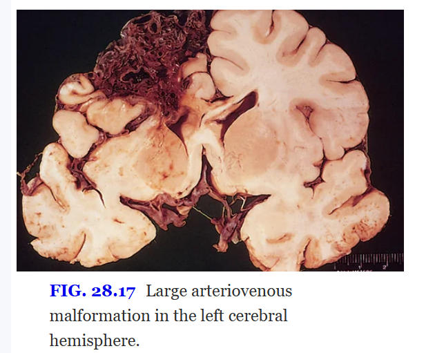
196
consist of distended, loosely organized vascular channels arranged back-to-back with collagenized walls of variable thickness; there is usually, but not invariably, no brain parenchyma between vessels in this type of malformation. They occur most often in the cerebellum, pons, and subcortical regions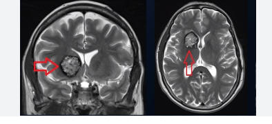
cavernous malformations
197
currently the most frequently diagnosed major cancer and the most common cause of cancer mortality worldwide.
lung cancer
198
lung cancers are broadly classified into ________________- types with the latter group including adrenocarcnoma and squamous cell carcinoma
small cell and non small cell
199
(females/males) are more susceptible to carcinogens in tobacco than the other sex
females
200
There is a linear correlation between the intensity of exposure to cigarette smoke and the appearance of ever more worrisome____________________ changes.
epithelial
201
industrail exposures that increase risk of developing lung cancer (x7)
asbestos, arsenic, chromium, uranium, nickel, vinyl chloride, and mustard gas
202
most frequent malignancy in individuals exposed to asbestos, particularly when coupled with smoking
lung cancer
203
Chronic exposure to air particulates in smog may cause lung irritation, inflammation, and repair, and you will recall that chronic inflammation and repair increases the risk of a variety of ________________
cancers
204
associated with tobacco smoking, but less so than other histologic subtypes; as a result, it is the most common subtype in never-smokers
adenocarcinoma
205
what type of mutations do adrenocarcinomas tend to be
oncogenic gain-of-function mutations involving components of growth factor receptor signaling pathways
-many involve TKRs
-many involve TKRs
206
highly associated with exposure to tobacco smoke and harbors diverse genetic aberrations, many of which are chromosome deletions involving tumor suppressor loci.
squamous cell carcinoma`
207
The _________- tumor supressor gene, which encodes the cyclin depednet kinase inhibitor P16 is mutated in 65% of tumors (squamous cell carcinoma)
CDKN2A
208
virtually always smoking related and has the highest mutational burden among lung cancers. There is almost universal inactivation of both TP53 and RB
small cell carcinoma
209
what chromosome is lost in small cell carcinoma
3p
210
4 types of morphological precursor epithelial lesions in lung cancer (x4)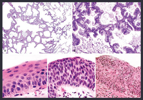
(1) atypical adenomatous hyperplasia (Fig. 15.33A), (2) adenocarcinoma in situ (Fig. 15.33B), (3) squamous metaplasia, dysplasia, and carcinoma in situ (Fig. 15.33C–E), and (4) diffuse idiopathic pulmonary neuroendocrine cell hyperplasia.
211
what does a show?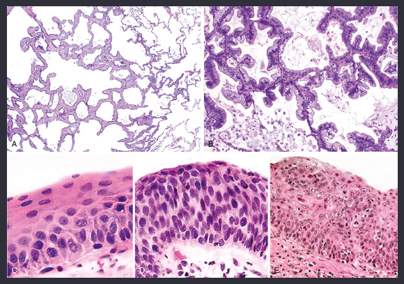
precursor lesions of lung cancer: Atypical adenomatous hyperplasia.
212
what does B show?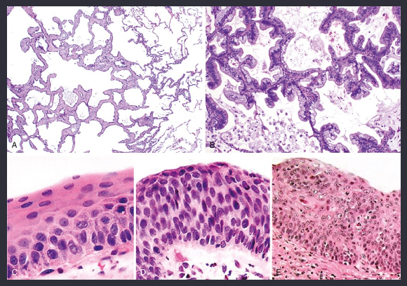
precursor lesion of lung cancer: Adenocarcinoma in situ, mucinous type.
213
squamous cell precursor: C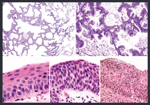
Squamous cell precursors; appearance of squamous dysplasia
214
squamous cell precursor: E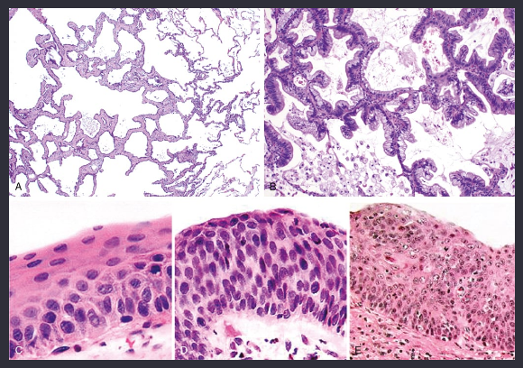
the stage immediately preceding invasive squamous carcinoma by definition has not penetrated the basement membrane and has cytologic features similar to those of carcinoma
215
most common form of lung cancer in adult males and feales
adrenocarcinoma
216
a small precursor lesion (≤5 mm) characterized by dysplastic pneumocytes lining alveolar walls that may be mildly fibrotic (see Fig. 15.33A). It can be single or multiple and can be close to or distant from an invasive tumor.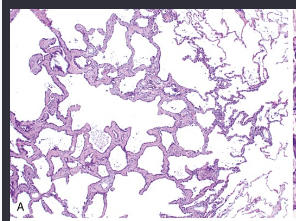
atypical adenomatous hyperplasia
217
a lesion that is less than 3 cm in size and is composed entirely of dysplastic cells growing along pre-existing alveolar septa in a so-called lepidic pattern 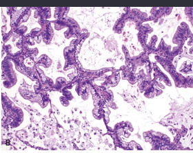
adenocarcinoma in situ
218
an invasive malignant epithelial tumor with glandular differentiation or mucin production by the tumor cells. Adenocarcinomas grow in various patterns, including lepidic (noninvasive), acinar, papillary, micropapillary, and solid. 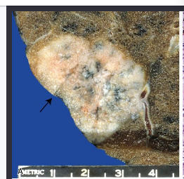
adrenocarcinoma
219
They vary histologically from well-differentiated tumors with obvious glandular elements (Fig. 15.34B), to papillary lesions resembling other papillary carcinomas, to solid masses with only occasional mucin-producing glands and cells. The Page 654majority express thyroid transcription factor-1 (TTF-1)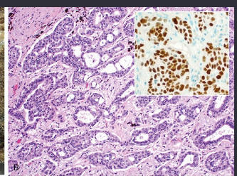
adrenocarcinomas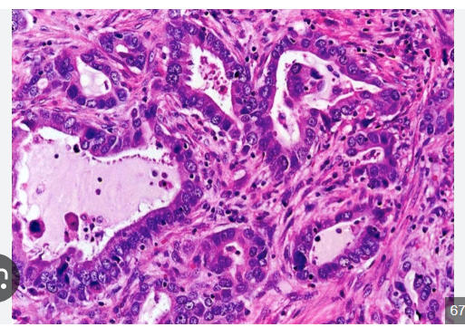
220
Smaller tumors (≤3 cm) with limited invasion (≤5 mm) associated with scarring and a lepidic-predominant growth pattern are called 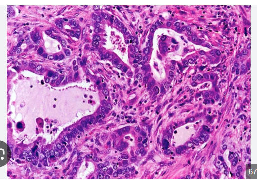
microinvasive adrenocarcinomas
221
adrenocarcinomas that tend to spread aerogenously, forming satellite tumors; thus, these are less likely to be cured by surgery.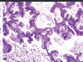
mucinous adrenocarcinomas
222
often antedated by squamous metaplasia or dysplasia in the bronchial epithelium, which then transforms to carcinoma in situ, a phase that may last for years (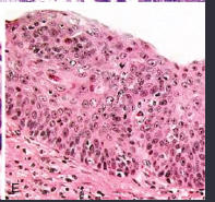
squamous cell carcinomas
223
how may squamous cell carcinomas invade the lung tissue?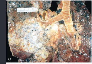
-grow into the bronchial lumen, producing an intraluminal mass.
- penetrate the wall of the bronchus and infiltrate along the peribronchial tissue into the adjavent carina or mediastinum
- penetrate the wall of the bronchus and infiltrate along the peribronchial tissue into the adjavent carina or mediastinum
224
squamous cell carcinoma is characterized by the presence of (x2) . Keratinization may take the form of squamous pearls or individual cells with markedly eosinophilic cytoplasm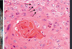
karatinization and intrecellular brdges
225
composed of relatively small cells with scant cytoplasm, ill-defined cell borders, finely granular nuclear chromatin (salt and pepper pattern), and absent or inconspicuous nucleoli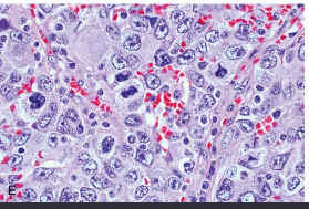
small cell carcinoma
226
small cell carcinoma: Basophilic staining of vascular walls due to encrustation by DNA from necrotic tumor cells (so-called _______________)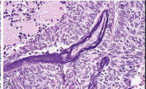
azzopardi effect
227
undifferentiated malignant epithelial tumor that lacks the cytologic and immunophenotypic features of other forms of lung cancer.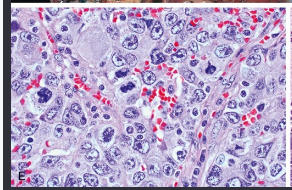
large cell carcinoma
228
Lung carcinomas have local effects that may cause several pathologic changes in the lung distal to the point of bronchial involvement. Partial obstruction may cause marked_____________; total obstruction may lead to_______________
focal emphysema
ateletasis
ateletasis
229
TNM staging: Carcinoma in situ
Adenocarcinoma in situ: adenocarcinoma with pure lepidic pattern, ≤3 cm
Squamous cell carcinoma in situ
Adenocarcinoma in situ: adenocarcinoma with pure lepidic pattern, ≤3 cm
Squamous cell carcinoma in situ
Tis
230
TNM staging: Tumor ≤3 cm without pleural or mainstem bronchus involvement (T1mi, minimally invasive adenocarcinoma; T1a, <1 cm; T1b, 1–2 cm; T1c, 2–3 cm)
T1
231
TNM staging: Tumor 3–5 cm or involvement of mainstem bronchus but not of carina, visceral pleural involvement, or lobar atelectasis (T2a, 3–4 cm, or <3 cm with pleural involvement; T2b, 4–5 cm)
T2
232
TNM staging: Tumor >5–7 cm or one with involvement of parietal pleura, chest wall (including superior sulcus tumors), diaphragm, phrenic nerve, mediastinal pleura, parietal pericardium, or separate tumor nodules in the same lobe
T3
233
TNM staging: Tumor >7 cm or any tumor with invasion of mediastinum, heart, great vessels, trachea, recurrent laryngeal nerve, esophagus, vertebral body, or carina, or separate tumor nodules in a different ipsilateral lobe
T4
234
TNM staging: No metastasis to regional lymph nodes
N0
235
TNM staging: Ipsilateral intraparenchymal or peribronchial or hilar nodal involvement
N1
236
TNM staging: Metastasis to ipsilateral mediastinal or subcarinal lymph nodes
N2
237
TNM staging: Metastasis to contralateral mediastinal or hilar lymph nodes, ipsilateral or contralateral scalene, or supraclavicular lymph nodes
N3
238
TNM staging: no distant metastiasis
M0
239
TNm staging: Distant metastasis (M1a, separate tumor nodule in contralateral lobe or pleural nodules or malignant pleural or pericardial effusion; M1b, single extrathoracic metastasis in a single organ; M1c, multiple extrathoracic metastases)
M1
240
pathological basis of cough lung tumor spread
involvement in central airway
241
path basis lung tumor spread: hemoptysis
Hemorrhage from tumor in airway
242
path basis lung tumor spreead: chest pain
Extension of tumor into mediastinum, pleura, or chest wall
243
path basis lung tumor spread: pnemonia, absecess, lobar collapse
Airway obstruction by tumor
244
path basis lung tumor spread: lipoid pnemonia
Tumor obstruction; accumulation of cellular lipid in foamy macrophages
245
path basis lung tumor spread: pleural effusion
tumor spread into pleura
246
path basis lunf tumor spread: hoarseness
recurrent laryngeal n invasion
247
path basis lung tumor spread dyusphagia
esophageal invasion
248
path basis lung tumor spread: diaphragm paralysis
phrenic n invasion
249
path basis lung tumor spread: rib distruction
chest wall invasion
250
path basis lung tumor sread: SVc syndrome
SVC compression by tumor
251
path basis lung tumor spread: horner syndrome
sympathetic ganglia invasion
252
path basis lung tumor spread pericarditis, tamponade
pericardial involvement
253
The normal lung contains _______________ within the epithelium as single cells or as clusters called neuroepithelial bodies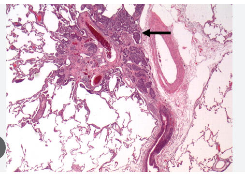
neuroendocrine cells
254
Carcinoid tumors are low-grade malignant epithelial neoplasms that are subclassified into (x2)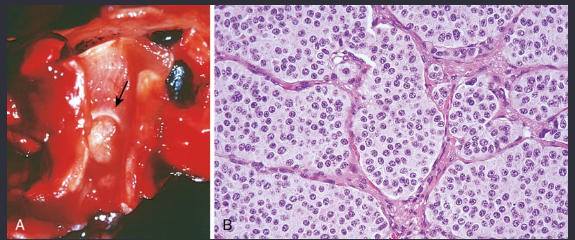
typical and atypical
255
whjat does this show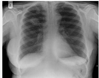
collar button (coin) lesion of a carcinoids
256
Persistent cough, hemoptysis, impairment of drainage of respiratory passages with secondary infections, bronchiectasis, emphysema, and atelectasis are all by-products of the intraluminal growth of these lesions.
carcinoids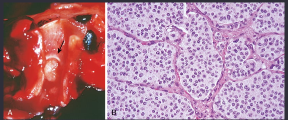
257
a pulmonary disorder that primarily affects young females of childbearing age. It is characterized by a proliferation of perivascular epithelioid cells that express markers of both melanocytes and smooth muscle cells. The proliferation distorts the involved lung, leading to cystic, emphysema-like dilation of terminal airspaces, thickening of the interstitium, and obstruction of lymphatic vessels.
lymphangioleiomyomatosis
258
a rare tumor that is more common in children, with an equal male-to-female ratio. Presenting symptoms include fever, cough, chest pain, and hemoptysis. It may also be asymptomatic. Imaging studies usually show a round, well-defined, peripheral mass. Calcification is present in about a quarter of cases.
inflammatoru myofibroblastic tumor
259
how are karyotype described(naming)
total number of chromosomes is given first, followed Page 145by the sex chromosome complement, and finally the description of abnormalities in ascending numerical order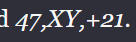
260
karyotype naming: the short arm of a chromosome is designated ____ and the long arm ____
p; q
261
what does this mean (karyotype naming)
a chromosomal segment located on the short arm of the X chromosome, in region 2, band 1, and sub-band 2.
262
may take the form of an abnormal number of chromosomes or alterations in the structure of one or more chromosomes.
cytogenic disorders
263
Any exact multiple of the haploid number of chromosomes (23) is called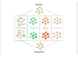
euploid
264
If an error occurs in meiosis or mitosis and a cell acquires a chromosome complement that is not an exact multiple of 23, it is referred to as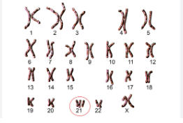
aneuploidy
265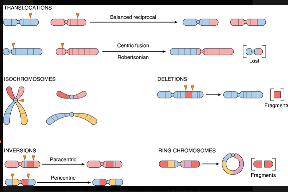
Image occlusion: name the hidden labels
266
Image occlusion: name the hidden labels
267
Image occlusion: name the hidden labels
268
Image occlusion: name the hidden labels
269
Image occlusion: name the hidden labels
270
Image occlusion: name the hidden labels
271
Image occlusion: name the hidden labels
272
Image occlusion: name the hidden labels
273
Image occlusion: name the hidden labels
274
results when one arm of a chromosome is lost and the remaining arm is duplicated, resulting in a chromosome consisting of only two short arms or two long arms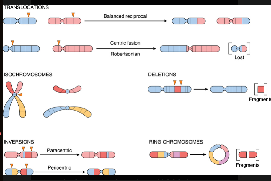
isochromosome formation
275
occur when there are two breaks within a chromosome arm, followed by loss of the chromosomal material between the breaks and fusion of the broken ends.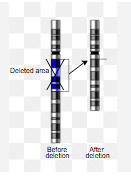
interstitial deletions
276
result from a single Page 146break in a chromosome arm, producing a fragment with no centromere, which is then lost at the next cell division. 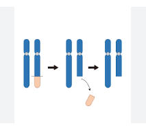
terminal deletions
277
most common cause of trisomy and therefore of Down syndrome is ...
meiotic nondisjunction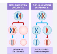
278
the failure of homologous chromosomes to separate during meiosis I, or the failure of sister chromatids to separate during meiosis II, resulting in gametes with an abnormal number of chromosomes
meiotic nondisjunction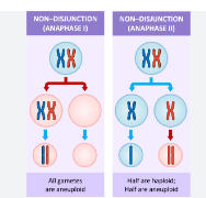
279
risks that come with having triosomy 21
-40% of the patients have congenital heart disease. The most frequent forms of congenital heart disease in Down syndrome are atrioventricular septal defects constituting 43% of cases, followed by ventricular septal defects (32%), atrial septal defects (19%), and tetralogy of Fallot (6%).
-increased risk of developing leukemia.
-alzheimers by 40yrs age
-abnormal immune responses that predispose them to serious infections, particularly of the lungs, and to thyroid autoimmunity
-increased risk of developing leukemia.
-alzheimers by 40yrs age
-abnormal immune responses that predispose them to serious infections, particularly of the lungs, and to thyroid autoimmunity
280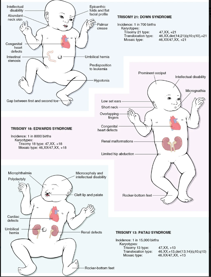
Image occlusion: name the hidden labels
281
Image occlusion: name the hidden labels
282
Image occlusion: name the hidden labels
283
trisomy 21: Most protein-coding genes mapped to chromosome 21 are overexpressed. Included among these is the gene for________________ can lead to alzheimers (kinda)
APP
284
trisomy 21: Approximately 10% of the genes overexpressed in Down syndrome are directly or indirectly involved in regulation of ______________
mitochondrial dysfunction
285
encompasses a spectrum of disorders that result from a small deletion of band q11.2 on the long arm of chromosome 22.
chromosome 22q11.2 deletion syndrome
286
what are the two disorders linked to chromosome 22q11.2 deletion syndrome? (x2)
-DiGeorge syndrome (thymic hypoplasia with diminished T-cell immunity and parathyroid hypoplasia with hypocalcemia) and
-velocardiofacial syndrome (congenital heart disease involving outflow tracts, facial dysmorphism, and developmental delay).
-velocardiofacial syndrome (congenital heart disease involving outflow tracts, facial dysmorphism, and developmental delay).
287
states that (1) only one of the X chromosomes is genetically active, (2) the other X chromosome of either maternal or paternal origin undergoes heteropyknosis and is rendered inactive, (3) inactivation of either the maternal or the paternal X chromosome occurs at random among all the cells of the blastocyst on or about day 5.5 of embryonic life, and (4) inactivation of the same X chromosome persists in all the cells derived from each precursor cell.
lyon hypothesis
288
defined as male hypogonadism that occurs when there are two or more X chromosomes and one or more Y chromosomes
kleinfelter syndrome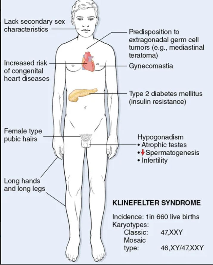
289
risks with kleinfelter disease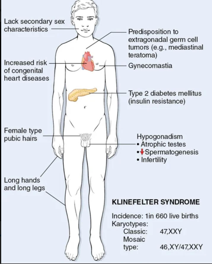
increased incidence of type 2 diabetes and the metabolic syndrome that gives rise to insulin resistance. Patients are at a higher risk for congenital heart disease, particularly mitral valve prolapse, which is seen in about 50% of adults. In addition, there is a higher prevalence of atrial and ventricular septal defects. There is also an increased incidence of osteoporosis and fractures due to sex hormonal imbalance
290
results from complete or partial monosomy of the X chromosome and is characterized by hypogonadism in phenotypic females. It is the most common sex chromosome abnormality in females, affecting about 1 in 2000 live-born females.
turner syndrome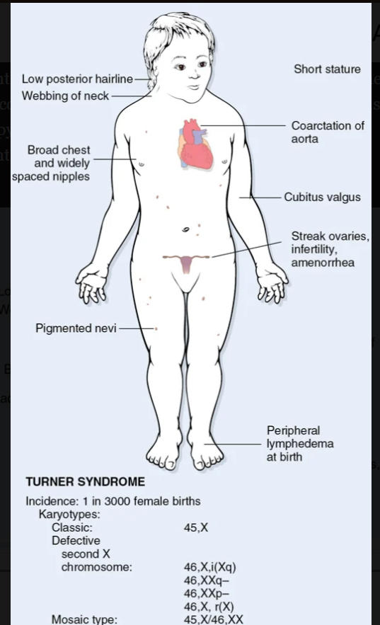
291
risks with turner syndrome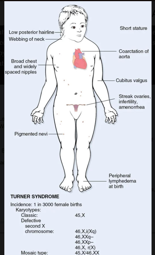
ilateral neck webbing and persistent looseness of skin on the back of the neck. Congenital heart disease is also common, affecting 25% to 50% of patients. Left-sided cardiovascular abnormalities, particularly preductal coarctation of the aorta and bicuspid aortic valve, Page 152are seen most frequently. Approximately 5% of young females initially diagnosed with coarctation of aorta have Turner syndrome. Aortic root dilation is present in 30% of cases
292
implies the presence of both ovarian and testicular tissue.
true hermaphrodite
293
has ovaries but male external genitalia or has testicular tissue but female-type genitalia
pseudohermaphrodite