Neuromuscular N3 Lecture ORIGINAL
Study these flashcardsFree · spaced repetition · iPhone, Android, Mac & web
First 300 of 986 cards
1
What structures make up the central nervous system (CNS)?
Brain
Spinal cord
Spinal cord
2
What is the primary function of the brain in the CNS?
the brain acts as the interpretation center
3
What role does the spinal cord play in the CNS?
the spinal cord functions as the transmission pathway
4
What structures make up the peripheral nervous system (PNS)?
Cranial nerves
Spinal (peripheral) nerves
Spinal (peripheral) nerves
5
Where do cranial nerves emerge from?
Cranial nerves emerge directly from the brain 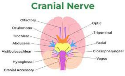
6
Where are the peripheral nerves located?
outside the brain and spinal cord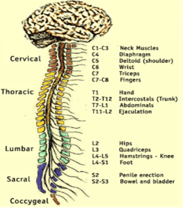
7
What is the function of the peripheral nerves?
Relay information between the brain and the rest of the body
8
What types of impulses do nerves carry?
Sensory impulses to the brain
Motor responses from the brain to the body
Both
Motor responses from the brain to the body
Both
9
What happens if a nerve itself is damaged?
Transmission of impulses to or from the brain can be affected, leading to sensory or motor dysfunction
10
What are the consequences of damage to the spinal cord?
Disrupt pathways for nerve impulses --> impairs both sensory and motor functions
11
What can result from brain damage related to nerve impulse processing?H
Brain may lose ability to properly interpret sensory information or send motor commands to the body
12
What is the primary role of the cranial nerves s
responsible for the sensory and motor functions of the head and neck
13
What is the longest cranial nerve?
Vagus nerve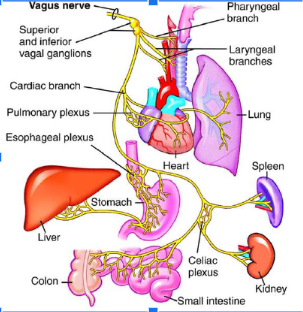
14
What effect does vagal stimulation have on the heart?
slows the formation of impulses in the sinus node
15
How is the vagus nerve involved therapeutically?
Can terminate SVT
16
What happens to the intracranial pressure during a vagal maneuver?
Cerebral blood volume will INCREASE, outflow will DECREASE --> increased intracranial pressure
17
How does the vagal maneuver affect the heart rate?
increased intracranial pressure stimulates the vagal nerve --> slows down formation of impulses in the SA node
Bearing down as with a bowel movement or even bending over --> increases thoracic pressure to slow the heart rate
Bearing down as with a bowel movement or even bending over --> increases thoracic pressure to slow the heart rate
18
What cranial nerves are assessed when checking vision and eye movements?
Optic (CN II) --> visual acuity
Oculomotor (CN III) --> pupil constriction, eye movement
Trochler (CN IV) --> able to look down, towards, and away from the nose
Abducens (CN VI) --> able to look left and right
Oculomotor (CN III) --> pupil constriction, eye movement
Trochler (CN IV) --> able to look down, towards, and away from the nose
Abducens (CN VI) --> able to look left and right
19
What cranial nerve is assessed during a smell test?
Olfactory (CN I)
20
What cranial nerve is assessed during a hearing test?
Acoustic (CN VIII)
21
What cranial nerve is assessed when checking facial and head movement?
Trigeminal (CN V) --> clench teeth, close eyes, light touch to face
Facial (CN VII) --> facial movement, close eyes, smile
Spinal accessory (CN XI) --> turn head, lift shoulders
Facial (CN VII) --> facial movement, close eyes, smile
Spinal accessory (CN XI) --> turn head, lift shoulders
22
What cranial nerve is assessed when checking gag reflex?
Glossopharyngeal (CN IX)
23
What cranial nerve is assessed when saying "ahh"?
Vagus (CN X) --> uvular and palate movement
24
What cranial nerve is used when sticking out your tongue?
Hypoglossal (CN XII)
25
What is an mnemonic for the 12 cranial nerves?
On --> Olfactory (CN 1)
Occasion --> Optic (CN 2)
Our --> Ocular (CN 3)
Trusty --> Trochlear (CN 4)
Truck --> Trigeminal (CN 5)
Acts --> Abducens (CN 6)
Funny --> Facial (CN 7)
Very --> Vestibulocochlear (CN 8)
Good --> Glossopharyngeal (CN 9)
Vehicle --> Vagus (CN 10)
Any --> Accessory (CN 11)
How --> Hypoglossal (CN 12)
Occasion --> Optic (CN 2)
Our --> Ocular (CN 3)
Trusty --> Trochlear (CN 4)
Truck --> Trigeminal (CN 5)
Acts --> Abducens (CN 6)
Funny --> Facial (CN 7)
Very --> Vestibulocochlear (CN 8)
Good --> Glossopharyngeal (CN 9)
Vehicle --> Vagus (CN 10)
Any --> Accessory (CN 11)
How --> Hypoglossal (CN 12)
26
What is a mnemonic for whether a cranial nerve is sensory, motor, or both?
Some Say Marry Money But My Brother Says Bad Business Marry Money
27
Where does the spinal cord begin and end?
Begins at the bottom of the brainstem (medulla oblongata)
Ends in the lower back
Ends in the lower back
28
What vertebrae mark the upper and lower boundaries of the spinal cord?
The spinal cord runs from the C1 vertebra to approximately the L1 vertebra
29
How long is the spinal cord?
about 18 inches
30
What fluid surrounds the spinal cord?
CSF (cerebrospinal fluid)
31
What are the three protective layers that shield the brain and spinal cord?
The meninges, which consist of the dura mater, arachnoid mater, and pia mater
32
Where does the spinal cord lie?
Inside the spinal column, which is made up of 33 vertebrae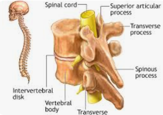
33
How many vertebrae are fused to form the sacrum?
Five vertebrae are fused to form the sacrum (part of the pelvis)
34
How many vertebrae are fused to form the coccyx?
Four small vertebrae are fused to form the coccyx (tailbone)
35
How many pairs of spinal nerves and roots are there?
There are 31 pairs of spinal nerves and roots
36
How many pairs of cervical nerves are there, and where do they pass through?
Eight pairs of cervical nerves (C1-C8) pass through the cervical (neck) region.
37
How many pairs of thoracic nerves are there, and where do they pass through?
Twelve pairs of thoracic nerves (T1-T12) pass through the thoracic region.
38
How many pairs of lumbar nerves are there, and where do they pass through?
Five pairs of lumbar nerves (L1-L5) pass through the lumbar region.
39
How many pairs of sacral nerves are there, and where are they located?
Five pairs of sacral nerves (S1-S5) are located in the sacral (buttock) region.
40
How many pairs of coccygeal nerves are there, and where do they pass through?
One pair of coccygeal nerves (C0) passes through the coccyx.
41
What is a dermatome?
A dermatome is an area on the body that relies on a specific spinal nerve.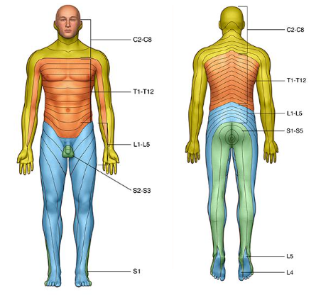
42
Why is the specific spinal nerve important for its dermatome?
The spinal nerve is critical because it carries all the nerve signals traveling between the dermatome and the brain.
43
Where is an epidural administered?
in the epidural space 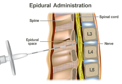
44
What is the cerebral cortex?
The cerebral cortex is the outermost layer of the brain that gives it a characteristic wrinkly appearance.
45
How is the cerebral cortex divided?
Divided lengthways into two cerebral hemispheres: the right and left
46
How many lobes are each hemisphere divided into, and what are they?
Each hemisphere is divided into four lobes: frontal, parietal, temporal, and occipital, connected by the corpus callosum.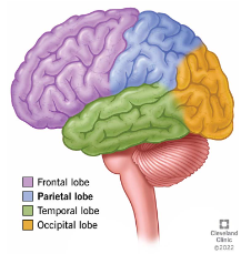
47
How do brain functions typically operate?
Most brain functions rely on different regions working together, yet each lobe carries out the bulk of certain specific functions.
48
What functions are the parietal lobe responsible for?
Touch perception
Movement control
Manipulation of objects
Movement control
Manipulation of objects
49
What functions are the occipital lobe responsible for?
Visual reception
Local orientation
Shape perception
Local orientation
Shape perception
50
What functions are the cerebellum responsible for?
Coordination and balance
Relex motor acts
Relex motor acts
51
What functions are the fronal lobe responsible for?
Voluntary movement
Planning
Intellect
Problem solving, abstract reasoning
Planning
Intellect
Problem solving, abstract reasoning
52
What functions are the temporal lobe responsible for?
Hearing and speech
Long term memory
Face recognition
Object perception
Long term memory
Face recognition
Object perception
53
What is the function of the brain stem?
conduction tract for pain, temperature, and pressure sensation
54
Where is temperature control regulated?
Temperature is regulated by the hypothalamus
55
What are the three sections of the brain stem, in descending order?
Midbrain
Pons
Medulla Oblongata
Pons
Medulla Oblongata
56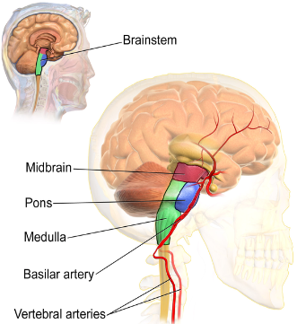
Image occlusion: name the hidden labels
57
Image occlusion: name the hidden labels
58
Image occlusion: name the hidden labels
59
What is the primary function of the midbrain?
The midbrain is crucial for eye movement
60
What functions does the pons coordinate?
The pons coordinates facial movements, hearing, and balance
61
What vital functions does the medulla oblongata regulate?
The medulla oblongata regulates breathing, heart rhythm, blood pressure, and swallowing.
62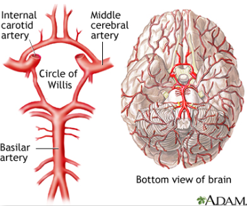
Image occlusion: name the hidden labels
63
Image occlusion: name the hidden labels
64
Image occlusion: name the hidden labels
65
Why are most cerebral aneurysms found around the circle of Willis?
Several bifurcations (where a bunch of blood vessels intersect), which make it more vulnerable to aneurysm formation
66
What is the blood-brain barrier (BBB)?
a filtering mechanism of the capillaries that carry blood to the brain and spinal cord tissue, blocking the passage of certain substances.
67
What is the primary function of the BBB?
The BBB keeps substances in the bloodstream out of the cerebrospinal circulation and brain tissue.
68
Which substances can pass through the blood-brain barrier?
Oxygen and CO2 (no shit sherlock)
Alcohol
Anesthetics
Water
Alcohol
Anesthetics
Water
69
What types of substances are prevented from passing through the BBB?
Large molecules such as albumin, anything attached to albumin, and many antibiotics are prevented from passing.
70
How does fentanyl interact with the BBB?
Fentanyl rapidly crosses the blood-brain barrier
71
How is fentanyl transferred in relation to the placenta?
Fentanyl is rapidly transferred by the human early placenta
72
How does CSF (cerebrospinal fluid) contribute to brain buoyancy?
CSF reduces the brain's net weight from 1400g to just 50g by creating a fluid bath
73
How does CSF protect the brain?
CSF acts as a shock absorber, preventing damage from the brain hitting the cranium
74
How does CSF help maintain homeostasis in the brain?
CSF regulates the distribution of metabolites, keeping the brain's external environment stable
75
How does CSF assist in clearing waste from the brain?
Waste products produced by brain cells are excreted into the CSF, which then drains into the bloodstream
76
Where is CSF produced?
CSF is produced by specialized cells in the ventricles of the brain
77
How much CSF is produced daily?
Approximately 500 ml of CSF is produced daily, ensuring continuous circulation around the CNS
78
How much CSF can the brain contain at a time?
The brain can only contain 120-150 ml of CSF
79
How is excess CSF removed from the brain?
Excess CSF is drained into the bloodstream through the arachnoid villi
80
How often is CSF recycled?
CSF is recycled about 3 times a day
81
Describe the movement of CSF through the ventricles
CSF moves from the lateral ventricle --> third ventricle --> fourth ventricle
82
Where does CSF go after passing through the fourth ventricle?
It moves into the subarachnoid space and/or the central canal of the spinal cord
83
What color should normal CSF be?
It should be clear
84
What should NOT be found in CSF?
Red blood cells
Bacteria
Bacteria
85
What is the normal specific gravity of CSF?
the normal specific gravity is 1.007
86
What is the normal protein count in CSF?
the protein count is 14-45 mg/100 ml
87
How many WBCs are normally present in CSF?
0.5 - 5 cells/mm³
88
What is the normal glucose concentration in CSF?
50-75 mg/100 ml
89
What other components may be tested for in CSF?
Immunoglobulins and electrolyte levels
90
What are some causes of elevated glucose levels in CSF?
Hyperglycemia
Infection --> bacteria or fungi
Inflammation of the CNS
Tumor
Lumbar puncture
Infection --> bacteria or fungi
Inflammation of the CNS
Tumor
Lumbar puncture
91
How can a lumbar puncture lead to elevated CSF glucose?
Damage to small vessels during a lumbar puncture can cause increased glucose levels in CSF
92
What are the three main components within the cranium, and what percentage of volume does each occupy?
Brain (80%)
Blood (10%)
CSF (10%)
Blood (10%)
CSF (10%)
93
How do the three components work together in the cranium?
They work together to maintain a stable intracranial pressure (ICP)
94
What happens when the volume of any cranial component increases?
Increase in intracranial pressure --> reduced cerebral blood flow, nerve compression
95
What is the consequence of medulla compression due to elevated ICP?
It disrupts hemodynamic balance, impairing critical functions like heart rate and respiration.
96
What are common causes of increased ICP?
Brain swelling from infection, stroke, or injury
Bleeding from stroke or trauma
Increased CSF production
Bleeding from stroke or trauma
Increased CSF production
97
How does an increase in CSF affect its absorption?
An increase in CSF caused impaired absorption --> reduced absorption into the blood stream = increase in ICP
98
What is the normal range for intracranial pressure (ICP)?
Normal ICP is 10-15 mmHg.
99
Why is increased intracranial pressure (IICP) considered a medical emergency?
IICP can impair oxygen delivery and perfusion to the brain, leading to severe damage.
100
How can elevated ICP further damage the central nervous system?
It can decrease blood flow to the brain or cause the brain to herniate through the opening at the back of the skull.
101
What are early signs of increased ICP (IICP)?
Rise in systolic blood pressure --> compensatory mechaism to maintain cerebral blood flow
Slow, bounding pulse
Respiratory irregularities
Slow, bounding pulse
Respiratory irregularities
102
What regulates cerebral blood flow?
Concentration of PaCO2 in the blood and brain tissue
103
What effect does PaCO2 have on cerebral blood flow?
Increased PaCO2 = vasodilation --> increased cerebral blood flow and higher ICP
Decreased PaCO2 = vasoconstriction --> limits blood flow to the brain, decreased venous outflow
Decreased PaCO2 = vasoconstriction --> limits blood flow to the brain, decreased venous outflow
104
What effect does decreased venous outflow have on ICP?
Decreased venous outflow --> increased cerebral blood volume, raising ICP
Ex: Valsalva maneuver
Ex: Valsalva maneuver
105
What is the body's natural response to maintaining PaCO2 levels?
Ventilation helps regulate PaCO2 levels to maintain cerebral blood flow
106
What is cerebral perfusion pressure (CPP)?
CPP is the net pressure gradient that drives oxygen delivery to cerebral tissue
Like blood pressure, but for the brain --> maintains perfusion to the brain
Like blood pressure, but for the brain --> maintains perfusion to the brain
107
What happens to cerebral blood vessels when CPP increases or decreases?
Greater pressure = vasoconstriction
Less pressure = vasodilation to maintain blood flow
Less pressure = vasodilation to maintain blood flow
108
What are the consequences of decompensation due to uncorrected increased ICP (IICP)?
Decompensation --> hypoxia and cell death
109
What is the normal range for CPP?
Normal CPP is 70 - 100 mmHg
110
How is CPP calculated?
CPP = MAP (Mean Arterial Pressure) - ICP
111
How is MAP calculated?
(Systolic + 2*Diastolic)/3
112
What is the optimal CPP (cerebral perfusion pressure)?
above 70 mmHg
113
What CPP indicates ischemia?
if CPP is between 50 - 70 mmHg
114
What CPP indicates irreversible brain damage?
if CPP is below 50 mmHg
115
What are the priority interventions for increased ICP?
Provide oxygen
Decrease cerebral edema
Maintain patient's head in midline and elevate HOB 30-45 degrees
Fluid restriction
Decrease cerebral edema
Maintain patient's head in midline and elevate HOB 30-45 degrees
Fluid restriction
116
What is the purpose of providing oxygen for increased ICP?
IICP compresses the brain stem, which contains the medulla, the respiratory center
117
How is cerebral edema decreased?
Fluid restriction
Hypertonic solution --> brings fluids back to the vascular space
Administer diuretics --> Mannitol
Drain excess CSF
Hypertonic solution --> brings fluids back to the vascular space
Administer diuretics --> Mannitol
Drain excess CSF
118
What procedure may be performed to decrease ICP? What are its risks?
Decompressive hemicraniectomy --> literally remove a part of the skull to allow the brain to swell
Risk for infection, damage to exposed brain
Risk for infection, damage to exposed brain
119
What is the purpose of stool softeners in treating increased ICP?
Avoiding any vagal stimulation as it can result in increased ICP
120
What are some considerations for sedation in increased ICP?
They may reduce stimulation to the brain, decreasing the ICP
Prevents neuro assessments
Vital signs, ICP monitoring, and ventilator therapy are essential
Prevents neuro assessments
Vital signs, ICP monitoring, and ventilator therapy are essential
121
How does mannitol work to decrease ICP?
Reduces cerebral edema by affecing osmotic gradient between plasma and brain cells
To summarize: moves fluids from brain --> vascular space
To summarize: moves fluids from brain --> vascular space
122
What is the purpose of furosemide in decreasing ICP?
removes excess fluid in the vascular space (remember that mannitol and 3% saline moves fluids from brain to vascular)
123
What is the purpose of administering glucocorticoids in decreasing ICP?
reduces inflammation and swelling
124
Why are antiepileptic drugs given when a patient has increased ICP?
Increased ICP has serious consequences, including seizures
125
Why is acetaminophen given in patients with increased ICP?
Reduce fever --> Fever can increase cerebral metabolic demand, increasing ICP and the risk of worsening neurological injury
Decrease headache
Decrease headache
126
What is the earliest sign of increased ICP?
Change in LOC (level of consciousness) --> decreased LOC (lethargy to coma, behavioral and speech changes)
127
Why might there be vomiting in increased intracranial pressure?
presure on the vomiting center located in the medulla oblongata
128
What are late manifestations of increased ICP?
Cushing's_Triad
- Bradycardia
- Increased systolic blood pressure and widening pulse pressure (difference between systolic and diastolic)
- Slowed, irregular respirations
- Bradycardia
- Increased systolic blood pressure and widening pulse pressure (difference between systolic and diastolic)
- Slowed, irregular respirations
129
When does Cushing's Triad manifest?
does not appear until ICP is increased for some time
Indicates brain stem compression
Indicates brain stem compression
130
What are the late neuro manifestations of increased ICP?
Decreased LOC
Decreased motor response to commands
Decreased sensory response to painful stimuli
Alterations in pupil size and reactivity
Decreased motor response to commands
Decreased sensory response to painful stimuli
Alterations in pupil size and reactivity
131
What respirations are typically seen in late manifestations of increased ICP?
Cheyne-Stokes_Respirations
a type of abnormal breathing characterized by a pattern of alternating periods of shallow and deep breathing, followed by pauses (apneas)
a type of abnormal breathing characterized by a pattern of alternating periods of shallow and deep breathing, followed by pauses (apneas)
132
What are the clinical manifestations of increased ICP in infants?
Bulging fontanels
Macewen sign --> tapping on skull to hear fluid
Irritability --> poor feeding, high-pitched cry
Setting-sun sign (eyes) --> see picture
Projectile vomiting unrelated to feeding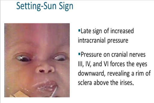
Macewen sign --> tapping on skull to hear fluid
Irritability --> poor feeding, high-pitched cry
Setting-sun sign (eyes) --> see picture
Projectile vomiting unrelated to feeding
133
How does manifestation of symptoms relate to intracranial pressure in infants?
Early signs are subtle
As pressure increases, the manifestation of symptoms also increases
As pressure increases, the manifestation of symptoms also increases
134
What neurological changes are seen with increased ICP in children?
Irritability, restlessness --> increased restlessness indicates lack of oxygen to brainstem
Behavioral and personality changes
Motor weakness, discoordination
Seizures
Behavioral and personality changes
Motor weakness, discoordination
Seizures
135
Is vomiting present in increased ICP in children?
Vomiting occurs with or without nausea
136
What visual manifestation is present with increased ICP in children?
Diplopia --> double vision
137
What sleep alterations are present with increased ICP in children?
somnolence
138
How should environmental noise be managed for children with IICP?
Environmental noise should be eliminated or minimized to reduce stress and prevent increases in ICP.
139
What is the caution regarding suctioning in children with IICP?
Suctioning should be questioned and used with caution, as it can increase ICP.
140
What are the nursing considerations for nutrition and hydration in children with IICP?
IV fluids and parenteral nutrition should be used initially --> caution to avoid overhydration
Later, gastric feeding via NG or G-tube can be introduced
Later, gastric feeding via NG or G-tube can be introduced
141
Why is it important to monitor the risk of aspiration during feeding in children with IICP?
Risk of aspiration increased due to impaired swallowing or neurological changes
142
Why is lumbar puncture contraindicated in the presence of IICP?
Lumbar puncture can cause a rapid shift in pressure, potentially leading to brain herniation.
143
What imaging techniques are used to diagnose increased intracranial pressure (IICP)?
CT (Computed Tomography)
MRI (Magnetic Resonance Imaging)
MRI (Magnetic Resonance Imaging)
144
How does cerebral angiography assist in diagnosing IICP?
It provides detailed images of blood vessels in the brain to identify blockages or abnormalities affecting ICP.
145
What do PET (Positron Emission Tomography) and SPECT (Single Photon Emission Computed Tomography) scans assess in cases of IICP?
They evaluate cerebral metabolism and blood flow
146
How is a transcranial doppler used in IICP diagnostics?
It measures cerebral blood flow velocity to detect changes related to ICP.
147
What is electrophysiologic monitoring, and how does it relate to IICP?
It directly observes cerebral blood flow using sensors to detect alterations in perfusion.
148
What is evoked potential monitoring used for in IICP cases?
It measures the brain’s electrical response to external stimuli, assessing neural function and integrity
149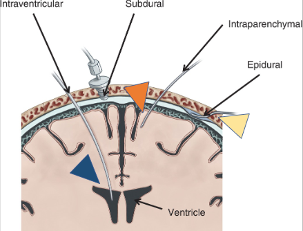
Image occlusion: name the hidden labels
150
Image occlusion: name the hidden labels
151
Image occlusion: name the hidden labels
152
Image occlusion: name the hidden labels
153
What are the indications for ICP monitoring?
GCS (Glasgow Coma Scale) score of 8 or less
A GCS score of <8 with respiratory assistance
Deteriorating neurologic condition
A GCS score of <8 with respiratory assistance
Deteriorating neurologic condition
154
What role does subjective clinical judgment play in deciding to monitor ICP?
Clinicians may use their discretion based on patient status, even without meeting strict criteria.
155
What is a critical sign of increased ICP related to Cushing’s triad?
Widening pulse pressure
156
What are the advantages and disadvantages of using an Intraventricular Catheter Device (IVC) to monitor intracranial pressure?
It allows for sampling of CSF
Most invasive
May allow leakage of CSF
Most invasive
May allow leakage of CSF
157
What methods of ICP monitoring do NOT cause leaks?
Subarachnoid bolt or screw
Subdural/epidural catheter censor
Fiber-optic Transducer-tipped catheter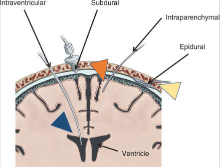
Subdural/epidural catheter censor
Fiber-optic Transducer-tipped catheter
158
What is the priority assessment for a patient on ICP monitoring?
Ongoing pupil checks
Select cranial nerve assessment
Frequent vitals and ICP
GCS (Glasgow Coma Scale) evaluations
Select cranial nerve assessment
Frequent vitals and ICP
GCS (Glasgow Coma Scale) evaluations
159
Why is the patient’s history of events leading to the present illness important in ICP monitoring?
It helps identify any injury or infection that could have caused IICP.
160
Why is it important to review the patient’s past medical history when monitoring ICP?
It can reveal conditions or risks that may predispose the patient to increased ICP.
161
What neurological assessments are performed when monitoring ICP?
Complete neurological exams, including:
- mental status
- level of consciousness (LOC)
- cranial nerve function.
- mental status
- level of consciousness (LOC)
- cranial nerve function.
162
What aspect of brain function is evaluated to assess balance and coordination?
Cerebellar function
163
Why are reflexes evaluated during ICP monitoring?
Assess the integrity of the nervous system
Detect neurological changes
Detect neurological changes
164
Why is it important to assess BOTH motor and sensory functions during ICP monitoring?
To identify any deficits that might indicate worsening neurological status or localized damage.
165
GCS (Glasgow Coma Scale) scores
Score of 15 = unaltered LOC
Score of 3 = extremely decreased LOC --> worst possible score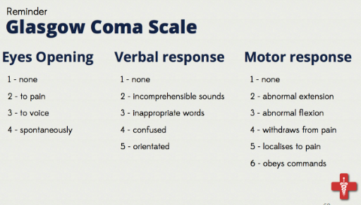
Score of 3 = extremely decreased LOC --> worst possible score
166
What is the normal range for pupil size?
2.0 - 5.0 mm
167
Which cranial nerve is assessed through pupillary response to light?
oculomotor nerve (CN III)
168
Why is any change in pupil size or inequality significant?
It may indicate neurological compromise, such as increased ICP or brain herniation, and should be reported immediately.
169
Where is the oculomotor nerve located, and why is this significant in pupil assessment?
It lies at the junction of the midbrain and tentorial notch, making it vulnerable to compression from brain herniation or changes in ICP.
170
What does a dilated, nonreactive pupil indicate?
Oculomotor nerve compression
171
What does a constricted, nonresponsive pupil indicate?
Sympathetic pathway disruption from brainstem involvement or certain drugs like morphine.
172
What can cause large pupils apart from neurological issues?
atropine
extreme stress
extreme stress
173
What is the normal shape of the pupil, and what does an oval pupil indicate?
Pupils are normally round
Oval pupil may indicate elevated ICP
Oval pupil may indicate elevated ICP
174
How should pupil size be documented?
In millimeters, often using a pupil gauge available on flowsheets.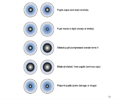
175
What does decerebrate posture usually indicate?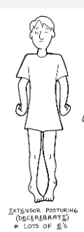
Indicates deterioration of the upper brainstem (particularly the midbrain and upper pons)
176
What is the definition of decorticate posture?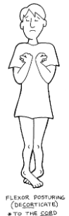
It is an abnormal posturing with rigidity, flexed arms, clenched fists, extended legs, and the arms bent inward toward the body with wrists and fingers held on the chest.
177
What does the presence of decorticate posture indicate?
It suggests severe damage to the brain, specifically to the corticospinal tract, and requires immediate medical attention.
178
How is decorticate posture generally viewed compared to decerebrate posture?
Decorticate posture is usually more favorable than decerebrate posture, although both are serious signs.
179
Can decorticate posture progress to decerebrate posture?
Yes, decorticate posture may progress to decerebrate posture, or the two may alternate.
180
In what patterns can decerebrate posture present?
It can occur on one or both sides of the body, or only in the arms, and may alternate with decorticate posture.
181
Is it possible to have both postures present simultaneously?
Yes, a person can display decorticate posture on one side and decerebrate posture on the other.
182
What does abnormal posturing indicate in a patient?
Pathological sign of neurological injury
Medical emergency
Medical emergency
183
What is the prognosis for patients with abnormal posturing following head injury?
Only 37% of patients with decorticate posturing survive after head injury
Only 10% of patients with decerebrate posturing survive after head injury
Only 10% of patients with decerebrate posturing survive after head injury
184
What are the typical outcomes for patients displaying abnormal posturing?
Death
Poor neurological outcomes
Poor neurological outcomes
185
What are the components of a neurological exam?
LOC --> Glasgow coma scale
Pupillary reaction
Motor and sensory function --> ROM, push/pull against pressure
Vital signs
Reflexes
Pupillary reaction
Motor and sensory function --> ROM, push/pull against pressure
Vital signs
Reflexes
186
What is the difference between an open fracture and a closed fracture in terms of head injury?
Open = injury broke skin over the fracture
Closed = injury didn't break skin over the fracture --> Most common
Closed = injury didn't break skin over the fracture --> Most common
187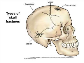
Image occlusion: name the hidden labels
188
Image occlusion: name the hidden labels
189
Image occlusion: name the hidden labels
190
Image occlusion: name the hidden labels
191
What is a linear skull fracture?
A break in the bone that does not cause movement of the bone
Simplest type of fracture
Simplest type of fracture
192
What characterizes a depressed skull fracture?
It involves the bone being pressed inward
May occur with or without a scalp laceration
May occur with or without a scalp laceration
193
What is a basilar skull fracture, and why is it serious?
Involves a fracture at the base of the skull
Often accompanied by bruises around the eyes ("raccoon eyes") and behind the ear ("Battle's sign")
Often accompanied by bruises around the eyes ("raccoon eyes") and behind the ear ("Battle's sign")
194
What happens in a comminuted skull fracture?
the bone is shattered into multiple pieces
195
What defines an open head injury?
Involves a skull fracture or penetration by an object that violates the integrity of the brain and dura, exposing them to environmental contaminants
196
What is the risk associated with an open head injury?
Infection (either from the penetrating object or from the environment)
197
What types of damage can occur with an open head injury?
Damage may occur to:
- underlying vessels
- dural sinuses
- brain tissue
- cranial nerves.
- underlying vessels
- dural sinuses
- brain tissue
- cranial nerves.
198
What are the most common causes of penetrating injuries to the skull?
Gunshot wounds (GSWs)
Knife injuries
Knife injuries
199
What are the hallmark signs of a basilar skull fracture?
Presence of:
- Battle's sign (bruise behind the ears)
- Raccoon eyes (periorbital ecchymosis)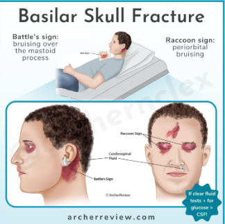
- Battle's sign (bruise behind the ears)
- Raccoon eyes (periorbital ecchymosis)
200
What is a significant sign that CSF is leaking in a basilar skull fracture?
CSF draining from the nose (rhinorrhea) or ears (otorrhea)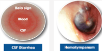
201
What is a concussion?
A type of traumatic brain injury caused by a bump, blow, or jolt to the head, or by a hit to the body that causes rapid movement of the head and brain.
202
What is a contusion (traumatic brain injury type)?
Bruising of the brain tissue
Scattered areas of bleeding on the surface of the brain
Most common closed head injury
Scattered areas of bleeding on the surface of the brain
Most common closed head injury
203
What are common cognitive symptoms of a concussion?
loss of consciousness
retrograde amnesia (loss of memory for events before injury)
anterograde amnesia (inability to form new memories after the injury)
retrograde amnesia (loss of memory for events before injury)
anterograde amnesia (inability to form new memories after the injury)
204
What are some physical symptoms of a concussion?
Headache , vomiting
Psychomotor impairment
Tinnitus
Sensitivity to light and sound
Psychomotor impairment
Tinnitus
Sensitivity to light and sound
205
What is a key management step for a concussion?
Cervical spine immobilization until spinal injury ruled out --> use log roll to move patient
206
What types of medications are commonly used for pain management in concussion cases?
Nonopioid analgesics --> acetaminophen, ketorolac
207
Why is it important to assess for cerebrospinal fluid leakage in concussion management?
CSF leakage --> more serious injury (like a skull fracture or brain injury)
208
What assessments should be performed for a client with a concussion?
Neuro and mental status assessments should be performed --> monitor cognitive function and any changes in condition
209
What precautions should be initiated for patients with concussions?
Seizure precautions --> due to risk of seizures following a traumatic brain injury
210
What is a coup injury?
Damage to brain tissue directly beneath point of impact 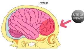
211
What is a contrecoup injury?
Occurs when a force strikes one side of the head hard enough to cause the brain to shift in the opposite direction, leading to injury on the opposite side of the impact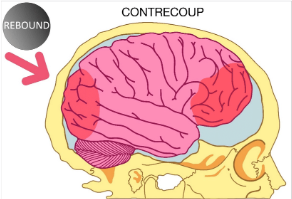
212
What is a laceration (closed head injury)?
Tears in brain tissue caused by a foreign object or pushed-in bone fragments from a skull fracture --> can lead to secondary hemorrhage and cerebral edema
213
What is diffuse axonal injury (DAI)?
AKA brain shearing
Occurs when brain is subjected to rapid acceleration and deceleration forces, causing widespread damage to the axons
Occurs when brain is subjected to rapid acceleration and deceleration forces, causing widespread damage to the axons
214
How does acceleration/deceleration contribute to brain injury?
Acceleration/deceleration forces can lead to the brain rebounding or rotating on the brain stem, resulting in diffuse axonal injury and functional impairment.
215
What is an example of an acceleration injury?
An acceleration injury occurs when an external force, such as a vehicle colliding with another, suddenly places the head in motion, such as being rear-ended in a motor vehicle accident (MVA).
216
What happens during a deceleration injury?
Occurs when a moving head is suddenly stopped or strikes a stationary object, leading to potential injury from the sudden halt
217
How can acceleration-deceleration forces affect the brain?
These forces can cause the cerebrum to rotate about the brainstem, resulting in shearing, straining, and distortion of brain tissue, particularly affecting the axons in the brainstem and cerebellum.
218
What are potential consequences of acceleration-deceleration injuries?
Small areas of hemorrhage may develop near impact site
Adjacent brain tissue can be destroyed, leading to significant neurological impairment
Adjacent brain tissue can be destroyed, leading to significant neurological impairment
219
What characterizes a mild brain injury?
A mild brain injury (such as a concussion), has no evidence of significant brain injury
May present with headaches and dizziness
May present with headaches and dizziness
220
How is a mild brain injury managed?
Management may include administering acetaminophen q.4h for headaches
221
What defines a moderate brain injury?
A moderate brain injury involves a loss of consciousness lasting from 30 minutes to 6 hours, with a Glasgow Coma Scale score of 9-12.
222
How is a moderate brain injury managed?
Patients typically require a short ICU stay to prevent complications such as brain edema, intracranial bleeding, and inadequate tissue perfusion
223
What are the characteristics of a severe brain injury?
A severe brain injury is characterized by a loss of consciousness for more than 6 hours and a Glasgow Coma Scale score of 3 to 8.
224
How is a severe brain injury managed?
Patients with a severe brain injury require intensive management in the ICU
225
What are common clinical manifestations of a brain injury?
Amnesia
Headaches
Dizziness
Seizures --> MOST SERIOUS
Loss of consciousness
Sleepiness/drowsiness
Restlessness or irritability
Disorientation or confusion
Headaches
Dizziness
Seizures --> MOST SERIOUS
Loss of consciousness
Sleepiness/drowsiness
Restlessness or irritability
Disorientation or confusion
226
Why are fluids frequently restricted in the first 24 hours after a head injury?
Fluids are often restricted in the first 24 hours following a head injury to manage and reduce the risk of cerebral swelling.
227
What are some severe clinical manifestations of a head injury?
Pupil changes
Papilledema (eyeball swelling)
Cushing's triad --> HTN, widened pulse pressure, bradycardia
Hypovolemic shock --> hypotension, tachycardia
Nuchal rigidity --> neck stiffness
Papilledema (eyeball swelling)
Cushing's triad --> HTN, widened pulse pressure, bradycardia
Hypovolemic shock --> hypotension, tachycardia
Nuchal rigidity --> neck stiffness
228
What does nuchal rigidity suggest in a patient with a brain injury?
Nuchal rigidity can indicate a CSF leak, which may be associated with a severe brain injury
229
Why should drainage from the nose or ear need to be tested for glucose?
Drainage that is positive for glucose indicates leakage of CSF
Provider should be notified STAT if drainage tests positive for glucose
Provider should be notified STAT if drainage tests positive for glucose
230
What are some potential complications following a brain injury?
Hypotension and hypoxia
Increased intracranial pressure
Cerebral edema
Hydrocephalus
Hemorrhage leading to hematomas
Brain herniation
Increased intracranial pressure
Cerebral edema
Hydrocephalus
Hemorrhage leading to hematomas
Brain herniation
231
What is the significance of hematomas in brain injuries?
ALL hematomas are potentially life-threatening because they act as space-occupying lesions and are surrounded by edema
232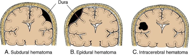
Image occlusion: name the hidden labels
233
Image occlusion: name the hidden labels
234
Image occlusion: name the hidden labels
235
What is a subdural hematoma?
a collection of blood located between the dura and arachnoid layer
236
Where is a subarachnoid hematoma found?
A subarachnoid hematoma refers to blood located in the subarachnoid space (below the arachnoid layer)
237
What defines an epidural hematoma?
Blood accumulation between the dura mater and the skull
238
What is an intracerebral hematoma?
Blood that accumulates within the brain tissue itself
239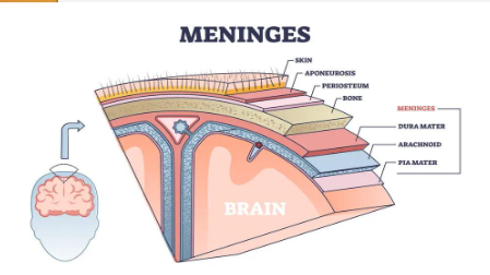
Image occlusion: name the hidden labels
240
Image occlusion: name the hidden labels
241
Image occlusion: name the hidden labels
242
What is hydrocephalus?
Too much CSF in the brain (specifically the ventricles)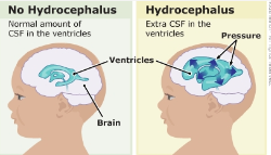
243
What are some causes of hydrocephalus?
Decreased reabsorption of CSF at the arachnoid villi
Interference or blockage with CSF outflow from the ventricular system
Interference or blockage with CSF outflow from the ventricular system
244
What happens if hydrocephalus is not treated?
If not treated --> further ICP elevations
Excess fluid puts harmful pressure on the brain
Excess fluid puts harmful pressure on the brain
245
What diagnostics are used for hydrocephalus?
Imaging --> CT, MRI, ultrasound
Lumbar puncture
ICP monitoring
Lumbar puncture
ICP monitoring
246
What is the main treatment for hydrocephalus?
The main treatment involves inserting a shunt into the brain, which is connected to a flexible tube that drains excess CSF into the chest cavity or abdomen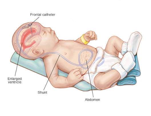
247
What is Endoscopic Third Ventriculostomy (EVT)
a procedure where a hole is made in the third ventricle in order to divert CSF and alleviate hydrocephalus
248
What are the signs and symptoms of shunt malfunction?
Headache
Double vision
Nausea and vomiting
Low grade fever
Seizures
Tenderness along shunt track
Increased sleepiness or exhaustion
Literally just s/s of increased ICP --> may be due to shunt blockage or ineffective CSF drainage
Double vision
Nausea and vomiting
Low grade fever
Seizures
Tenderness along shunt track
Increased sleepiness or exhaustion
Literally just s/s of increased ICP --> may be due to shunt blockage or ineffective CSF drainage
249
How does a brain herniation occur?
increased ICP --> decreased cerebral perfusion --> brain ischemia and edema
If left untreated --> brain herniates downward towards brainstem OR laterally from a unilateral lesion to cerebral hemisphere
Can lead to irreversible brain damage and possibly death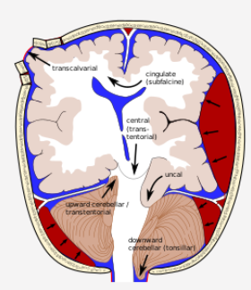
If left untreated --> brain herniates downward towards brainstem OR laterally from a unilateral lesion to cerebral hemisphere
Can lead to irreversible brain damage and possibly death
250
What are the key questions to ask about the history of a TBI (traumatic brain injury)?
When, where, and how did the injury occur?
Was there a loss of consciousness involved? If yes, how long?
Has there been any change in the level of consciousness?
Was there a loss of consciousness involved? If yes, how long?
Has there been any change in the level of consciousness?
251
What additional history points are important in assessing traumatic brain injury?
Hand dominance (left or right)
Any eye injuries
Allergies
Was the patient a victim of violence?
Any eye injuries
Allergies
Was the patient a victim of violence?
252
What is the first priority in the physical assessment of a traumatic brain injury ?
ABC's --> airway, breathing, circulation
253
Why is it important to assess level of consciousness (LOC) in TBI?
A change in LOC is an early indicator of brain injury and helps detect neurological deterioration.
254
What are key signs of increased ICP that should be assessed in traumatic brain injury?
Headache
Vomiting
Altered mental status
Pupil changes
Cushing's triad --> bradycardia, widened pulse pressure, irregular respiration
Vomiting
Altered mental status
Pupil changes
Cushing's triad --> bradycardia, widened pulse pressure, irregular respiration
255
What must be considered regarding spinal cord injury in TBI patients?
Treat all traumatic brain injury patients as if they have a spinal cord injury unless ruled out by imaging
256
Why are older adults more prone to life-threatening cervical spine injuries in TBI?
Greater risk for C1 and C2 spinal cord injuries, which can be life-threatening
257
What is the primary imaging modality used in the diagnosis of TBI?
CT scan --> quickly identifies fractures, bleeding, or swelling
258
When is Magnetic Resonance Imaging (MRI) preferred in TBI evaluation?
MRI is used to detect soft tissue damage, small contusions, and diffuse axonal injuries (DAI) that may not appear on a CT scan.
259
How is ultrasound utilized in traumatic brain injury diagnostics?
non-invasive assessment of raised intracranial pressure, cerebral autoregulation and diagnosis of intracranial haematoma and other space-occupying lesions
260
What do evoked potentials help diagnose in TBI patients?
Assess function of optic nerves and electrical response to external stimuli, revealing damage to sensory pathways
261
Which lab tests are crucial in the diagnostic workup of TBI?
ABGs --> evaluate oxygenation
CBCs --> check for infection or bleeding
CMP --> electrolytes and organ function
ECG --> monitor cardiac functioning
Toxicology screen --> detect drug or ETOH involvement
CBCs --> check for infection or bleeding
CMP --> electrolytes and organ function
ECG --> monitor cardiac functioning
Toxicology screen --> detect drug or ETOH involvement
262
What is a key goal in treating traumatic brain injury regarding intracranial pressure?
Preventing increased ICP to avoid further brain injury and maintain cerebral perfusion
263
How is fluid and electrolyte balance managed in TBI patients?
Careful monitoring of electrolytes (esp. sodium) and fluid intake to prevent dehydration or cerebral edema
264
What are the guidelines for positioning TBI patients?
Elevate HOB 30-45 degrees --> promote venous drainage
Avoid neck flexion or extreme hip flexion --> can increase ICP
Avoid neck flexion or extreme hip flexion --> can increase ICP
265
When is the induction of a barbiturate coma considered in TBI?
used to reduce cerebral metabolism and ICP in severe brain injuries when other interventions fail
266
What are the priorities for pulmonary management in TBI patients?
Maintain airway patency and oxygenation
Prevent aspiration pneumonia and monitor for respiratory distress
Prevent aspiration pneumonia and monitor for respiratory distress
267
Why is it important to prevent complications of immobility in TBI patients?
Reduce risk of DVT, pressure ulcers, and pneumonia
268
What are some surgical management options for TBI?
Decompressive craniectomy --> relieve ICP
Hematoma evacuation --> stop bleeding
Placement of ventricular drains --> remove excess CSF
Hematoma evacuation --> stop bleeding
Placement of ventricular drains --> remove excess CSF
269
What is the role of intracranial pressure (ICP) monitoring in TBI?
To track ICP levels continuously, guiding treatment decisions to prevent brain herniation.
270
What should always be questioned in TBI management?
Isometric exercises --> can increase ICP by raising intrathoracic and venous pressure
271
What is the role of Mannitol (Osmitrol) in TBI management?
Mannitol is an osmotic diuretic that reduces ICP by pulling fluid from brain tissues into the bloodstream.
272
How does furosemide (Lasix) help in TBI treatment?
As a loop diuretic, it supports Mannitol by removing excess fluid and reducing cerebral edema.
273
Why are glucocorticoids like dexamethasone and methylprednisolone used in TBI?
Reduce inflammation and cerebral edema
Use is controversial due to risk of infection and hyperglycemia
Use is controversial due to risk of infection and hyperglycemia
274
What is the purpose of Dexmedetomidine (Precedex) and Propofol (Diprivan) in TBI patients?
These sedative agents manage agitation, anxiety, and ventilator asynchrony to promote healing.
275
Why should opioids like morphine sulfate and fentanyl be used cautiously in TBI?
Can suppress respiration
Can obscure neurological assessments
Can obscure neurological assessments
276
What is the role of Phenytoin (Dilantin) in TBI management?
antiepileptic used to prevent or control seizures following brain injury
277
When are acetaminophen (Tylenol) and aspirin (ASA) used in TBI?
Helps control fever and mild pain
not used for fevers caused by HYPOTHALAMIC INJURY due to risk of bleeding
not used for fevers caused by HYPOTHALAMIC INJURY due to risk of bleeding
278
What is a key intervention in managing both TBI and increased intracranial pressure (IICP)?
Review interventions for IICP, such as positioning, diuretics, and ICP monitoring, to prevent further damage.
279
How frequently should vital signs be monitored in TBI patients?
every 1-2 hours or as ordered by the provider
280
Why is it important to monitor and document cardiac dysrhythmias and blood pressure in TBI patients?
Cardiac and blood pressure changes can indicate autonomic dysfunction, cerebral perfusion issues, or a deteriorating neurological status.
281
How are blood pressure and heart rate managed in TBI patients?
Target ranges for blood pressure (to ensure cerebral perfusion) and heart rate are established and adhered to, adjusting interventions accordingly.
282
How is fever caused by hypothalamic injury treated in TBI patients?
It is managed with cool-air hypothermia techniques, such as sponge baths, cooling blankets, or ice packs.
283
What is the purpose of therapeutic hypothermia in TBI management?
Reduces brain metabolism and prevents secondary brain injury by lowering the body's temperature
284
What additional care considerations are required when using therapeutic hypothermia?
Monitor for shivering (can increase metabolism) and electrolyte imbalances that can occur during rewarming
285
Why should hyperventilation be avoided in the first 24 hours after brain surgery?
Hyperventilation can lead to cerebral vasoconstriction, reducing cerebral perfusion and worsening brain injury during this critical period.
286
What is the target PaCO2 range for TBI patients?
PaCO2 should be maintained between 35-38 mm Hg to avoid vasodilation or vasoconstriction that could affect ICP and cerebral blood flow.
287
What should the PaO2 level be maintained at in TBI management?
PaO2 should remain between 80-100 mm Hg to ensure adequate oxygenation of the brain.
288
What precaution should be taken during ET suctioning for intubated TBI patients?
Avoid aggressive hyperventilation, which can lead to excessive reductions in PaCO2, impairing cerebral blood flow.
289
What is the purpose of administering IV lidocaine during suctioning in TBI patients?
IV lidocaine suppresses the cough reflex and reduces cerebral metabolic demands by inhibiting sodium channels, potentially lowering ICP.
290
What interventions should be taken to support respiratory care in TBI patients?
Collaborate with respiratory therapy to provide humidified air and chest physiotherapy to prevent complications like pneumonia.
291
What is the recommended head of bed (HOB) elevation for TBI patients, and why?
Elevate the HOB to 30˚ to promote venous drainage and reduce ICP.
Ensure log rolling during repositioning to maintain spinal alignment.
Ensure log rolling during repositioning to maintain spinal alignment.
292
What is the target cerebral perfusion pressure (CPP) for patients with TBI?
Maintain CPP > 70 mm Hg to ensure adequate oxygen delivery and prevent secondary brain injury.
293
What monitoring technique is used to assess ICP and cerebral perfusion in TBI patients?
ICP waveform monitoring helps guide interventions and optimize CPP by tracking pressure changes in real-time.
294
What is the prognosis for patients with severe TBI?
Many patients with severe TBI do not survive due to the extensive brain damage
295
What are the three key prerequisites for diagnosing brain death?
1. Persistent coma
2. Absence of brainstem reflexes
3. Inability to breathe independently
2. Absence of brainstem reflexes
3. Inability to breathe independently
296
How many neurologic examinations are required to confirm brain death?
At least one neurologic examination is required, but protocols may vary based on institutional or national guidelines.
297
What reflexes must be absent to confirm brainstem death?
Reflexes such as pupillary, corneal, gag, and oculocephalic reflexes must be absent.
298
What test can confirm the absence of spontaneous breathing during brain death evaluation?
An apnea test evaluates the ability to breathe independently by monitoring CO₂ levels without ventilator support.
299
Which spinal nerve level is crucial for diaphragmatic innervation?
the C4 spinal nerve
300
Why is immediate airway management critical in high cervical spine injuries?
High cervical injuries (C3-C5) can impair the ability to breath
remember C4 innervates the diaphragm
remember C4 innervates the diaphragm
686 more cards in OutLearn. Open the full deck