OXY/CIRC N3 Lecture ORIGINAL
Study these flashcardsFree · spaced repetition · iPhone, Android, Mac & web
First 300 of 323 cards
1
Endocarditis
Inflammation of the heart's inner lining and valves
Usually caused by an infective agent (bacteria, fungi, viruses)
Usually caused by an infective agent (bacteria, fungi, viruses)
2
Endocarditis: Main Causitive Organism
Streptococcus
Staphylococcus
Staphylococcus
3
Endocarditis: Risk Factors
IV drug abusers
Valve replacement recipients
Structural cardiac defects
Pharyngitis
Valve replacement recipients
Structural cardiac defects
Pharyngitis
4
Endocarditis: Criteria for Diagnosis
Positive blood culture (usually strep)
Murmurs
S3 and S4 present
Murmurs
S3 and S4 present
5
Endocarditis: Manifestations
Fever/chills
Malaise, fatigue
Anorexia, weight loss
S3, S4 sounds
Chest pain
Petechiae
Splinter hemorrhages (see top left in pic)
Osler's nodes (see top right)
Janeway's lesions (see middle pics)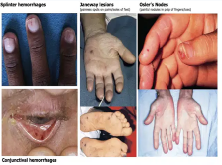
Malaise, fatigue
Anorexia, weight loss
S3, S4 sounds
Chest pain
Petechiae
Splinter hemorrhages (see top left in pic)
Osler's nodes (see top right)
Janeway's lesions (see middle pics)
6
Endocarditis: Heart murmurs
murmurs may be present in infective endocarditis due to damage to the heart valves
7
Endocarditis: Complications
Heart failure
Systemic embolization
Sepsis
Heart blocks
Systemic embolization
Sepsis
Heart blocks
8
Endocarditis Complications: Heart failure
Presents with:
- Edema
- Fatigue
- Shortness of breath
- Crackles
- Edema
- Fatigue
- Shortness of breath
- Crackles
9
Endocarditis Complications: Systemic Embolization
Left-sided heart embolizations:
- Brain
- Limbs
- Kidney
- Liver
- Spleen
Right-sided heart embolizations:
- Pulmonary embolism
- Brain
- Limbs
- Kidney
- Liver
- Spleen
Right-sided heart embolizations:
- Pulmonary embolism
10
Endocarditis: Treatment
Antibiotics --> high dose IV 4-6 weeks (not for viral infections UNLESS there is a secondary bacterial infection)
Adequate pain control
Surgical management if antibiotics don't work
Adequate pain control
Surgical management if antibiotics don't work
11
Endocarditis: Surgical Management
Remove infected valves
Repair damaged valves
Repair shunts
Drain abcesses
Repair damaged valves
Repair shunts
Drain abcesses
12
Endocarditis: Nursing Management
Assess for HF: tachycardia, fatigue, SOB, edema, crackles
Balance activities --> adequate rest vs mobility
Monitor for the presence of murmurs or additional murmurs
Adequate pain control
Balance activities --> adequate rest vs mobility
Monitor for the presence of murmurs or additional murmurs
Adequate pain control
13
Endocarditis Patient Education
Report to provider STAT: recurrent fever, fatigue, SOB, weight loss or gain
Antibiotic prophylaxis --> 4-6 weeks of therapy (some may have to take for life)
Antibiotic prophylaxis --> 4-6 weeks of therapy (some may have to take for life)
14
Myocarditis
Focal or diffuse inflammation of the myocardium
15
Myocarditis: Pathophysiology
Inflammation of the heart muscle (caused by virus, bacteria, radiation, pharmacologic, or chemical factors)
↓
May lead to hypertrophy, fibrosis (hardening), and inflammatory changes to the myocardium and conduction system
↓
Heart muscle weakens --> Contractility decreases
↓
Left ventricular dysfunction leads to pulmonary venous and arterial hypertension, pulmonary edema (think left-sided heart failure)
↓
May lead to hypertrophy, fibrosis (hardening), and inflammatory changes to the myocardium and conduction system
↓
Heart muscle weakens --> Contractility decreases
↓
Left ventricular dysfunction leads to pulmonary venous and arterial hypertension, pulmonary edema (think left-sided heart failure)
16
Myocarditis: Complications
Cardiomyopathy
17
Myocarditis: Early Manifestations
Fever
Fatigue, malaise
Myalgias (muscle pain)
Pharyngitis
Dyspnea
Lymphadenopathy (swollen lymph nodes)
Nausea/Vomiting
Fatigue, malaise
Myalgias (muscle pain)
Pharyngitis
Dyspnea
Lymphadenopathy (swollen lymph nodes)
Nausea/Vomiting
18
Myocarditis: Manifestations 7-10 days Post-Infection
Pleuritic chest pain
Pericardial friction rub
Pericardial effusion
Pericardial friction rub
Pericardial effusion
19
Myocarditis: Late Manifestions
S3 heart sound
Dysrhythmias
Crackles
JVD
Syncope --> due to not enough oxygenated blood reaching brain
Peripheral edema
Angina
Think HEART FAILURE
Dysrhythmias
Crackles
JVD
Syncope --> due to not enough oxygenated blood reaching brain
Peripheral edema
Angina
Think HEART FAILURE
20
Myocarditis: Diagnostics
Assess cardiac function:
- ECG --> ST changes
- Nuclear scans (MRI)
- Echocardiography
Confirm Diagnosis:
- Endomyocardial biopsy --> DEFINITIVE
- ECG --> ST changes
- Nuclear scans (MRI)
- Echocardiography
Confirm Diagnosis:
- Endomyocardial biopsy --> DEFINITIVE
21
Myocarditis: General Supportive Measures
O2
Bed rest, restricted activity (space activity and rest) --> don't overwork heart
Semi-Fowler's position --> help with breathing
Bed rest, restricted activity (space activity and rest) --> don't overwork heart
Semi-Fowler's position --> help with breathing
22
Myocarditis: Pharmacological Treament
ACE Inhibitors/Beta Blockers --> decrease preload
Diuretics --> decrease fluid volume, decrease preload
nitroprusside and milrinone --> reduces afterload, improve cardiac output
digoxin --> improve contraction, reduce HR
Anticoagulants --> for patients with low ejection fraction (EF)
AVOID NSAIDs FOR PAIN CONTROL
Diuretics --> decrease fluid volume, decrease preload
nitroprusside and milrinone --> reduces afterload, improve cardiac output
digoxin --> improve contraction, reduce HR
Anticoagulants --> for patients with low ejection fraction (EF)
AVOID NSAIDs FOR PAIN CONTROL
23
Myocarditis Drugs - ACE Inhibitors
MOA: prevent conversion of angiotensin I to angiotensin II (which is a potent vasoconstrictor) --> decreased vasopressor activity and decreased aldosterone secretion
Side Effects: hypotension, hyperkalemia, cough
Contraindications: allergy, renal impairment (excreted by kidneys)
Side Effects: hypotension, hyperkalemia, cough
Contraindications: allergy, renal impairment (excreted by kidneys)
24
Myocarditis Drugs - Beta Blockers
MOA - competitive antagonism of catecholamines at peripheral and cardiac adrenergic receptors --> reduces HR, cardiac contractility, and at higher doses, relaxes vascular smooth muscle, lowering the BP
Side Effects: hypotension, bradycardia, dizziness
Contraindications: severe bradycardia, cardiogenic shock, systolic BP <100 mmHg
Side Effects: hypotension, bradycardia, dizziness
Contraindications: severe bradycardia, cardiogenic shock, systolic BP <100 mmHg
25
Myocarditis Drugs - Vasodilators
nitroprusside and milrinone
Reduces afterload, improves cardiac output
Caution for Hypotension
Reduces afterload, improves cardiac output
Caution for Hypotension
26
Myocarditis Drugs - Digoxin
Improves contraction and reduces HR
Side Effects: yellow-green visual discoloration, nausea, vomiting, cardiac dysrhythmias
Digoxin Toxicity is exacerbated by hypokalemia
Check apical pulse for 60 seconds before administration
Side Effects: yellow-green visual discoloration, nausea, vomiting, cardiac dysrhythmias
Digoxin Toxicity is exacerbated by hypokalemia
Check apical pulse for 60 seconds before administration
27
Myocarditis Drugs - Anticoagulants
For patients with low ejection fraction (for the blood that doesn't get pumped out and pools in the heart)
Heparin for acute myocarditis
Warfarin for chronic
Heparin for acute myocarditis
Warfarin for chronic
28
Preload
Volume of blood in the ventricles at the end of diastole (heart relaxation)
29
Frank Starling Law
When the volume of blood in the ventricles increases, the heart's stroke volume increases in response.
More cardiac muscle stretch = stronger contraction force
More cardiac muscle stretch = stronger contraction force
30
Afterload
Resistance left ventricle must overcome to circulate blood
31
Pericarditis
Condition in which pericardium around the heart becomes inflammed
32
Pericarditis Pathophysiology
Pericardial tissue is damaged by bacteria, fungi, virus, or another substance that releases chemical mediators of inflammation into surrounding tissue
↓
Chemical mediators dilate blood vessels and increase vessel permeability
↓
Vessel walls leak fluids and proteins, causing extracellular edema
↓
Chemical mediators dilate blood vessels and increase vessel permeability
↓
Vessel walls leak fluids and proteins, causing extracellular edema
33
Pericarditis: Causes
Bacteria, virus, fungi infectie agent (usually respiratory)
Post MI syndromes
Connective tissue disease
Post MI syndromes
Connective tissue disease
34
Pericarditis: Post MI Syndromes
Acute Pericarditis: 48-72h post MI
Dressler's Syndrome: 4-6 weeks post MI
Dressler's Syndrome: 4-6 weeks post MI
35
Pericarditis: Manifestations
Pericardial friction rub --> hallmark sign
Pericardial effusion --> presents as dyspnea/tachypnea
Sharp, severe radiating chest pain (made worse by inspiration and lying down, relieved by sitting)
Elevated WBC, high fever
ST elevation, Afib
Pericardial effusion --> presents as dyspnea/tachypnea
Sharp, severe radiating chest pain (made worse by inspiration and lying down, relieved by sitting)
Elevated WBC, high fever
ST elevation, Afib
36
Pericarditis: Positioning
Sitting upright
Leaning forward
Leaning forward
37
Pericarditis: Treatment
Depends on the cause
- If bacterial: antibiotics 4-6 weeks
- if not baterial: corticosteroids
- if due to malignancy: radiation or chemo
Pericardiocentesis
- If bacterial: antibiotics 4-6 weeks
- if not baterial: corticosteroids
- if due to malignancy: radiation or chemo
Pericardiocentesis
38
Pericarditis: Nursing Management
Monitor for worsening pericardial effusion:
- Risk for cardiac tamponade
- Ineffective heart pumps
- Risk for cardiac tamponade
- Ineffective heart pumps
39
Pericardiocentesis: Nursing Management
ECG monitoring
Hemodynamic measurements
Post op: watch for recurrence of tamponade
Hemodynamic measurements
Post op: watch for recurrence of tamponade
40
Cardiac Tamponade
MEDICAL EMERGENCY
Accumulation of fluid in the pericardial space = reduced ventricular filling --> decreased CO, shock, death
Accumulation of fluid in the pericardial space = reduced ventricular filling --> decreased CO, shock, death
41
Cardiac Tamponade Manifestations
Beck's Triad:
1. Hypotension with narrowed pulse pressure
2. JVD
3. Muffled heart sounds
Pulsus paradoxus - BP drops on inspiration
Tachycardia --> bradycardia (if not treated fast enough)
Dyspnea
Electrical alternans (alternating QRS amplitude from high-low-high-low)
1. Hypotension with narrowed pulse pressure
2. JVD
3. Muffled heart sounds
Pulsus paradoxus - BP drops on inspiration
Tachycardia --> bradycardia (if not treated fast enough)
Dyspnea
Electrical alternans (alternating QRS amplitude from high-low-high-low)
42
Cardiac Tamponade Treatment
Pericardiocentesis
Pericardial drain (if there isn't that much fluid)
Hemodynamic monitoring
Supportive therapy: O2, vasopressors, close monitoring for reoccurence
Pericardial drain (if there isn't that much fluid)
Hemodynamic monitoring
Supportive therapy: O2, vasopressors, close monitoring for reoccurence
43
Left Sided Valvular Heart Diseases
Aortic Stenosis (can't open fully)
Aortic Regurgitation (can't close fully)
Mitral Stenosis (can't open fully)
Mitral Regurgitation (can't close fully)
Aortic Regurgitation (can't close fully)
Mitral Stenosis (can't open fully)
Mitral Regurgitation (can't close fully)
44
Aortic Stenosis Etiology
Congenital Bicuspid Valve Defect
Rheumatic Fever
Atherosclerosis degenerative disease
Rheumatic Fever
Atherosclerosis degenerative disease
45
Aortic Regurgitation Etiology
Rheumatic fever
Endocarditis
Enlargement Aortic root
Endocarditis
Enlargement Aortic root
46
Mitral Stenosis Etiology
Rheumatic fever
Congenital defect
Congenital defect
47
Mitral Regurgitation Etiology
Infective Endocarditis
Rheumatoid arthritis
Myocardial Infarction
SLE (systemic lupus erythematosus)
Rheumatoid arthritis
Myocardial Infarction
SLE (systemic lupus erythematosus)
48
Mitral Stenosis - Manifestations
Fatigue
Dyspnea on exertion, Orthopnea (difficulty breathing while flat)
Paroxysmal nocturnal dyspnea (patient suddenly wakes up with SOB)
Hemoptysis (coughing blood)
Hepatomegaly
JVD
Pitting Edema
Rumbling, apical diastolic murmur
Dyspnea on exertion, Orthopnea (difficulty breathing while flat)
Paroxysmal nocturnal dyspnea (patient suddenly wakes up with SOB)
Hemoptysis (coughing blood)
Hepatomegaly
JVD
Pitting Edema
Rumbling, apical diastolic murmur
49
Mitral Regurgitation Manifestations
Fatigue
Dyspnea on exertion, orthopnea
Palpitations
Atrial fibrillation
JVD
Pitting edema
High-pitched, holosystolic murmur @ apex
Dyspnea on exertion, orthopnea
Palpitations
Atrial fibrillation
JVD
Pitting edema
High-pitched, holosystolic murmur @ apex
50
Aortic Stenosis Manifestations
Fatigue
Dyspnea on exertion, orthopnea
Paroxysmal nocturnal dyspnea
Angina
Syncope on exertion --> due to reduced CO
Harsh, systolic crescendo-decrescendo murmur
Dyspnea on exertion, orthopnea
Paroxysmal nocturnal dyspnea
Angina
Syncope on exertion --> due to reduced CO
Harsh, systolic crescendo-decrescendo murmur
51
Aortic Regurgitation Manifestations
Fatigue
Paroxysmal nocturnal dyspnea
Othopnea
Palpitations
Angina
Sinus Tachycardia
Blowing, decrescendo diastolic murmur
Paroxysmal nocturnal dyspnea
Othopnea
Palpitations
Angina
Sinus Tachycardia
Blowing, decrescendo diastolic murmur
52
Auscultation Locations
Aortic --> second right intercostal space, right sternal border
Pulmonic --> Second left intercostal space, left sternal border
Erb's point --> Third left intercostal, left sternal border
Tricuspid --> Fourth left intercostal, left sternal border
Mitral --> Fifth intercostal, left mid-clavicular line
Pulmonic --> Second left intercostal space, left sternal border
Erb's point --> Third left intercostal, left sternal border
Tricuspid --> Fourth left intercostal, left sternal border
Mitral --> Fifth intercostal, left mid-clavicular line
53
Valvular Heart Diseases: Pharmacological Treatment
Same meds for heart failure
- Diuretics
- Beta blockers
- ACE inhibitors
- Vasodilators
- Anticoagulants
- Digoxin
- Prophylactic antibiotics
- Diuretics
- Beta blockers
- ACE inhibitors
- Vasodilators
- Anticoagulants
- Digoxin
- Prophylactic antibiotics
54
Valvular Heart Diseases - Nursing Management
Oral health --> limit infections
Balance rest vs. exercise --> progressively increase activity to increase cardiac tolerance
Balance rest vs. exercise --> progressively increase activity to increase cardiac tolerance
55
Valvular Heart Diseases - Minimally Invasive Procedures
Balloon valvuloplasty
Transcatheter Aortic Valve Replacement (TAVR)
Transcatheter Aortic Valve Replacement (TAVR)
56
Valvular Heart Diseases - Surgical Intervention
Valvuloplasty
- Open fused valves --> allows for greater blood flow
- Does NOT reverse damage
- Does NOT prolong life, but provides symptom relief, improve ADLs and quality of life
- Indication: when ADLs are affected
Valve Replacement
- Requires lifelong anticoagulation --> check PT/INR, PTT
- Open fused valves --> allows for greater blood flow
- Does NOT reverse damage
- Does NOT prolong life, but provides symptom relief, improve ADLs and quality of life
- Indication: when ADLs are affected
Valve Replacement
- Requires lifelong anticoagulation --> check PT/INR, PTT
57
PT INR lab values
monitoring for warfarin therapy (lifetime treatment)
PT = 11-13.5 seconds
INR = 0.8 - 1.1
PT = 11-13.5 seconds
INR = 0.8 - 1.1
58
PTT lab values
monitoring for heparin therapy (short term)
PTT = 30-40 seconds
PTT (with DVT therapy) = 50-90 seconds
PTT = 30-40 seconds
PTT (with DVT therapy) = 50-90 seconds
59
Post Heart Valve Replacement Assessment
Monitor for bleeding
Monitor for signs of regurgitant valve --> heart murmur, cardiac output, heart rhythm
Monitor for signs of systemic emboli
Monitor respiratory status
Monitor renal function --> BUN/Creatinine, urine output > 30 ml/hr
Monitor for signs of regurgitant valve --> heart murmur, cardiac output, heart rhythm
Monitor for signs of systemic emboli
Monitor respiratory status
Monitor renal function --> BUN/Creatinine, urine output > 30 ml/hr
60
BUN Creatinine Lab Values
BUN = 6-20 mg/dL
Creatinine = 0.6-1.2 mg/dL
Creatinine = 0.6-1.2 mg/dL
61
Post Heart Valve Replacement Nursing Interventions
Assess for s/s of complications
encourage early ambulation
provide adequate pain control
encourage early ambulation
provide adequate pain control
62
Valvular Heart Diseases - Collaborative Management
Ensure home environment can provide rest
Increase ADLs as soon as pt can tolerate
Drug adherence - anticoagulant treatment
Home care aide to assist with ADLs
Incision care
Increase ADLs as soon as pt can tolerate
Drug adherence - anticoagulant treatment
Home care aide to assist with ADLs
Incision care
63
Valvular Heart Diseases - Patient Education
Notify provider if: history of defective heart valve, any invasive dental work
Notify provider STAT if: fever, petechiae, SOB
Lifetime anticoagulant treatment (Warfarin for prosthetic valves) --> bleeding precautions, avoid dark green leafy veg
Notify provider STAT if: fever, petechiae, SOB
Lifetime anticoagulant treatment (Warfarin for prosthetic valves) --> bleeding precautions, avoid dark green leafy veg
64
Aneurysms of the Central Arteries
Permanant localized dilation 2x its normal diameter
Common in males age 65+
Common locations include:
- Thoracic aortic aneurysm
- Abdominal aorta
Common in males age 65+
Common locations include:
- Thoracic aortic aneurysm
- Abdominal aorta
65
Causes of Aneurysms
Arteriosclerosis - hardening of arteries
Atherosclerosis - narrowing due to plaque buildup
Tobacco use, HTN, Hyperlipidemia
CAD
Congenital anomalies --> Marfan's syndrome (breakdown of vascular tissue), Ehlers Danlos syndrome (collagen defect)
Penetrating or blunt trauma --> less common
Atherosclerosis - narrowing due to plaque buildup
Tobacco use, HTN, Hyperlipidemia
CAD
Congenital anomalies --> Marfan's syndrome (breakdown of vascular tissue), Ehlers Danlos syndrome (collagen defect)
Penetrating or blunt trauma --> less common
66
Aortic Aneurysms Diagnostics
CT w/ contrast --> assess size and location
Ultrasound
MRI
Aortic arteriography
Chest X-ray --> "eggshell appearance" = aortic dissection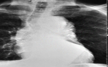
Ultrasound
MRI
Aortic arteriography
Chest X-ray --> "eggshell appearance" = aortic dissection
67
Thoracic Aortic Aneurysm - Manifestations
Asymptomatic --> usually found during routine exams
If present --> difuse chest pain that extends to interscapular area
If present --> difuse chest pain that extends to interscapular area
68
Aortic Arch Aneurysm - Manifestations
Hoarseness --> due to pressure in laryngeal nerve
Dysphagia --> pressure in esophagus makes it difficult to speak
Neck Vein Distention --> due to pressure in superior vena cava
Dysphagia --> pressure in esophagus makes it difficult to speak
Neck Vein Distention --> due to pressure in superior vena cava
69
Abdominal Aortic Aneurysm - Manifestations
Asymptomatic --> detected on routine exam
Pulsatile abdominal mass
Bruit over the aneurysm
Symptoms mimic abdominal and back disorder --> epigastric + back pain, altered bowel elimination
Pulsatile abdominal mass
Bruit over the aneurysm
Symptoms mimic abdominal and back disorder --> epigastric + back pain, altered bowel elimination
70
Aneurysm Complication
Rupture
Dissection
HTN --> MOST COMMON CAUSE OF RUPTURE
Dissection
HTN --> MOST COMMON CAUSE OF RUPTURE
71
Aneurysm Assessment
Assess for severe radiating back/flank pain (mimics kidney stone pain)
Assess for pulsation and bruit on xiphoid and umbilicus --> DO NOT PALPATE
Assess for s/s hemorrhagic shock --> hypotension, tachycardia, dysrhythmia, diaphoresis, oliguria, decreased LOC
Assess for pulsation and bruit on xiphoid and umbilicus --> DO NOT PALPATE
Assess for s/s hemorrhagic shock --> hypotension, tachycardia, dysrhythmia, diaphoresis, oliguria, decreased LOC
72
Aortic Aneurysms Assessment Findings
if AAA:
- Abdominal distention
- Grey Turner Sign (back and flank bruising)
if TAA:
- SOB
- Hoarseness
- Dysphagia
- Abdominal distention
- Grey Turner Sign (back and flank bruising)
if TAA:
- SOB
- Hoarseness
- Dysphagia
73
Nonsurgical Management of Asymptomatic Aneurysms
Monitor + Maintain BP --> antihypertensive drugs
Monitor size --> CT scan, ultrasound
Educate:
- S/S of impending rupture
- Medications and follow-up
Monitor size --> CT scan, ultrasound
Educate:
- S/S of impending rupture
- Medications and follow-up
74
Surgical Management of Aneurysms
Elective:
- Endovascular Aneurysm Repair (EVAR) procedure
Emergent:
- Resection or repair (aneurysmectomy)
- Endovascular stent grafts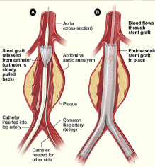
- Endovascular Aneurysm Repair (EVAR) procedure
Emergent:
- Resection or repair (aneurysmectomy)
- Endovascular stent grafts
75
Complications After Aneurysm Surgery
Graft Occlusion --> Monitor distal pulses (dorsalis pedis and post tib)
Graft Rupture --> Monitor for increasing abdominal girth, hypovolemic shock (↓BP, ↑HR)
Embolization
Paraplegia
Graft Rupture --> Monitor for increasing abdominal girth, hypovolemic shock (↓BP, ↑HR)
Embolization
Paraplegia
76
Post Aneurysm Repair Discharge Teaching
Activity restriction --> 6-12 weeks; no stair climbing, no heavy lifting
Incisional care
Pain management
Incisional care
Pain management
77
Vascular Emergencies of the Aorta
Aortic Dissection
Aortic Rupture
Aortic Rupture
78
Aortic Dissection
Inner wall of the aorta is damaged or torn --> can lead to less blood flowing to organs and limbs
Can lead to aortic rupture --> not good
Many people who have an ascending aortic dissection or severe descending aortic dissection do not survive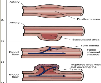
Can lead to aortic rupture --> not good
Many people who have an ascending aortic dissection or severe descending aortic dissection do not survive
79
Aortic Dissection: Etiologies
Atherosclerosis, chronic HTN, congenital heart defects
Hereditary Connective Tissue Disorder (Marfans, Ehlers-Danlos)
Vascular inflammation (syphillis, Takayasu's arteritis)
Trauma (such as motor vehicle crash, falls)
Iatrogenic events ( cardiac catherization, cardiopulmonary bypass)
Pregnancy (rare)
Crack cocaine use
Basically anything that threatens the integrity of the vessel wall
Hereditary Connective Tissue Disorder (Marfans, Ehlers-Danlos)
Vascular inflammation (syphillis, Takayasu's arteritis)
Trauma (such as motor vehicle crash, falls)
Iatrogenic events ( cardiac catherization, cardiopulmonary bypass)
Pregnancy (rare)
Crack cocaine use
Basically anything that threatens the integrity of the vessel wall
80
Aortic Dissection Manifestations
Varies with size, location, and stem of dissection --> most common site is ascending aorta
Sudden onset of severe tearing pain
- Substernal = ascending dissection
- Intrascapular = descending aorta
Hypertension --> Hypotension
Diastolic murmur secondary to aortic regurgitation
Sudden onset of severe tearing pain
- Substernal = ascending dissection
- Intrascapular = descending aorta
Hypertension --> Hypotension
Diastolic murmur secondary to aortic regurgitation
81
Ascending Aorta Dissection Impairments
Damage to aortic valve
Impaired coronary blood flow
Cardiac tamponade
Laryngeal nerve compression
Bleeding into pleural space
Impaired coronary blood flow
Cardiac tamponade
Laryngeal nerve compression
Bleeding into pleural space
82
Ascending Aorta Dissection Manifestations: Due to Damaged aortic valve
Diastolic murmur
83
Ascending Aorta Dissection Manifestations: Due to Impaired Coronary Blood Flow
Due to the associated impaired coronary blood flow:
- Chest pain indicative of MI
- Chest pain indicative of MI
84
Ascending Aorta Dissection Manifestations: Due to Cardiac Tamponade
Due to the associated impairment of cardiac tamponade:
- Muffled heart sounds
- Hypotension
- Tachycardia
- JVD
- Muffled heart sounds
- Hypotension
- Tachycardia
- JVD
85
Ascending Aorta Dissection Manifestations: Laryngeal Nerve Compression
Hoarseness associated with laryngeal nerve compression impairment
86
Ascending Aorta Dissection Manifestations: Due to Bleeding into Pleural Space
Dyspnea
Hemothorax
Hemothorax
87
Aortic Arch Dissection Impairments
Reduced blood flow to the brain
Interruption of cervical sympathetic ganglia
Impaired brachiocephalic flow
Interruption of cervical sympathetic ganglia
Impaired brachiocephalic flow
88
Aortic Arch Dissection Manifestations: Due to reduced blood flow to the brain
- Syncope
- Altered mental status
- Horner syndrome --> ptosis (eyelid drooping), miosis (pupil constriction), anhidrosis (absence of sweating on one side of the face)
- Altered mental status
- Horner syndrome --> ptosis (eyelid drooping), miosis (pupil constriction), anhidrosis (absence of sweating on one side of the face)
89
Aortic Arch Dissection Manifestations: Due to Impaired Brachiocephalic Flow
Blood pressure differential
Asymmetric pulses in upper extremities
Asymmetric pulses in upper extremities
90
Descending Aorta Dissection Impairments
Spinal Cord Ischemia
Mesenteric Artery Ischemia (the artery that supplies blood to ur intestines)
Mesenteric Artery Ischemia (the artery that supplies blood to ur intestines)
91
Descending Aorta Dissection Manifestations
Due to spinal cord ischemia
- Limb paresthesia/paralysis
Due to mesenteric artery ischemia
- Acute abdominal pain
- Melena (gi bleed)
- Hyperactive bowel sounds
- Limb paresthesia/paralysis
Due to mesenteric artery ischemia
- Acute abdominal pain
- Melena (gi bleed)
- Hyperactive bowel sounds
92
Thoracoabdominal Aorta Dissection Impairments
Renal artery ischemia
Lower limb ischemia
Lower limb ischemia
93
Thoracoabdominal Aorta Dissection Manifestations: Due to Renal Artery Ischemia
- Flank pain
- Oliguria
- Oliguria
94
Thoracoabdominal Aorta Dissection Manifestations: Due to Lower Limb Ischemia
Diminshed/absent pulses in lower extremities
95
Aortic Dissection Diagnostics
Chest X-ray --> wide mediastinum
12 Lead ECG --> lack of ischemic changes (unless dissection affects coronary arteries)
Troponin and Creatine Kinase --> elevated if coronary arteries affected
CT scan
Transesophageal electrography (TEE)
MRI
Smooth muscle myosin --> elevated
12 Lead ECG --> lack of ischemic changes (unless dissection affects coronary arteries)
Troponin and Creatine Kinase --> elevated if coronary arteries affected
CT scan
Transesophageal electrography (TEE)
MRI
Smooth muscle myosin --> elevated
96
Aortic Dissection Post Op Complications: Potential Extension of Dissection or Rupture
Establish baseline assessment
Serial assessments to detect changes in LOC, motor/sensory function, ischemic changes
Assess bilateral pulses, BP, urinary output --> priority (BP should be evaluated bilaterally to rule out "psuedo" hypotension created by dissection into brachiocephalic vessel)
Monitor Oxygen saturation --> hypoxemia is common post op
IV meds to decrease BP and HR
Administer analgesics and sedation PRN --> dissection creates pain, which can exacerbate BP and HR
Serial assessments to detect changes in LOC, motor/sensory function, ischemic changes
Assess bilateral pulses, BP, urinary output --> priority (BP should be evaluated bilaterally to rule out "psuedo" hypotension created by dissection into brachiocephalic vessel)
Monitor Oxygen saturation --> hypoxemia is common post op
IV meds to decrease BP and HR
Administer analgesics and sedation PRN --> dissection creates pain, which can exacerbate BP and HR
97
Aortic Dissection Post Op Complications: Hypothermia
Continuously monitor core temperature (use Pulmonary Artery catheter)
Warm air blankets for temperature < 36C
Meperidine prn for shivering
Fluid warmers may be useful (esp if blood products are administered)
Warm air blankets for temperature < 36C
Meperidine prn for shivering
Fluid warmers may be useful (esp if blood products are administered)
98
Aortic Dissection Post Op Complications: Bleeding
Control hemorrhage
Maintain patency of chest tubes
Maintain MAP of 70-80 --> increased pressure puts stress on suture lines and cannulation sites
Monitor H&H, platelet, PT/INR, PTT
Administer medications for bleeding (warfarin, heparin) --> have vitamin K, protamine sulfate on hand to reverse
Replace volume: blood products, volume expanders (dextran, albumin)
Maintain patency of chest tubes
Maintain MAP of 70-80 --> increased pressure puts stress on suture lines and cannulation sites
Monitor H&H, platelet, PT/INR, PTT
Administer medications for bleeding (warfarin, heparin) --> have vitamin K, protamine sulfate on hand to reverse
Replace volume: blood products, volume expanders (dextran, albumin)
99
Aortic Dissection Post Op Complications: Neurologic Dysfunction
Impaired perfusion to brain and spinal cord may occur during period of circulatory arrest, emboli, or debris from surgical procedure
Allow patient to awaken ASAP to evaluate neuro status
Assess cognitive function, as well as gross motor movement and sensation
Allow patient to awaken ASAP to evaluate neuro status
Assess cognitive function, as well as gross motor movement and sensation
100
Aortic Dissection Post Op Complications: Myocardial Ischemia
Potential for ischemic complications related to location of aortic injury, surgical approach, and preexisting disease
Continuous ECG monitoring (esp. ST segment monitoring)
Administer perioperative beta blockers as ordered --> decrease incidence of MI in pts with vascular disease
Continuous ECG monitoring (esp. ST segment monitoring)
Administer perioperative beta blockers as ordered --> decrease incidence of MI in pts with vascular disease
101
Aortic Dissection Post Op Complications: Spinal Cord Ischemia
Optimize perfusion to spinal cord by maintaining adequate BP --> MAP > 80 mmHg
If lumbar drain --> maintain system @ prescribed level and drain CSF pressure < 10 mmHg
Q.1h Neuro check --> numbness/tingling, weakness in lower extremities
If lumbar drain --> maintain system @ prescribed level and drain CSF pressure < 10 mmHg
Q.1h Neuro check --> numbness/tingling, weakness in lower extremities
102
Aortic Dissection Post Op Complications: Distal Embolization
Cross-clamping or emboli may compromise flow to lower extremities
Perform neurovascular checks --> motor function, sensation, capillary refill, distal pulses (palpation or Doppler)
Perform neurovascular checks --> motor function, sensation, capillary refill, distal pulses (palpation or Doppler)
103
Aortic Dissection Post Op Complications: Renal Dysfunction
Renal failure may occur secondary to perioperative hypotension, hypovolemia, use of contrast dye, cross-clamping, or preexisting renal disease
Monitor urine ouptut q.1H --> > 30 ml/hr
Monitor filling pressures and maintain preload with fluids PRN
Maintain adequate blood pressure for renal perfusion
Monitor urine ouptut q.1H --> > 30 ml/hr
Monitor filling pressures and maintain preload with fluids PRN
Maintain adequate blood pressure for renal perfusion
104
Aortic Dissection Post Op Complication: GI Dysfunction
Patients are at risk for an ileus postop, esp. w/ repairs of abdominal aorta
Maintain NG tube to low continuous suction until bowel sounds are present
Monitor pt for s/s of ischemic bowel, including abdomnial pain and distention, persistent acidosis, elevated WBCs, diarrhea
Maintain NG tube to low continuous suction until bowel sounds are present
Monitor pt for s/s of ischemic bowel, including abdomnial pain and distention, persistent acidosis, elevated WBCs, diarrhea
105
Aortic Dissection Post Op Complication: Pulmonary Dysfunction
Related to post surgery risk for atelectasis
Monitor O2 Sat PRN
Maintain mechanical ventilation PRN
Reposition frequently
If extubated: Deep breathing exercises and incentive spirometer q.1-2H
Splinting with pillow during cough
Provide adequate pain medication to promote activitiy without inducing respiratory depression
Monitor O2 Sat PRN
Maintain mechanical ventilation PRN
Reposition frequently
If extubated: Deep breathing exercises and incentive spirometer q.1-2H
Splinting with pillow during cough
Provide adequate pain medication to promote activitiy without inducing respiratory depression
106
Aortic Aneurysm: Management of inoperable cases
Goal is to stabilize to prevent rupture
Control BP (systolic 100-110 mmHg) --> beta blockers, ACE inhibitors, vasodilators
Management of hypercholesterolemia; smoking cessation
Regular follow up --> monitor size progression
ICU care for suspected dissection
- Aggressive BP control and monitoring
- Prep for surgery as warranted
- Psycho/emotional support for patient and family
Control BP (systolic 100-110 mmHg) --> beta blockers, ACE inhibitors, vasodilators
Management of hypercholesterolemia; smoking cessation
Regular follow up --> monitor size progression
ICU care for suspected dissection
- Aggressive BP control and monitoring
- Prep for surgery as warranted
- Psycho/emotional support for patient and family
107
Cardiomyopathy
Heart Enlargement = inefficient pump
Two types: Dilated Cardiomyopathy (DCM), Hypertrophic Cardiomyopathy (HCM)
Can lead to HF, dysrhythmias, sudden death if left untreated
Two types: Dilated Cardiomyopathy (DCM), Hypertrophic Cardiomyopathy (HCM)
Can lead to HF, dysrhythmias, sudden death if left untreated
108
Cardiomyopathy: Etiology
mostly unknown
Bombay university uno
Bombay university uno
109
Cardiomyopathy: Risk Factors
HTN
Chronic excessive alcohol intake
Pregnancy complications
Systemic infection
Chronic excessive alcohol intake
Pregnancy complications
Systemic infection
110
Cardiomyopathy: Diagnostics
Echocardiography
Angiocardiography
ECG --> T wave inversion, ST segment depression
Angiocardiography
ECG --> T wave inversion, ST segment depression
111
Dilated Cardiomyopathy
Fibrosis of myocardium/endocardium
Dilated chambers
Prevalent mural wall thrombi (thrombi that sticks to the wall)
Dilated chambers
Prevalent mural wall thrombi (thrombi that sticks to the wall)
112
Dilated Cardiomyopathy: Manifestations
Fatigue, Weakness, Dizziness, Syncope --> could be onset of deadl arrhythmia
Heart failure; left side
Dysrhythmias or heart block
Systemic or pulmonary emboli
S3 and S4 gallops
Moderate to severe cardiomegaly (enlarged heart)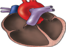
Heart failure; left side
Dysrhythmias or heart block
Systemic or pulmonary emboli
S3 and S4 gallops
Moderate to severe cardiomegaly (enlarged heart)
113
Hypertrophic Cardiomyopathy - Nonobstructed
Hypertrophy of all walls
Hypertrophied septum
Relatively small chamber size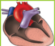
Hypertrophied septum
Relatively small chamber size
114
Hypertrophic Cardiomyopathy (Unobstructed): Manifestations
Dyspnea, fatigue --> lungs need to work harder to adequately oxygenate
Angina (kinda obvious ngl)
Syncope --> due to decreased cardiac output
Palpitations
Mild cardiomegaly (enlarged heart)
Ventricular dysrhythmias (i wonder why)
Heart failure (no shit)
Sudden death is common
Angina (kinda obvious ngl)
Syncope --> due to decreased cardiac output
Palpitations
Mild cardiomegaly (enlarged heart)
Ventricular dysrhythmias (i wonder why)
Heart failure (no shit)
Sudden death is common
115
Obstructed Hypertrophic Cardiomyopathy
Same as nonobstructed EXCEPT there is obstruction of left ventricular outflow tract associated with hypertrophied septum and mitral valve incompetence
116
Hypertrophic Cardiomyopathy (Obstructed): Manifestations
Same as nonobstructed
Mitral regurgitation murmur
Atrial fibrillation
Mitral regurgitation murmur
Atrial fibrillation
117
Dilated Cardiomyopathy: Management
O2 therapy
Drug Therapy:
- Diuretics --> insert foley, I&O
- Morphine --> reduces venous return (preload)
- Vasodilators
- Digoxin
Control of dysrhythmias
Report dizziness or fainting --> indication of onset of lethal dysrhythmia
Avoid alcohol, strenuous exercise
Surgery --> heart transplant
Drug Therapy:
- Diuretics --> insert foley, I&O
- Morphine --> reduces venous return (preload)
- Vasodilators
- Digoxin
Control of dysrhythmias
Report dizziness or fainting --> indication of onset of lethal dysrhythmia
Avoid alcohol, strenuous exercise
Surgery --> heart transplant
118
Hypertrophic Cardiomyopathy: Management
For both obstructive and nonobstructive
Drug Therapy:
- Beta Blockers
- Antidysrhythmic drugs
- Nitrates and other vasodilators are contraindicated in the obstructed form --> CO is already low, don't wanna make it worse
Conversion of afib
Automated implantable cardioverter defibrillator
Genetic Counseling
Surgical management:
- Ventriculomyotomy or muscle resection with mitral valve replacement
Drug Therapy:
- Beta Blockers
- Antidysrhythmic drugs
- Nitrates and other vasodilators are contraindicated in the obstructed form --> CO is already low, don't wanna make it worse
Conversion of afib
Automated implantable cardioverter defibrillator
Genetic Counseling
Surgical management:
- Ventriculomyotomy or muscle resection with mitral valve replacement
119
Left Sided Heart Failure Pathophysiology
The pumping ability of left ventricle fails and cardiac output decreases
↓
Blood backs up into the left atrium and lungs, causing pulmonary congestion
↓
If left untreated, left-sided heart failure leads to right-sided heart failure
↓
Blood backs up into the left atrium and lungs, causing pulmonary congestion
↓
If left untreated, left-sided heart failure leads to right-sided heart failure
120
Right Sided Heart Failure Pathophysiology
Ineffective contractile function of the right ventricle
↓
Blood backs up in right atrium and peripheral circulation
↓
Peripheral edema and engorgement of kidneys and other organs
↓
Blood backs up in right atrium and peripheral circulation
↓
Peripheral edema and engorgement of kidneys and other organs
121
Heart Failure Risk Factors
HTN
CAD
Cardiomyopathy
Valvular disease
Cardiac infections
DM
Family history
Acute myocardial infarction
Hyperthyroidism
Substance abuse
Congenital defects
Dysrhythmias
Alcohol/Tobacco use
Severe lung disease
CAD
Cardiomyopathy
Valvular disease
Cardiac infections
DM
Family history
Acute myocardial infarction
Hyperthyroidism
Substance abuse
Congenital defects
Dysrhythmias
Alcohol/Tobacco use
Severe lung disease
122
Compensatory Mechanism of HF: Decreased Cardiac Output
Decreased Cardiac Output and Systemic Blood Pressure
Baroreceptors in the left ventricle, aortic arch, and carotid sinus are activated
↓
Vasomotor regulatory center in medulla is stimulated
↓
Sympathetic nervous system activated
↓
Increase in catecholamines (epinephrine and norepinephrine
↓
Vasoconstriction --> increase in afterload, blood pressure, HR
↓
Impaired contractility = decreased cardiac output
Baroreceptors in the left ventricle, aortic arch, and carotid sinus are activated
↓
Vasomotor regulatory center in medulla is stimulated
↓
Sympathetic nervous system activated
↓
Increase in catecholamines (epinephrine and norepinephrine
↓
Vasoconstriction --> increase in afterload, blood pressure, HR
↓
Impaired contractility = decreased cardiac output
123
Compensatory Mechanisms of HF: Decreased Perfusion to the Kidneys
RAAS system activated
↓
Angiotensin II --> leads to vasoconstriction
↓
Aldosterone released into the kidneys --> retain more sodium and water
↓
Angiotensin II --> leads to vasoconstriction
↓
Aldosterone released into the kidneys --> retain more sodium and water
124
Compensatory Mechanisms of HF: Ventricular Remodeling
Hypertrophy and dilation of ventricles
↓
Impaired contractility = decreased cardiac output
↓
Impaired contractility = decreased cardiac output
125
Left Sided HF Manifestations
Crackles secondary to pulmonary edema
Increased HR, S3 and S4 present
Fatigue, Dyspnea, Orthopnea, Nocturnal Dyspnea --> Do NOT lay flat
Nocturia
Pulsus Alternans (alternating pulses: strong -> weak0
Dry, hacking cough
Increased HR, S3 and S4 present
Fatigue, Dyspnea, Orthopnea, Nocturnal Dyspnea --> Do NOT lay flat
Nocturia
Pulsus Alternans (alternating pulses: strong -> weak0
Dry, hacking cough
126
Right Sided HF Manifestations
Peripheral edema
Increased HR
Fatigue
Ascites (fluid in abdomen)
Anasarca (general swelling around body)
Weight Gain
JVD
Hepatomegaly --> right upper quadrant pain
Right pleural effusion
Anorexia, GI bloating, Nausea
Increased HR
Fatigue
Ascites (fluid in abdomen)
Anasarca (general swelling around body)
Weight Gain
JVD
Hepatomegaly --> right upper quadrant pain
Right pleural effusion
Anorexia, GI bloating, Nausea
127
Heart Failure Diagnostics
Detect underlying etiology
History and Physical
chest xray
12 Lead ECG
Detect Ejection Fraction --> echocardiogram, stress test, cardiac catherization
History and Physical
chest xray
12 Lead ECG
Detect Ejection Fraction --> echocardiogram, stress test, cardiac catherization
128
Heart Failure Labs
BNP (brain natriuretic peptide) --> increased in HF (should be under 100 pg/mL)
Cardiac enzymes (troponin and creatine kinase)
BMP, CBC
LFT
Thyroid function studies
Cardiac enzymes (troponin and creatine kinase)
BMP, CBC
LFT
Thyroid function studies
129
BNP (brain natriuretic peptide) Function
When the heart works harder to pump blood, it releases BNP
BNP signals blood vessels to widen and kidneys to remove water and salt
Increased in heart failure
BNP signals blood vessels to widen and kidneys to remove water and salt
Increased in heart failure
130
Heart Failure Treatment
Sodium and fluid restrictions; I&O
O2 therapy
Biventricular pacemaker
Implantable cardioverter-defibrillator
Circulatory Assistive Devices --> Intra-aortic balloon pump, ventricular assist device
Cardiac Transplantation
End of life care
O2 therapy
Biventricular pacemaker
Implantable cardioverter-defibrillator
Circulatory Assistive Devices --> Intra-aortic balloon pump, ventricular assist device
Cardiac Transplantation
End of life care
131
Heart Failure Drug Therapy: Diuretics
Decreases preload
132
Heart Failure Drug Therapy: ACE Inhibitors
Decreases systemic vascular resistance --> increases cardiac output and improves tissue perfusion
133
Heart Failure Drug Therapy: Beta Blockers
Decreases heart rate --> decreases heart O2 demand
134
Heart Failure Drug Therapy: Inotropics
Milrinone
Improves contractility --> improves cardiac output
Improves contractility --> improves cardiac output
135
Heart Failure Drug Therapy: Beta Agonists
Dopamine, Dobutamine, Levophed (norepinephrine)
136
Heart Failure Drug Therapy: Cardiac Glycosides
digoxin
Improve contractility, decrease HR
Improve contractility, decrease HR
137
Heart Failure Drug Therapy: Nitrates
Vasodilation
Decrease preload
Avoid Viagra
Decrease preload
Avoid Viagra
138
Heart Failure Drug Therapy: Morphine
Decrease preload, anxiety, work of breathing
Relaxes blood vessels, slows the heart, and slows breathing down
Relaxes blood vessels, slows the heart, and slows breathing down
139
Heart Failure Drug Therapy: Anticoagulants
Since they aren't as physically active and also blood is not effectively moving and is stagnant, anticoagulants may be required to prevent thromboembolisms
140
Heart Transplant Criteria
Life expectancy less than 1 year
Age < 65
Normal or slightly elevated pulmonary vascular resistance
Absence of active infection
Stable psychosocial status
No evidence of current drug or alcohol abuse
Age < 65
Normal or slightly elevated pulmonary vascular resistance
Absence of active infection
Stable psychosocial status
No evidence of current drug or alcohol abuse
141
Heart Transplant: Immediate Post Op care
monitor for bleeding --> s/s of cardiac tamponade
medications to support HR and maintain CO
Caution to change position slowly (orthostatic hypotension)
Pacemaker
Monitor for transplant protocol --> s/s of rejection (flu-like symptoms, swelling); immunosuppressants
medications to support HR and maintain CO
Caution to change position slowly (orthostatic hypotension)
Pacemaker
Monitor for transplant protocol --> s/s of rejection (flu-like symptoms, swelling); immunosuppressants
142
Heart Failure Nursing Management
Strict I&O
Monitor for signs of fluid overload --> daily weights (1-2 lb increase); breath sounds, SOB (how many pillows needed?)
Safety --> progressive mobility
Monitor for signs of fluid overload --> daily weights (1-2 lb increase); breath sounds, SOB (how many pillows needed?)
Safety --> progressive mobility
143
Heart Failure: Patient/Family Education
Energy conservation
Follow-up care
S/S to report --> SOB, swelling in legs, ankles, feet, chest pain, etc.
Follow-up care
S/S to report --> SOB, swelling in legs, ankles, feet, chest pain, etc.
144
Heart Failure: Effectiveness of drug treatment
Morphine --> decrease preload
Diuretics --> decrease preload
Vasodilators --> decrease afterload
Amiodarone --> control dysrhythmia
Digoxin --> improve contractility
Diuretics --> decrease preload
Vasodilators --> decrease afterload
Amiodarone --> control dysrhythmia
Digoxin --> improve contractility
145
Heart Failure: Support Devices
Intra-aortic Balloon Pump --> balloon is deflated during systole. inflated during diastole (rest)
- Balloon inflation during diastole allows the heart to fill
HeartMate II Ventricular Assist Device (Thoratec) --> used as bridge to heart transplant
- Balloon inflation during diastole allows the heart to fill
HeartMate II Ventricular Assist Device (Thoratec) --> used as bridge to heart transplant
146
Cardiogenic Shock
when your heart cannot pump enough blood and oxygen to the brain and other vital organs
147
Cardiogenic Shock Causes: Systolic Dysfunction
Inability of the heart to pump blood forward
Ex: MI, cardiomyopathy, blunt cardiac injury, severe systemic or pulmonary HTN, myocardial depression
Ex: MI, cardiomyopathy, blunt cardiac injury, severe systemic or pulmonary HTN, myocardial depression
148
Cardiogenic Shock Causes: Diastolic Dysfunction
Inability of the heart to fill
Ex: Cardiac tamponade, ventricular hypertrophy, cardiomyopathy
Ex: Cardiac tamponade, ventricular hypertrophy, cardiomyopathy
149
Cardiogenic Shock Causes: Dysrhythmias
Bradydysrhythmias
Tachydysrhythmias
Tachydysrhythmias
150
Cardiogenic Shock Causes: Structural factors
Vavular stenosis or regurgitation
Ventricular Septal Rupture
Tension pneumothorax
Ventricular Septal Rupture
Tension pneumothorax
151
Cardiogenic Shock Manifestations
Systolic BP < 90 mmHg, HR >100 bpm
Weak, thready pulse
Chest pain, dysrhythmias
Tachypnea
S3,S4, Crackles
JVD
Decreased O2 saturation
Cool, pale, moist skin
Urine output <30 mL
Decreased cardiac output
Change in sensorium
Weak, thready pulse
Chest pain, dysrhythmias
Tachypnea
S3,S4, Crackles
JVD
Decreased O2 saturation
Cool, pale, moist skin
Urine output <30 mL
Decreased cardiac output
Change in sensorium
152
Cardiogenic Shock: Improve Tissue Perfusion
Pharmacological agents to maintain cardiac output
- Inotropic agents (dobutamine, milrinone) --> increases contractility
- Vasopressors (dopamine)
- Diuretics --> reduce preload
- Vasodilators --> reduce preload and afterload
- Antidysrhythmic agents
Intubation and mechanical ventilation --> sedate + paralyze
- Inotropic agents (dobutamine, milrinone) --> increases contractility
- Vasopressors (dopamine)
- Diuretics --> reduce preload
- Vasodilators --> reduce preload and afterload
- Antidysrhythmic agents
Intubation and mechanical ventilation --> sedate + paralyze
153
Cardiogenic Shock: When Conventional Treatment Fails
IABP (intra-aortic balloon pump)
Ventricular Assist Device
Ventricular Assist Device
154
Cardiogenic Shock: Nursing Management
Early identification --> facilitate earlyl reperfusion treatment
Close monitoring (q.10-15 minutes)
Limit myocardial O2 demands --> administer pharmacological agents
Maintain respiratory status
Monitor for complications
Close monitoring (q.10-15 minutes)
Limit myocardial O2 demands --> administer pharmacological agents
Maintain respiratory status
Monitor for complications
155
Cardiogenic Shock: Norepinephrine
First choice for cardiogenic shock
Vasopressor support
Some positive inotropic effects
Minimum doses should be used and systemic vascular resistance should be monitored during administration
Vasopressor support
Some positive inotropic effects
Minimum doses should be used and systemic vascular resistance should be monitored during administration
156
Cardiogenic Shock: Dopamine
Vasopressor support with some positive inotropic effects
May increase pulmonary artery occlusion pressure
Minumum dose should be used; systemic vascular resistance should be monitored
May increase pulmonary artery occlusion pressure
Minumum dose should be used; systemic vascular resistance should be monitored
157
Cardiogenic Shock: Dobutamine
produces positive inotropic effect by increasing cardiac index
also decreases afterload (through vasodilation)
also decreases afterload (through vasodilation)
158
Cardiogenic Shock: Milrinone
Used to induce peripheral vasodilation and provide inotropic support
159
Cardiogenic Shock: Nitroglycerin
Decrease preload and afterload
160
Cardiogenic Shock: Antidysrhythmic Agents
Amiodarone --> for sustained ventricular or atrial tachyarrhythmias
161
Conduction System of the Heart
SA node (main pacemaker; 60-100 bpm)
↓
AV node (backup pacemaker; 40-60 bpm)
↓
Bundle of His (super backup pacemaker; 30-40 bpm)
↓
Purkinje fibers
↓
AV node (backup pacemaker; 40-60 bpm)
↓
Bundle of His (super backup pacemaker; 30-40 bpm)
↓
Purkinje fibers
162
P wave
represents depolarization (contraction) of the atria
163
QRS complex
represents depolarization (contraction) of the ventricles
164
T wave
Represents repolarization (back to resting state) of the ventricles
165
PR segment
amount of time it takes for electrical signal to move from SA to AV nodes
166
Junctional Dysrhythmias: ECG Characteristics
Rhythm is regular
P wave: abnormal; inverted or hidden
PR interval < 0.12 seconds when P wave precedes QRS
QRS is normal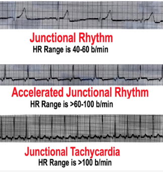
P wave: abnormal; inverted or hidden
PR interval < 0.12 seconds when P wave precedes QRS
QRS is normal
167
Junctional Dysrhytmias: Causes
CAD, HF, cardiomyopathy
Electrolyte imbalances
Inferior wall MI
Rheumatic heart disease
SA node disease (i.e. Sick Sinus Syndrome)
Drugs (such as digoxin, amphetamines)
Vagal simulation
Cardiac surgery
Electrolyte imbalances
Inferior wall MI
Rheumatic heart disease
SA node disease (i.e. Sick Sinus Syndrome)
Drugs (such as digoxin, amphetamines)
Vagal simulation
Cardiac surgery
168
Junctional Dysrhythmias: Manifestations
May be asymptomatic
Lightheadedness, palpitations
Effort intolerance, chest heaviness
Neck tightness or pounding
SOB, weakness
Hypotension, Bradycardia
Altered LOC, blurred vision
Lightheadedness, palpitations
Effort intolerance, chest heaviness
Neck tightness or pounding
SOB, weakness
Hypotension, Bradycardia
Altered LOC, blurred vision
169
Junctional Dysrhythmias: Nursing Diagnosis
Risk for reduced cardiac output
170
Junctional Dysrhythmias: Management
Treat the underlying cause
Discontinue meds causing the arrhythmia
If caused by Digitalis toxicity
- Administer Digoxin Immune Fab
- Check serum potassium (potentiates dig toxicity)
Atropine --> increase HR
Temporary or permanent pacemaker
Ablation
Discontinue meds causing the arrhythmia
If caused by Digitalis toxicity
- Administer Digoxin Immune Fab
- Check serum potassium (potentiates dig toxicity)
Atropine --> increase HR
Temporary or permanent pacemaker
Ablation
171
Atrioventricular Block
Interruption in conduction between atria and ventricles
differentiated by PR Interval:
- 1st degree --> impulses reach ventricles
- 2nd degree --> some impulses do not reach ventricles
- 3rd degree --> no sinus impulses reach ventricles (Complete Heart Block)
differentiated by PR Interval:
- 1st degree --> impulses reach ventricles
- 2nd degree --> some impulses do not reach ventricles
- 3rd degree --> no sinus impulses reach ventricles (Complete Heart Block)
172
1st Degree AV Block
R is far from P --> PR interval greater than 0.2 seconds (one box)
Most common form of heart block
Impulse conduction between atria and Bundle of His is delayed at the level of the AV node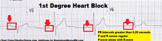
Most common form of heart block
Impulse conduction between atria and Bundle of His is delayed at the level of the AV node
173
1st Degree AV Block: Causes
MI or ischemia
Medications: digoxin, Ca Channel Blockers (amlodipine, diltiazem), beta blockers
Myocarditis
Degenerative changes of the heart
Medications: digoxin, Ca Channel Blockers (amlodipine, diltiazem), beta blockers
Myocarditis
Degenerative changes of the heart
174
1st Degree AV Block: Manifestations
Asymptomatic
Dizziness, syncope
Dizziness, syncope
175
2nd Degree Mobitz I
Longer, Longer, Drop --> PR interval continues to increase untill impulse is not conducted (QRS complex is dropped)
Risk: Reduced cardiac output, progression to 3rd degree
Risk: Reduced cardiac output, progression to 3rd degree
176
2nd Degree Mobitz I: Causes
CAD, Inferior wall MI
Rheumatic fever
Increased vagal stimulation
Medications: propanolol, digitalis, verapamil
Rheumatic fever
Increased vagal stimulation
Medications: propanolol, digitalis, verapamil
177
2nd Degree Mobitz II
Some P's don't get through --> atrial rhythm is regular, ventricular rhythm is irregular (dropped/non-conducted beats)
More lethal than Mobitz I
Risk: Reduced cardiac output, Progression to 3rd Degree Heart Block
More lethal than Mobitz I
Risk: Reduced cardiac output, Progression to 3rd Degree Heart Block
178
2nd Degree Mobitz II: Causes
Anterior MI
Medications (like digoxin)
Changes of the heart
CAD
Lyme disease
Medications (like digoxin)
Changes of the heart
CAD
Lyme disease
179
3rd Degree Complete AV Block
P's and Q's don't agree --> impulses from atria are completely blocked at AV node (can't be conducted to the ventricles
Most serious type
Risks: Progression to asystole; rate is too slow and ineffective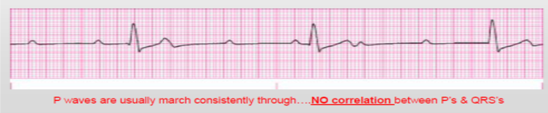
Most serious type
Risks: Progression to asystole; rate is too slow and ineffective
180
3rd Degree Complete AV Block: Causes
Congenital condition
CAD
Acute MI, inferior wall MI
Degenerative changes of the heart
Digitalis toxicity
CAD
Acute MI, inferior wall MI
Degenerative changes of the heart
Digitalis toxicity
181
3rd Degree Complete AV Block: Manifestations
Severe fatigue
Dyspnea
Chest pain
Decreased LOC
Hypotension
Pallor
Diaphoresis
Dyspnea
Chest pain
Decreased LOC
Hypotension
Pallor
Diaphoresis
182
Heart Block Diagnostics
ECG
Echocardiogram
Electrolytes
Drug Level
Purpose is to figure out the cause
Echocardiogram
Electrolytes
Drug Level
Purpose is to figure out the cause
183
Heart Block Treatment: 1st Degree, 2nd Degree Mobitz I
If asymptomatic --> continue to monitor, treat the underlying cause
184
Heart Block Management
Goal: Raise HR to improve cardiac output, improve ventricular rate
Oxygen therapy; bed rest
Medications: atropine, epinephrine, Isoproterenol, Dopamine
Temporary or transcutaneous pacemaker --> until rhythm resolves
Holter Monitor
Oxygen therapy; bed rest
Medications: atropine, epinephrine, Isoproterenol, Dopamine
Temporary or transcutaneous pacemaker --> until rhythm resolves
Holter Monitor
185
Holter Monitor Considerations
think of it as a "portable ECG monitor"
Pt Education:
- Keep diary of activities and symptoms
- Do NOT shower while on monitor
Pt Education:
- Keep diary of activities and symptoms
- Do NOT shower while on monitor
186
Pacemaker
Stimulate a heartbeat if HR drops too low
Usually initiates pacing stimulus if ventricular rate drops below the preset level (usually 72 bpm)
Two types: Temporary (transcutaneous) and Permanent
Usually initiates pacing stimulus if ventricular rate drops below the preset level (usually 72 bpm)
Two types: Temporary (transcutaneous) and Permanent
187
Temporary Pacemaker Indications
Treat temporary problems (duh)
- Heart surgery
- Overdose of medicine
- Emergencies
- Heart surgery
- Overdose of medicine
- Emergencies
188
Permanent Pacemaker Indications
Long-term rhythm problems
189
Permanent Pacemaker: Patient Teaching
Keep site dry and clean for a week
Bed rest for first 12 hours
Avoid lifting for one week
Avoid high output generators (like an MRI)
Teach patient how to take a pulse --> if set at 72 bpm, pulse should be at 72 bpm
Low HR = pacemaker failure
Carry a pacemaker info card
Watch for pre-implementation S/S --> chest pain, diaphoresis, dizziness
Bed rest for first 12 hours
Avoid lifting for one week
Avoid high output generators (like an MRI)
Teach patient how to take a pulse --> if set at 72 bpm, pulse should be at 72 bpm
Low HR = pacemaker failure
Carry a pacemaker info card
Watch for pre-implementation S/S --> chest pain, diaphoresis, dizziness
190
Normal Atrial Pacing
Atrial pacing "spikes" followed by P waves 
191
Normal Ventricular Pacing
Ventricular pacing "spikes" followed by wide, bizarre QRS complexes
QRS will look wide and bizzare --> this is because impulse is generated in the ventricle (does not follow normal pathways from atria)
QRS will look wide and bizzare --> this is because impulse is generated in the ventricle (does not follow normal pathways from atria)
192
Pacemaker Risks
Danger is potential for pacer spike to land on T wave
Although unlikely, it can cause Ventricular Fibrillation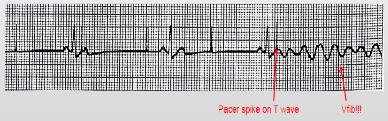
Although unlikely, it can cause Ventricular Fibrillation
193
Paroxysmal Supraventricular Tachycardia (PSVT)
Originates above Bundle of His
Triggered by a Premature Atrial Contraction (PAC)
HR: 100 - 300 bpm
P wave is hidden in preceding T wave; PR interval is shortened or normal; QRS is usually normal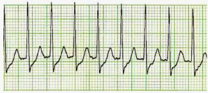
Triggered by a Premature Atrial Contraction (PAC)
HR: 100 - 300 bpm
P wave is hidden in preceding T wave; PR interval is shortened or normal; QRS is usually normal
194
PSVT Concerns
If HR > 180 bpm and sustained --> low cardiac output, hypotension
195
PSVT Risk Factors
Overexertion
Emotional stress
Stimulants (like caffeine)
Rheumatic Heart Disease
Digitalis toxicity
CAD
Emotional stress
Stimulants (like caffeine)
Rheumatic Heart Disease
Digitalis toxicity
CAD
196
PSVT Treatment
Vagal stimulation --> Valsalva, coughing
Cardioversion
Ablation --> radiofrequency catheter ablation
Implantable Cardioverter Defibrillator
Medications (adenosine, diltiazem, amiodarone)
Cardioversion
Ablation --> radiofrequency catheter ablation
Implantable Cardioverter Defibrillator
Medications (adenosine, diltiazem, amiodarone)
197
PSVT Pharmacological Management
IV adenosine --> Administer IV rapidly (1-2 seconds), causes brief asystole (place on heart monitor)
Antiarrhythmics --> Diltiazem, amiodarone
Antiarrhythmics --> Diltiazem, amiodarone
198
Premature Ventricular Contractions (PVCs)
More life-threatening than atrial dysrhythmias
Result from increased irritability of the ventricles
Result from increased irritability of the ventricles
199
PVCs Risk Factors
CHF, COPD, MI (esp. anterior MI)
Anemia
Hypokalemia --> #1 cause
- PVC's, prolonged PR interval, T wave inversion
- Patient complains of N/V
Hypomagnesemia
Anesthesia, Infection, Surgery
Stress, Nicotine, Caffeine, Alcohol
Anemia
Hypokalemia --> #1 cause
- PVC's, prolonged PR interval, T wave inversion
- Patient complains of N/V
Hypomagnesemia
Anesthesia, Infection, Surgery
Stress, Nicotine, Caffeine, Alcohol
200
PVC Assessment
Assess the patient --> are they symptomatic or asymptomatic
Check how many PVCs per minute --> if less than 6, continue to monitor
Are the PVCs the same shape?
If >10/min with symptoms --> treatment time
Check how many PVCs per minute --> if less than 6, continue to monitor
Are the PVCs the same shape?
If >10/min with symptoms --> treatment time
201
PVC treatment
Oxygen
Prevent progression to VTach --> assess and treat cause
Lidocaine IVP --> 1 mg/kg
Prevent progression to VTach --> assess and treat cause
Lidocaine IVP --> 1 mg/kg
202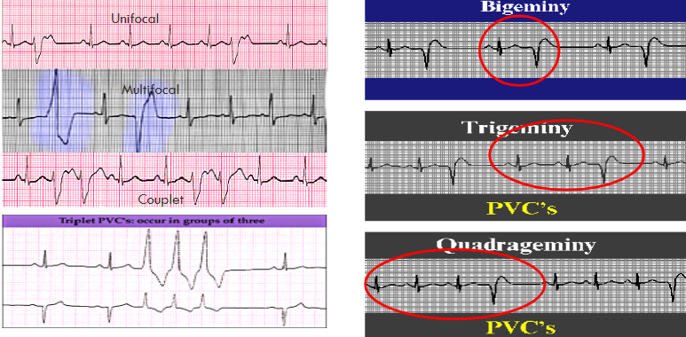
Types of PVCs
203
R on T Phenomenon
PVC occuring on a T wave
Can trigger a lethal arrhythmia
Can trigger a lethal arrhythmia
204
Ventricular Tachycardia
Repetitive firing at 150-250 bpm
Can be preceded by PVCs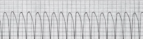
Can be preceded by PVCs
205
VTach Causes
MI, CAD, HF
Hypokalemia, Hypomagnesemia
Valve defects
Digitalis toxicity
Cardiomyopathy
Hypokalemia, Hypomagnesemia
Valve defects
Digitalis toxicity
Cardiomyopathy
206
Stable vs. Nonstable VTach
Determined by a presence of a pulse
207
Stable VTach Treatment
Lidocaine + Amiodarone --> be aware of seizures for lidocaine
Synchronized cardioversion --> avoid R on T phenomenon
Ablation --> for chronic cases
Synchronized cardioversion --> avoid R on T phenomenon
Ablation --> for chronic cases
208
Pulseless VTach Treatment
Defibrillation - Unsynchronized
CPR/ACLS
CPR/ACLS
209
Cardioversion
Elective procedure --> Obtain informed consent
Patient is awake and sedated
Synchronized with QRS
50-200 Joules
ECG Monitor
Patient is awake and sedated
Synchronized with QRS
50-200 Joules
ECG Monitor
210
Defibrillation
Emergency --> pulseless VTach or VFib
Patient has no cardiac output; patient is unconscious
Begin with 200 Joules; may go up to 360 Joules
ECG monitor
Patient has no cardiac output; patient is unconscious
Begin with 200 Joules; may go up to 360 Joules
ECG monitor
211
Amiodarone
Used for sustained and nonsustained VTach, VFib
Also used for atrial dysrhythmias
IV or PO
Many side effects --> hypotension, corneal microdeposits (visual disturbances; most common)
Most serious concern is Pulmonary Toxicity
Also used for atrial dysrhythmias
IV or PO
Many side effects --> hypotension, corneal microdeposits (visual disturbances; most common)
Most serious concern is Pulmonary Toxicity
212
Ventricular Fibrillation
Result of electrical chaos in ventricles --> no CO = no systemic perfusion
Rhythm is fatal if not successfuly ended in 3-5 minutes
Rhythm is fatal if not successfuly ended in 3-5 minutes
213
Ventricular Fibrillation Causes
Accidental elective shock (R on T phenomenon)
Hyper/Hypo Kalemia
Hypoxemia --> esp from MI, CAD, cardiomyopathy, etc.
Acidosis
Drug toxicity
Hyper/Hypo Kalemia
Hypoxemia --> esp from MI, CAD, cardiomyopathy, etc.
Acidosis
Drug toxicity
214
Ventricular Fibrillation Management
Defibrillation
ACLS measures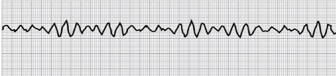
ACLS measures
215
ACLS Measures
1. 30:2 ratio compressions to ventilations
2. Shock once at effective setting (~360 Joules) --> resume CPR after shock
3. Start IV and put on O2
4. If still no pulse, IVP epinephrine 1mg q. 3-5 minutes
5. When ready, intubate or LMA (laryngeal mask airway)
6. Rhythm check q.2min; Epinephrine 1mg Q.3min
2. Shock once at effective setting (~360 Joules) --> resume CPR after shock
3. Start IV and put on O2
4. If still no pulse, IVP epinephrine 1mg q. 3-5 minutes
5. When ready, intubate or LMA (laryngeal mask airway)
6. Rhythm check q.2min; Epinephrine 1mg Q.3min
216
Ventricular Asystole
AKA ventricular standstill
Complete absence of any ventricular rhythm
No electrical activity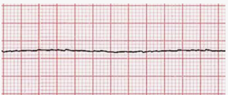
Complete absence of any ventricular rhythm
No electrical activity
217
Pulseless Electrical Activity (PEA)
Electrical activity on ECG monitor but no ventricular activity (no pulse)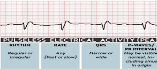
218
Causes of Asystole and PEA 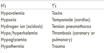
219
Thoracic Injuries
Account for 50% of trauma deaths
Blunt trauma (non-penetrating), penetrating (like stabs or gunshot wounds)
Emergency approach --> Remember the ABC's (airway, breathing, circulation)
Blunt trauma (non-penetrating), penetrating (like stabs or gunshot wounds)
Emergency approach --> Remember the ABC's (airway, breathing, circulation)
220
Pulmonary Contusion
Bruising of lung secondary to blunt trauma or rib fracture 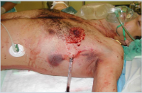
221
Pulmonary Contusion Pathophysiology
Intra-alveolar hemorrhage = Interstitial edema = ↓ pulmonary compliance, ↓ gas exchange, ↓ lung perfusion = ventilation perfusion imbalance
222
Pulmonary Contusion S/S
May be asymptomatic first 24-48 hours
Hypoxemia, SOB
Blood-tinged sputum
Crackles, wheezes
Atelectasis
Hypoxemia, SOB
Blood-tinged sputum
Crackles, wheezes
Atelectasis
223
Pulmonary Contusion: Diagnostics
Chest X-Ray --> pulmonary infiltrate, hazy opacity 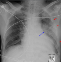
224
Pulmonary Contusion: Management
Goal: Aggressive Respiratory Care
- O2, deep breathing exercises, incentive spirometry
- Adequate pain control
- Semi Fowlers
- If side lying, good lung down
- Noninvasive Positive-Pressure Vent (NPPV)
- Mechanical ventilation with PEEP (positive end-expiratory pressure)
- O2, deep breathing exercises, incentive spirometry
- Adequate pain control
- Semi Fowlers
- If side lying, good lung down
- Noninvasive Positive-Pressure Vent (NPPV)
- Mechanical ventilation with PEEP (positive end-expiratory pressure)
225
Pulmonary Contusion: Complications
ARDs
Emphysema
Pneumonia
Lung abcess
Pulmonary Embolism
Emphysema
Pneumonia
Lung abcess
Pulmonary Embolism
226
Flail Chest
due to fracture of 2+ ribs
Characterized by paradoxical chest wall movement --> sucking inward on inspiration; puffing outward on expiration
Absent breath sounds = pneumothorax (air leak between lungs and chest wall)
Severly impairs ability to expand lungs and thus gas exchange
Characterized by paradoxical chest wall movement --> sucking inward on inspiration; puffing outward on expiration
Absent breath sounds = pneumothorax (air leak between lungs and chest wall)
Severly impairs ability to expand lungs and thus gas exchange
227
Flail Chest: Management
Goal: Vigilant respiratory care - assess for other injuries
Humidified O2
Pain control
Promote lung expansion --> chest tube
Effective oxygenation --> mechanical ventilation (PEEP), Monitor ABGs and vital capacity (how much air they can exhale)
Surgical stabilization of rib fracture
Humidified O2
Pain control
Promote lung expansion --> chest tube
Effective oxygenation --> mechanical ventilation (PEEP), Monitor ABGs and vital capacity (how much air they can exhale)
Surgical stabilization of rib fracture
228
Pneumothorax
air in the pleural space
Closed = no external wound
Open = penetrating chest wound
Closed = no external wound
Open = penetrating chest wound
229
Pneumothorax Etiology
Mechanical ventilation
Insertion of subclavian catheter
Perforation of esophagus
Rib Fracture
Blebs or bullae rupture (COPD)
Insertion of subclavian catheter
Perforation of esophagus
Rib Fracture
Blebs or bullae rupture (COPD)
230
Pneumothorax Manifestations
Dyspnea, chest pain (no shit sherlock)
Decreased movement that is involved with chest wall
Diminished/Absent breath sounds on affected side
Hyperresonance to percussion
Decreased movement that is involved with chest wall
Diminished/Absent breath sounds on affected side
Hyperresonance to percussion
231
Pneumothorax Management
Vented Dressing (only on 3 sides) --> open the pneumothorax to release air
Stabilize impaling object --> Do NOT remove
Insertion of chest tube
Heimlich (Flutter valve) --> attached to chest tube to release air
Stabilize impaling object --> Do NOT remove
Insertion of chest tube
Heimlich (Flutter valve) --> attached to chest tube to release air
232
Complication of Pneumothorax
Tension Pneumothorax
233
Tension Pneumothorax
a life-threatening condition that occurs when air accumulates in the pleural space, putting pressure on the lungs and heart
Open: thoracic cavity is exposed to outside air
Closed: no disruption in thoracic cage
Open: thoracic cavity is exposed to outside air
Closed: no disruption in thoracic cage
234
Tension Pneumothorax Pathophysiology
Air gets trapped --> causing lung collapse
↓
Interferes with venous return
↓
Decreased diastolic filling
↓
Compromised cardiac output --> lung is being pushed and the ventricles cannot contract properly
↓
Interferes with venous return
↓
Decreased diastolic filling
↓
Compromised cardiac output --> lung is being pushed and the ventricles cannot contract properly
235
Tension Pneumothorax Etiology
Idiopathic: chest tube is clamped or blocked
Mechanical ventilation, resuscitive efforts
Perforation/trauma
Spontaneous: COPD with ruptured bullae or bleb (literally just pimples of air on the lung)
Mechanical ventilation, resuscitive efforts
Perforation/trauma
Spontaneous: COPD with ruptured bullae or bleb (literally just pimples of air on the lung)
236
Tension Pneumothorax: Manifestations
Mediastinal shift --> asymmetry of thorax, tracheal deviation
Subcutaneous emphysema --> infiltration of air under the dermal layer
Tachypnea, Tachycardia, Hypotension --> hypotension due to decreased CO
Respiratory distress (no shit)
Absent breath sounds on affected side
Distended neck veins, cyanosis
Subcutaneous emphysema --> infiltration of air under the dermal layer
Tachypnea, Tachycardia, Hypotension --> hypotension due to decreased CO
Respiratory distress (no shit)
Absent breath sounds on affected side
Distended neck veins, cyanosis
237
Tension Pneumothorax: Management
Needle decompression
O2 therapy
Chest tube insertion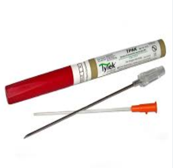
O2 therapy
Chest tube insertion
238
Hemothorax
Serious condition in which blood collects in the pleural space
>1500 mL of blood in chest cavity
Arterial bleeding requires surgical intervention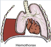
>1500 mL of blood in chest cavity
Arterial bleeding requires surgical intervention
239
Hemothorax Etiology
Blunt or penetrating chest trauma
Immediate post-op CABG
Immediate post-op CABG
240
Hemothorax Manifestations
Hypotension associated with absence of breath sounds
Dullness to percussion on affected side of the chest
Diminished/Absent breath sounds on affected side
Hypovolemic shock
Dullness to percussion on affected side of the chest
Diminished/Absent breath sounds on affected side
Hypovolemic shock
241
Hemothorax Treatment
Chest Tube
Auto-transfusion --> transfuse own blood
Thoracotomy --> indicated if >200 ml/hr for 2-4 hours, injuries to major cardiovascular structures
Auto-transfusion --> transfuse own blood
Thoracotomy --> indicated if >200 ml/hr for 2-4 hours, injuries to major cardiovascular structures
242
Hemothorax Complication
Cardiac Tamponade
243
Acute Cardiac Tamponade
Accumulation of fluid in pericardial space --> MEDICAL EMERGENCY
Reduced ventricular filling, hemodynamic compromise = shock, death
Reduced ventricular filling, hemodynamic compromise = shock, death
244
Cardiac Tamponade Manifestations
Beck's Triad --> Hypotension, JVD, Muffled heart sounds
Pulsus Paradoxus --> sys. BP increases on expiration rather than inspiration
Electrical alternans (QRS alternates going high, low, high)
Tachycardia --> can eventually lead to bradycardia
Dyspnea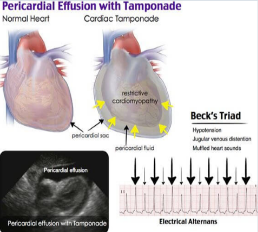
Pulsus Paradoxus --> sys. BP increases on expiration rather than inspiration
Electrical alternans (QRS alternates going high, low, high)
Tachycardia --> can eventually lead to bradycardia
Dyspnea
245
Cardiac Tamponade: Management
Hemodynamic monitoring
Fluid resuscitation
Pericardiocentesis
Pericardial drain --> if there isn't that much fluid to drain
Monitor for recurrence
Fluid resuscitation
Pericardiocentesis
Pericardial drain --> if there isn't that much fluid to drain
Monitor for recurrence
246
Chest Drains: Air Leak
Presence of bubbles in the water seal
Intermittent air leak = injury to lungs or airway
Continuous air leak = either large injury to lung or airways or a leak in the tubing
Intermittent air leak = injury to lungs or airway
Continuous air leak = either large injury to lung or airways or a leak in the tubing
247
Management of Chest Tube Drainage System
Ensure occlusive dressing is tight and intact
Assess for difficulty breathing --> pulse ox
Assess breath sounds for each lung
Check alignment of trachea
Check tube insertion site --> puffiness or crackling may indicate subcutaneous emphysema
Observe site for infection
Pain control
Deep breathing exercises and incentive spirometry
Assess for difficulty breathing --> pulse ox
Assess breath sounds for each lung
Check alignment of trachea
Check tube insertion site --> puffiness or crackling may indicate subcutaneous emphysema
Observe site for infection
Pain control
Deep breathing exercises and incentive spirometry
248
Thoracic Injuries: Emergency Management
Administer high flow O2 w/ nonrebreather mask
Establish 2 large bore (20 g or 18g) IV catheters --> fluid resuscitation as appropriate
Remove clothing to assess injury --> cover sucking chest wound w/ nonporous dressing taped on 3 sides
Stabilize, don't remove impaling object
Stabilize flail rib segment
After cervical spine injury is ruled out, place in Semi-Fowler's
Monitor vitals, LOC, O2 sat, cardiac rhythm, resp. status, urinary output
If tension pneumothorax develops, release dressing
Establish 2 large bore (20 g or 18g) IV catheters --> fluid resuscitation as appropriate
Remove clothing to assess injury --> cover sucking chest wound w/ nonporous dressing taped on 3 sides
Stabilize, don't remove impaling object
Stabilize flail rib segment
After cervical spine injury is ruled out, place in Semi-Fowler's
Monitor vitals, LOC, O2 sat, cardiac rhythm, resp. status, urinary output
If tension pneumothorax develops, release dressing
249
Pulmonary Empyema
Collection of pus, necrotic tissue in pleural space --> Infection
250
Pulmonary Empyema: Common Causes
Pneumonia
Lung abcess
Infected pleural effusion
Lung abcess
Infected pleural effusion
251
Pulmonary Empyema: Other Causes
Penetrating chest trauma
Iatrogenic factors --> chest surgery, thoracentesis
Iatrogenic factors --> chest surgery, thoracentesis
252
Pulmonary Empyema: Manifestations
Fever > 100.4F
Chest pain, dyspnea
Weak cough, inability to clear secretions
Presence of crackles
Weight loss
Night sweats
Chest pain, dyspnea
Weak cough, inability to clear secretions
Presence of crackles
Weight loss
Night sweats
253
Pulmonary Empyema: Diagnostics
Chest XRay
CT scan
CT scan
254
Pulmonary Empyema: Management
Treatment: Same for anyone with infection
- antibiotics (if indicated)
Chest tube, thoracentesis --> empty empyema cavity
Decortication (if not resolved with above) --> remove fibrous/infected tissue on lung surgically
- antibiotics (if indicated)
Chest tube, thoracentesis --> empty empyema cavity
Decortication (if not resolved with above) --> remove fibrous/infected tissue on lung surgically
255
Pulmonary Empyema: Goal of treatment
Lung re-expansion
Resolution of effective breathing
Improvement of pulmonary function
Resolution of effective breathing
Improvement of pulmonary function
256
Acute Respiratory Distress Syndrome (ARDs)
a serious and potentially fatal lung condition that occurs when the lungs are severely injured, making it difficult for them to supply the body with enough oxygen
257
ARDs Pathophysiology
Damage to the alveoli reduces blood flow to the lungs, allowing platelets to aggregate. The platelets release substances that inflame and damage the alveolar membrane and increase capillary permeability
↓
Increased permeability of alveolocapillary membranes allow fluids to accumulate in the lung interstitium, alveoli, and small airways, causing them to stiffen
↓
Ventilation is impaired, reducing oxygenation of pulmonary capillary blood
↓
Pulmonary edema worsens. Inflammation leads to fibrosis, which further impedes gas exchange
↓
Increased permeability of alveolocapillary membranes allow fluids to accumulate in the lung interstitium, alveoli, and small airways, causing them to stiffen
↓
Ventilation is impaired, reducing oxygenation of pulmonary capillary blood
↓
Pulmonary edema worsens. Inflammation leads to fibrosis, which further impedes gas exchange
258
ARDs: Causes
Pulmonary aspiration
Shock; trauma; serious infection
Embolisms
Sepsis
Drugs OD (heroin, opioids, ASA)
Hemolytic disorders (e.g. bood transfusions)
Cardiopulmonary bypass
Drowning
Shock; trauma; serious infection
Embolisms
Sepsis
Drugs OD (heroin, opioids, ASA)
Hemolytic disorders (e.g. bood transfusions)
Cardiopulmonary bypass
Drowning
259
ARDs Features
Persistent hypoxemia despite 100% O2 --> refractory hypoxemia, a CARDINAL feature
Decreased pulmonary compliance
Dyspnea, tachypnea
Noncardiac bilateral pulmonary edema
Life-threatening
Decreased pulmonary compliance
Dyspnea, tachypnea
Noncardiac bilateral pulmonary edema
Life-threatening
260
Early Signs of ARDs
Diaphoresis
Refractory hypoxemia --> low O2 sat despite high oxygen
Restlessness, anxiety, confusion
Dyspnea, Tachypnea
Tachycardia
Refractory hypoxemia --> low O2 sat despite high oxygen
Restlessness, anxiety, confusion
Dyspnea, Tachypnea
Tachycardia
261
Late Signs of ARDs
Cyanosis
Somnolence -->drowsiness
Blood tinged frothy sputum
Pulmonary edema
Somnolence -->drowsiness
Blood tinged frothy sputum
Pulmonary edema
262
ARDs: Diagnostic Assessment
PaO2 < 60 mmHg on ABG; refractory hypoxemia
Sputum culture (bronchoscopy)
Chest Xray --> diffuse haziness/white-out/Ground glass (see image)
no cardiac involvement on ECG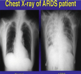
Sputum culture (bronchoscopy)
Chest Xray --> diffuse haziness/white-out/Ground glass (see image)
no cardiac involvement on ECG
263
ARDs Management
*Early recognition of high-risk patients*--> patients who aspirated
Mechanical ventilation with PEEP or CPAP
Sedation
Postioning --> Prone, kinetic beds
ECMO for severe ARDs
Nutrition --> can't heal without food (think Maslow's)
Prepare patient and family --> 35-60% mortality rate
Mechanical ventilation with PEEP or CPAP
Sedation
Postioning --> Prone, kinetic beds
ECMO for severe ARDs
Nutrition --> can't heal without food (think Maslow's)
Prepare patient and family --> 35-60% mortality rate
264
ARDs Pharmacological Therapy
Antibiotics
Vasopressors --> support vital signs (like BP)
Vitamin C and E
N-acetylcysteine --> break up mucus
Nitric Acid --> relaxes smooth muscle in pulmonary vessels
Surfactants --> prevent alveolar collapse
Vasopressors --> support vital signs (like BP)
Vitamin C and E
N-acetylcysteine --> break up mucus
Nitric Acid --> relaxes smooth muscle in pulmonary vessels
Surfactants --> prevent alveolar collapse
265
Extracorporeal Membrane Oxygenation 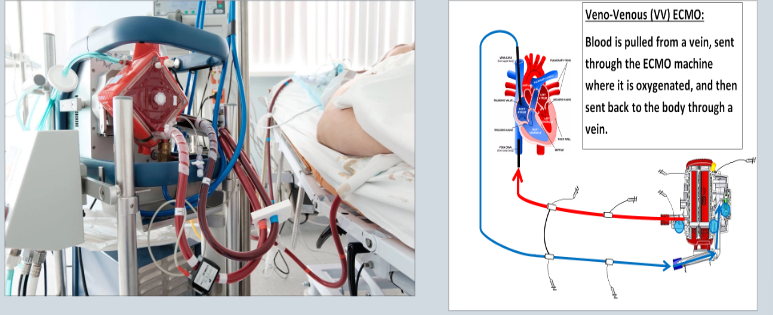
266
Hemodynamic Monitoring 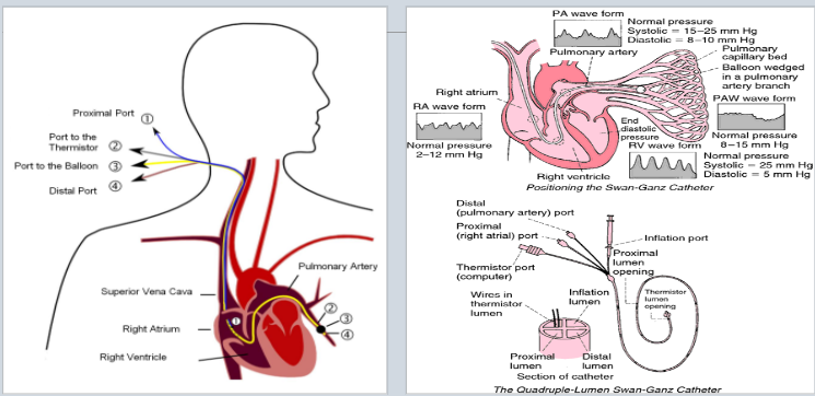
267
Acute Respiratory Failure (ARF)
Inability to maintain adequate oxygenation for tissues OR adequate removal of CO2 from tissues
Classified as extrapulmonary or intrapulmonary
Can be Ventilation or Oxygenation failure or both
Patient is hypoxemic/hypercapnic due to impaired gas exchange
Classified as extrapulmonary or intrapulmonary
Can be Ventilation or Oxygenation failure or both
Patient is hypoxemic/hypercapnic due to impaired gas exchange
268
V/Q Mismatch
Most common cause of hypoxemia
Occurs when either the ventilation (airflow) or perfusion (blood flow) in the lungs is impaired, preventing the lungs from optimally delivering oxygen to the blood.
Low V/Q ratio may occur from: decrease of ventilation; overperfusion in presence of normal ventilation
Occurs when either the ventilation (airflow) or perfusion (blood flow) in the lungs is impaired, preventing the lungs from optimally delivering oxygen to the blood.
Low V/Q ratio may occur from: decrease of ventilation; overperfusion in presence of normal ventilation
269
Acute Respiratory Failure: Etiology
Defect in respiratory control center in brain
Poor respiratory muscle function (esp. diaphragm)
Extrapulmonary causes: CNS depression, CV disorders, Endocrine disorders, Metabolic disorders, airway irritants, thoracic abnormalities
Disorders of the respiratory tract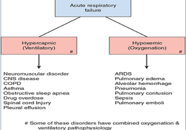
Poor respiratory muscle function (esp. diaphragm)
Extrapulmonary causes: CNS depression, CV disorders, Endocrine disorders, Metabolic disorders, airway irritants, thoracic abnormalities
Disorders of the respiratory tract
270
Acute Respiratory Failure: Manifestations
Hypoxemia, Hypercapnia
Acidosis, dyspnea
O2 sat may be normal but elevated end-tidal CO2 (amount of CO2 on expiration)
Acidosis, dyspnea
O2 sat may be normal but elevated end-tidal CO2 (amount of CO2 on expiration)
271
Hypoxic Respiratory Failure S/S
Restlessness
Irritability
Agitation
Confusion
Tachycardia
Irritability
Agitation
Confusion
Tachycardia
272
Hypercapnic Respiratory Failure S/S
CO2 Narcosis:
Decreased LOC, drowsiness
Lethargy
Seizure
Headache
Decreased LOC, drowsiness
Lethargy
Seizure
Headache
273
Intubation
Performed emergently on patient in cardiopulmonary arrest
Performed electively with impending respiratory failure --> establishes airway, assist in removal of secretions, protects airway from aspiration
Performed electively with impending respiratory failure --> establishes airway, assist in removal of secretions, protects airway from aspiration
274
Verification of Endotracheal Tube Placement
Assess breath sounds bilaterally
Symmetrical chest expansion
End Tidal CO2 Detector --> most accurate
Portable Chest X-ray --> most accurate
Symmetrical chest expansion
End Tidal CO2 Detector --> most accurate
Portable Chest X-ray --> most accurate
275
Endotracheal Tube Suction
Preoxygenate with 100% O2 for one minute before
Based on standard procedure to prevent:
- hypoxemia
- airway trauma
- infection
- increased intracranial pressure
In line suction vs. single use suction catheter
Based on standard procedure to prevent:
- hypoxemia
- airway trauma
- infection
- increased intracranial pressure
In line suction vs. single use suction catheter
276
Ventilator Controls and Settings: Tidal Volume
Tidal Volume (Vt) --> amount of air delivered with each preset breath
277
Ventilator Controls and Settings: Rate
breaths/min
278
Ventilator Controls and Settings: Fraction of Inspired Oxygen (FiO2)
amount of O2 delivered with each breath
279
Ventilator Controls and Settings - I:E ratio
duration of inspiration in comparison to expiration
280
Ventilator Controls and Settings: PEEP (positive expiratory end pressure)
amount of positive pressure added with each expiration
281
Ventilator Alarms: High Pressure Alarm
May be due to:
- Increased secretions
- Mucous plug
- Pt coughing, gagging, biting tube
- Pt fighting ventilator
- Condensation in tube
- Increased secretions
- Mucous plug
- Pt coughing, gagging, biting tube
- Pt fighting ventilator
- Condensation in tube
282
Ventilator Alarms: Low Pressure Alarm
May be due to:
- Leak in tubing (produces a sound) --> check cuff inflation
- Pt stops spontaneous breathing in CPAP/SIMV mode
- Cuff leak in endotracheal tube
- Leak in tubing (produces a sound) --> check cuff inflation
- Pt stops spontaneous breathing in CPAP/SIMV mode
- Cuff leak in endotracheal tube
283
Intubation - Care
Assess tube placement
Breath sounds; chest wall movement
Prevent accidental extubation --> soft wrist restraints, sedation
Check pilot balloon
Nutrition status --> so they can heal up
VAP protocol --> prevent infection
Monitor Urine Output --> ventilators can lead to decreased kidney perfusion
Breath sounds; chest wall movement
Prevent accidental extubation --> soft wrist restraints, sedation
Check pilot balloon
Nutrition status --> so they can heal up
VAP protocol --> prevent infection
Monitor Urine Output --> ventilators can lead to decreased kidney perfusion
284
Mechanical Ventilation: Nursing Management
Assess patient FIRST, ventilator SECOND
Monitor patient response
Manage ventilator system
Prevent complications of prolonged intubation
Monitor patient response
Manage ventilator system
Prevent complications of prolonged intubation
285
Complications of Prolonged intubation
Vocal cord paralysis --> hoarseness
Aspiration --> ventilator associated pneumonia (VAP)
Aspiration --> ventilator associated pneumonia (VAP)
286
VAP protocol
Maintain HOB at 30-45 degrees
Interrupt sedation to assess readiness to wean from ventilator --> "sedation vacation"
Provide prophylaxis for DVT
Administer medications for peptic ulcer disease prophylaxis
Daily oral care with chlorhexidine --> prevent infection
Interrupt sedation to assess readiness to wean from ventilator --> "sedation vacation"
Provide prophylaxis for DVT
Administer medications for peptic ulcer disease prophylaxis
Daily oral care with chlorhexidine --> prevent infection
287
Tidal Volume (TV)
amount of air moving in/out of lungs during a breath
288
Modes of Ventilation: Assist-Control Ventilation (AC)
Preset Tidal volume, respiratory rate
Pt triggers breath between vent-initiated breaths
Ensures pt receives adequate ventilation regardless of spontaneous efforts
Pt triggers breath between vent-initiated breaths
Ensures pt receives adequate ventilation regardless of spontaneous efforts
289
Modes of Ventilation: Synchronized Intermittent Mandatory Vent (SIMV)
Similar to AC --> preset tidal volume, respiratory rate
If pt initiates a breath --> delivery of the mandatory breath is synchronized with spontaneousu effort
If pt initiates a breath --> delivery of the mandatory breath is synchronized with spontaneousu effort
290
Modes of Ventilation: Continuous Positive Airway Pressure (CPAP)
Keep awake --> must have adequate respiratory drive and tidal volume
Vent has NO mandatory breaths --> Patient performs all work of breathing
Vent has NO mandatory breaths --> Patient performs all work of breathing
291
Modes of Ventilation: Bi-Level Positive Airway Pressure (BiPAP)
Noninvasive pressure support ventilation
292
Mechanical Ventilation - Cardiac Complications
Hypotension --> monitor I&O, weight, hydration, S/S of hypovolemia
293
Mechanical Ventilation - Respiratory Complications
COPD --> bullae or blebs
Barotrauma --> pneumothorax
Ventilator associated pneumonia (VAP) --> VAP protocol
Ventilator Dependence - wean asap
Barotrauma --> pneumothorax
Ventilator associated pneumonia (VAP) --> VAP protocol
Ventilator Dependence - wean asap
294
Mechanical Ventilation - GI Complications
Nutrition --> Enteral feedings/TPN within 48 hours
Stress Ulcer --> antacids, PPIs (-prazoles)
Stress Ulcer --> antacids, PPIs (-prazoles)
295
Mechanical Ventilation - Musculoskeletal Complications
Muscle Decondition/Atrophy --> passive exercise
296
Mechanical Ventilation - Other Considerations
Fluid/electrolyte imbalance
Pressure sores
UTIs
Pressure sores
UTIs
297
Mechanical Ventilation Weaning
Process of going from ventilator dependence to spontaneous breathing
298
Tracheostomy Suction
Notice the different suction pressures for age groups?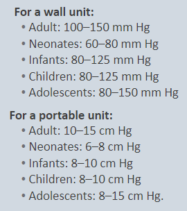
299
New Tracheostomy: Dislodgement
Tube dislodgement in the first 72 hours after surgery is an EMERGENCY
- Tracheostomy tract --> not matured, replacement is difficult
- Trach tube may end up in subcutaneous tissue --> "false passage" of oxygen
Place trach tube of same type/size (or one size smaller) at bedside
Place trach tray at bedside
O2 and suction units at bedside (if wall unit isn't available)
- Tracheostomy tract --> not matured, replacement is difficult
- Trach tube may end up in subcutaneous tissue --> "false passage" of oxygen
Place trach tube of same type/size (or one size smaller) at bedside
Place trach tray at bedside
O2 and suction units at bedside (if wall unit isn't available)
300
New Tracheostomy: Subcutaneous Emphysema
Air escapes into tissues of neck, chest, and face
Crepitus - puffy with crackling when pressing area involved (like bubble wrap)
Notify physician immediately --> EMERGENCY
Crepitus - puffy with crackling when pressing area involved (like bubble wrap)
Notify physician immediately --> EMERGENCY
23 more cards in OutLearn. Open the full deck