PRO/REG N2 Lecture ORIGINAL
Study these flashcardsFree · spaced repetition · iPhone, Android, Mac & web
All cards
1
Autoimmune
When your body doesn't recognize its own cells and attacks them
2
5 Signs of inflammation
Redness
Warmth
Swelling
Pain
Loss of function
Think: "Red hot swelling + pain and loss of function"
Warmth
Swelling
Pain
Loss of function
Think: "Red hot swelling + pain and loss of function"
3
Juvenile Idiopathic Arthritis (Juvenile Rheumatoid Arthritis)
Idiopathic = unknown cause
Chronic childhood arthritis
Inflammation of joint synovium and surrounding tissues (leads to joint erosion, destruction, and fibrosis of articular cartilage)
Adhesions and ankyloses (stiffness) of joints may occur
Chronic childhood arthritis
Inflammation of joint synovium and surrounding tissues (leads to joint erosion, destruction, and fibrosis of articular cartilage)
Adhesions and ankyloses (stiffness) of joints may occur
4
Who is affected by JIA?
Starts before age 16 with peak onset between ages 1-3 years
Twice as many girls as boys are affected
Twice as many girls as boys are affected
5
systemic juvenile idiopathic arthritis
a type of JIA where children have periods of arthritis along with fever and rash
6
Oligoarthritis
Most common form of JIA
Affects four or fewer joints, typically the large ones (knees, ankles, elbows)
Affects four or fewer joints, typically the large ones (knees, ankles, elbows)
7
Polyarthritis
A form of JIA that affects five or more joints, often on both sides of the body (both wrists, both knees, etc.)
8
JIA Clinical Manifestations
Joint swelling + loss of motion
May not report any pain
Growth disturbances (overgrowth or undergrowth)
Exacerbations and remissions (can be unpredictable)
Less joints affected = greater chance of remission
inflammation can occur in other tissues
May not report any pain
Growth disturbances (overgrowth or undergrowth)
Exacerbations and remissions (can be unpredictable)
Less joints affected = greater chance of remission
inflammation can occur in other tissues
9
JIA diagnosis
DIAGNOSIS BY EXCLUSION
Classification by:
1. Age of onset before age 16
2. Arthritis in 1+ joints for 6 weeks or longer
3. Exclusion of other causes (such as inflammation, fractures, tumors, infection, or congenital defects)
Classification by:
1. Age of onset before age 16
2. Arthritis in 1+ joints for 6 weeks or longer
3. Exclusion of other causes (such as inflammation, fractures, tumors, infection, or congenital defects)
10
Labs used in JIA diagnosis
ANA (antinuclear antibodies)
ESR (erythrocyte sedimentation rate)
CRP (C-reactive protein)
CBC (check for leukocytosis, or elevated WBCs)
ESR (erythrocyte sedimentation rate)
CRP (C-reactive protein)
CBC (check for leukocytosis, or elevated WBCs)
11
Imaging used in JIA diagnosis
X-Rays can indicate joint deterioration, joint space narrowing, bone erosion, osteoporosis, ankylosis, etc.
MRI and CT scans can look at tissue for swelling
MRI and CT scans can look at tissue for swelling
12
Ankylosis
a stiffness of a joint due to abnormal adhesion and rigidity of the bones of the joint, which may be the result of injury or disease.
13
ANA (antinuclear antibodies) lab test
measures titer of group of antibodies that destroy cell nuclei & cause cell death in those with autoimmune disease
used to diagnose autoimmune disorders
used to diagnose autoimmune disorders
14
ESR (erythrocyte sedimentation rate) lab test
Measures how quickly RBCs settle to the bottom of a test tube
Non-specific measure of inflammation
Non-specific measure of inflammation
15
CRP (C-reactive protein) lab test
tests for a protein produced by the liver in response to inflammation
More sensitive to inflammatory changes
More sensitive to inflammatory changes
16
JIA therapeutic management
NO CURE
Major goals of therapy are:
1. Control pain
2. Preserve joint ROM and function
3. Minimize effects of inflammation
4. Promote normal growth and development
Major goals of therapy are:
1. Control pain
2. Preserve joint ROM and function
3. Minimize effects of inflammation
4. Promote normal growth and development
17
JIA Treatment Plan
Outpatient care is mainstay of treatment
Treatment plan includes:
1. Medications
2. Physical/Occupational therapy
3. Eye exams
4. Splints (to avoid muscle contracture)
5. Dietary management
6. Comfort measures, psychosocial support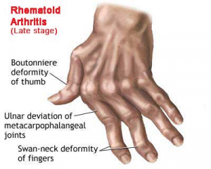
Treatment plan includes:
1. Medications
2. Physical/Occupational therapy
3. Eye exams
4. Splints (to avoid muscle contracture)
5. Dietary management
6. Comfort measures, psychosocial support
18
Dietary Management of JIA
Calcium and Vitamin D rich foods (corticosteroids and methotrexate may inhibit calcium absorption)
High fiber foods (can help with inflammation)
Fruits and vegetables (for vitamins and nutrients)
Omega-3s (heart healthy and anti-inflammatory)
Protein (for muscles)
High fiber foods (can help with inflammation)
Fruits and vegetables (for vitamins and nutrients)
Omega-3s (heart healthy and anti-inflammatory)
Protein (for muscles)
19
JIA drug therapy
NSAIDs such as ibuprofen and naproxen (suppress inflammation and pain)
DMARDs (Disease-modifying antirheumatic drugs) such as methotrexate
Glucorticoids such as prednisone and methylprednisolone (acutely active exacerbations or systemic features)
DMARDs (Disease-modifying antirheumatic drugs) such as methotrexate
Glucorticoids such as prednisone and methylprednisolone (acutely active exacerbations or systemic features)
20
NSAIDs patient teaching
Potential side effects such as GI irritation (take with food), hepatic and renal (drug is metabolized in the liver with excretion by the kidneys), and bleeding
We don't use opioids as the pain is inflammation-based (also don't want to create more problems)
We don't use opioids as the pain is inflammation-based (also don't want to create more problems)
21
DMARDs (disease modifying anti-rheumatic drugs) patient teaching
Monitor CBC (can decrease blood counts) and LIVER function (excreted through liver)
GI disturbances (take with food)
AVOID live vaccines and alcohol
Teratogenic effects (can cause birth defects, so use birth control)
Need negative TST (tuberculin skin test) before starting therapy (results can be compromised by DMARDs)
GI disturbances (take with food)
AVOID live vaccines and alcohol
Teratogenic effects (can cause birth defects, so use birth control)
Need negative TST (tuberculin skin test) before starting therapy (results can be compromised by DMARDs)
22
Glucocorticoids patient teaching
Monitor blood sugar if diabetic (can cause hyperglycemia)
Side effects (increased risk of infection, weight gain, Cushingoid features, osteoporosis)
Low salt and calorie diet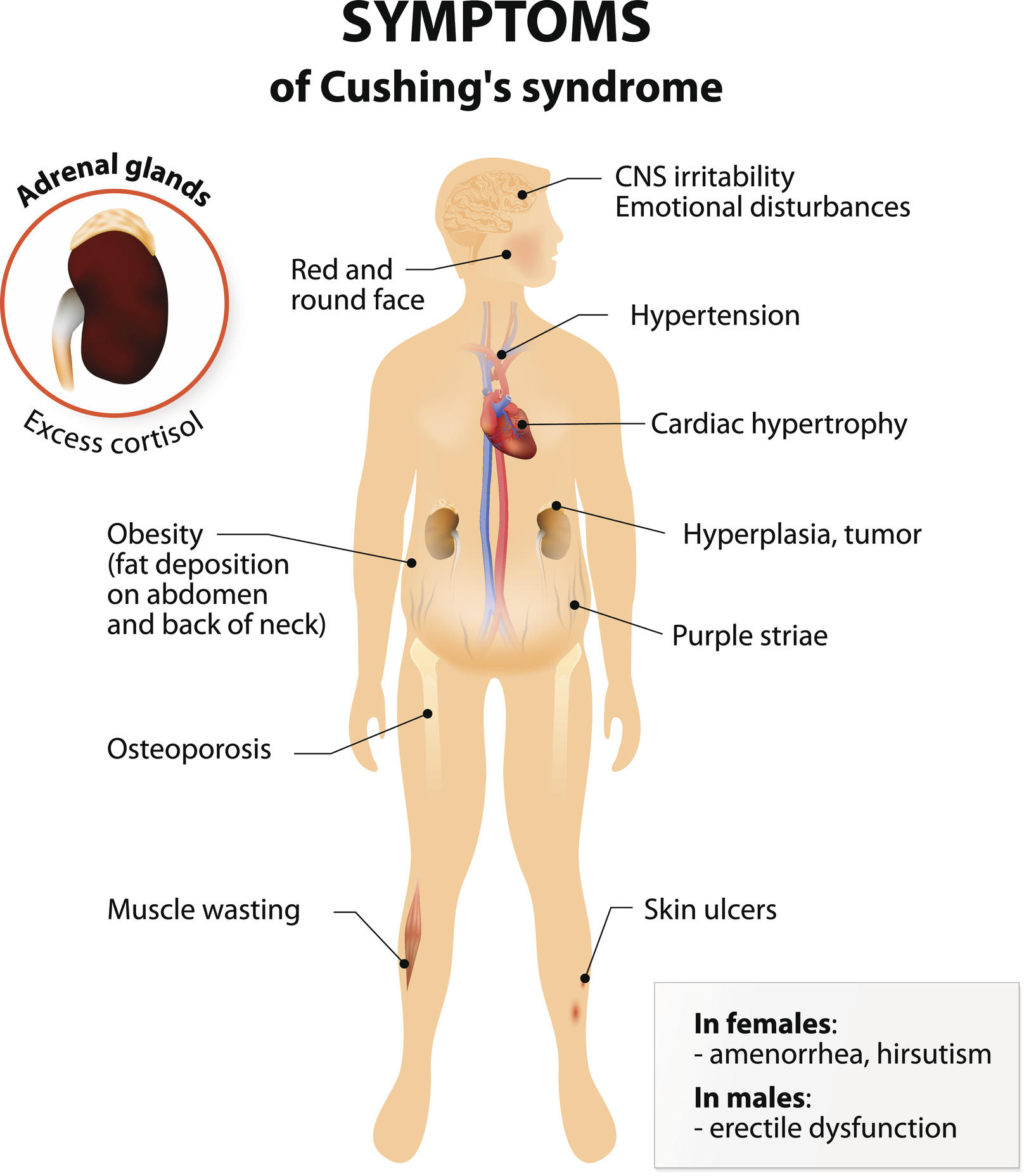
Side effects (increased risk of infection, weight gain, Cushingoid features, osteoporosis)
Low salt and calorie diet
23
JIA Physical/Occupational therapy
Goal is to preserve function and prevention of deformity
Physical therapy = focus on ROM exercises, muscle strength. etc.
Occupational therapy = focus on ADLs
Non weightbearing exercises (water therapy)
Physical therapy = focus on ROM exercises, muscle strength. etc.
Occupational therapy = focus on ADLs
Non weightbearing exercises (water therapy)
24
JIA Nursing care
Assessment (physical, social, psychological)
Pain relief
Adherence to treatment
Promote general health
Involve the family in plan of care
Pain relief
Adherence to treatment
Promote general health
Involve the family in plan of care
25
Rheumatoid Arthritis (RA) pathophysiology
Chronic, progressive, systemic inflammatory autoimmune disease
Transformed autoantibodies (Rheumatoid factors [RFs]) attack healthy tissues, particularly synovium, causing inflammation
Articular cartilage, joint capsule, ligaments and tendons are affected and then eventually the bone is invaded and destroyed
Affects primarily synovial joints
Transformed autoantibodies (Rheumatoid factors [RFs]) attack healthy tissues, particularly synovium, causing inflammation
Articular cartilage, joint capsule, ligaments and tendons are affected and then eventually the bone is invaded and destroyed
Affects primarily synovial joints
26
RA characteristics
Natural remissions and exacerbations (disease is controlled by decreasing the intensity and # of exacerbations)
Systemic disorder (can affect other organs or body systems with connective tissue such as heart, eyes, lungs, etc.)
Women are more likely than men to get it
Systemic disorder (can affect other organs or body systems with connective tissue such as heart, eyes, lungs, etc.)
Women are more likely than men to get it
27
RA early manifestations
Joint inflammation
Systemic effects: low grade fever, fatigue, weakness, anorexia, paresthesias
Systemic effects: low grade fever, fatigue, weakness, anorexia, paresthesias
28
RA late manifestations
Joints: deformities, moderate to severe pain, morning stiffness
Systemic: osteoporosis, severe fatigue, anemia, weight loss, *subcutaneous nodules*, peripheral neuropathy, vasculitis, pericarditis, kidney disease
Systemic: osteoporosis, severe fatigue, anemia, weight loss, *subcutaneous nodules*, peripheral neuropathy, vasculitis, pericarditis, kidney disease
29
RA assessment findings (early)
Hands typically involved
slightly reddened, warm, stiff, swollen, painful (esp. on palpation)
Bilateral and symmetric joint involvement
# of joints involved increases as disease progresses
slightly reddened, warm, stiff, swollen, painful (esp. on palpation)
Bilateral and symmetric joint involvement
# of joints involved increases as disease progresses
30
RA assessment findings (late)
Joint deformities (swan neck, ulnar deviation, Boutonniere deformity)
TMJ (temporomandibular joint) involvement: pain on chewing or opening mouth
Moderate to severe pain
Decreased ROM
Muscle atrophy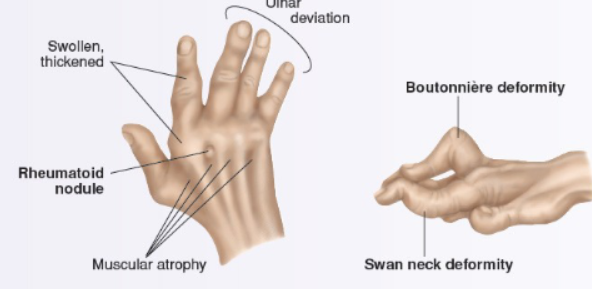
TMJ (temporomandibular joint) involvement: pain on chewing or opening mouth
Moderate to severe pain
Decreased ROM
Muscle atrophy
31
What to do during RA assessment for spine?
Palpate spine for tenderness, pain, loss of motion
Subluxation (cracking) of 1st and 2nd vertebrae may be life threatening
- keep neck straight and neutral, then notify PCP stat
Subluxation (cracking) of 1st and 2nd vertebrae may be life threatening
- keep neck straight and neutral, then notify PCP stat
32
Rheumatoid nodules are associated with _____
severe destructive disease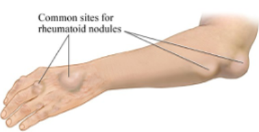
33
Why do nurses assess for peripheral neuropathy with RA?
RA affects connective tissue, not just in the joints but also in the heart, lungs, and the blood vessels
B/c of that, it is important to assess if pt is receiving enough circulation
B/c of that, it is important to assess if pt is receiving enough circulation
34
RA diagnostics
Rheumatoid factor (RF) and anti-cyclic citrullinated peptide (anti-CCP) can help differentiate similar disease processes from RA
antinuclear antibody (ANA)
ESR and CRP (for inflammation)
CBC
Arthrocentesis
antinuclear antibody (ANA)
ESR and CRP (for inflammation)
CBC
Arthrocentesis
35
Why is CBC lab important for RA diagnosis?
there is anemia and thrombocytosis (platelets) in late disease RA
36
Normal WBC count
5000-10000 WBCs per microliter
37
Normal platelet count
150,000 to 450,000 platelets per microliter of blood
38
Normal Hemoglobin and Hematocrit (H&H)
Men: 14-18 g/dL
Women: 12-16 g/dL
Women: 12-16 g/dL
39
Normal RBC count
Men: 4.5 - 6.0 million
Women: 4.0 to 5.0 million
Women: 4.0 to 5.0 million
40
RA imaging
X-ray: assess for joint damage
MRI/CT scan: determine presence and degree of cervical spine involvement
MRI/CT scan: determine presence and degree of cervical spine involvement
41
Anti-cyclic citrullinated peptide (anti-CCP) lab testWhy
specific and sensitive for early RA detection
can be used as a marker for aggressive, erosive late-stage disease
can be used as a marker for aggressive, erosive late-stage disease
42
Arthrocentesis
Aspirate synovial fluid to relieve pressure & analyze fluid cells for WBC, cloudiness, volume, rheumatoid factor.
43
Arthrocentesis pt teaching
Teach pt to ice and rest joint for 24 hrs after.
If increased pain or swelling – see PCP immediately
If increased pain or swelling – see PCP immediately
44
RA drug therapy
NSAIDs
DMARDs
BRMs (Biological Response Modifiers)
Glucocorticosteroids
DMARDs
BRMs (Biological Response Modifiers)
Glucocorticosteroids
45
DMARD drugs used to treat RA
methotrexate (Otrexup)
Leflunomide (Arava)
Hydroxychloroquine (Plaquenil)
Leflunomide (Arava)
Hydroxychloroquine (Plaquenil)
46
Methotrexate (Otrexup)
immunosuppresive DMARD
low dose (25mg PO weekly)
takes 4-6 weeks to begin to decrease inflammation
Monitor for bone marrow suppression (WBC, platelets)
teratogenic (birth defects)
low dose (25mg PO weekly)
takes 4-6 weeks to begin to decrease inflammation
Monitor for bone marrow suppression (WBC, platelets)
teratogenic (birth defects)
47
Leflunomide (Arava)
Immunosuppressive DMARD
Reduces swelling, stiffness, improves mobility.
Takes 4-6 weeks; may be up to 3 months to see benefits.
Monitor for bone marrow suppression – WBC & platelets
Teratogenic (Birth defects)
Reduces swelling, stiffness, improves mobility.
Takes 4-6 weeks; may be up to 3 months to see benefits.
Monitor for bone marrow suppression – WBC & platelets
Teratogenic (Birth defects)
48
Hydroxychloroquine (Plaquenil)
antimalarial drug that helps decrease joint and muscle pain
SE: mild stomach discomfort, light-headedness, headache.
Report blurred vision as it can cause retinal damage.
Eye exam before starting drug and every 6 months
SE: mild stomach discomfort, light-headedness, headache.
Report blurred vision as it can cause retinal damage.
Eye exam before starting drug and every 6 months
49
Biological Response Modifiers (BRMs) used for RA
decreases immune response and inflammation
Enbrel (etanercept)
Remicade (infliximab)
Humira (adalimumab)
Enbrel (etanercept)
Remicade (infliximab)
Humira (adalimumab)
50
Enbrel (etanercept) patient teaching
twice weekly SQ
teach patient to self administer, assess for injection site reaction
teach patient to self administer, assess for injection site reaction
51
Remicade (infliximab) patient teaching
IV infusion then again week 2 & week 6. If there is a response, give maintenance infusions every 8 weeks.
Teach pt to report chest pain or difficulty breathing during infusion.
Monitor blood pressure & infusion site.
Teach pt to report chest pain or difficulty breathing during infusion.
Monitor blood pressure & infusion site.
52
Humira (adalimumab) patient teaching
SQ every two weeks
Monitor for injection site reaction
Monitor for injection site reaction
53
Glucocorticoid patient teaching for RA
given for fast-acting anti-inflammatory & immunosuppressive effects
Prednisone: high dose = short duration, can also be given as a low chronic dose
Chronic use can cause infection, hypertension, weight gain, osteoporosis, glaucoma, and hyperglycemia (in pts with DM)
Prednisone: high dose = short duration, can also be given as a low chronic dose
Chronic use can cause infection, hypertension, weight gain, osteoporosis, glaucoma, and hyperglycemia (in pts with DM)
54
RA surgical interventions
Synovectomy (remove inflamed synovium)
Total joint arthroplasty
Total joint arthroplasty
55
RA nonpharmacological interventions
ice for acute inflammation (small bag of corn or frozen peas can be used as ice pack)
heat for stiffness, comfort (hot shower to relieve morning stiffness)
Heated paraffin (wax) dips
Finger & hand exercises
heat for stiffness, comfort (hot shower to relieve morning stiffness)
Heated paraffin (wax) dips
Finger & hand exercises
56
plasmapheresis
plasma is treated to remove antibodies causing the autoimmune reaction causing the disease
used in severe, life-threatening cases
used in severe, life-threatening cases
57
RA nursing interventions
Collaboration with PT/OT, dietician
Managing fatigue (pacing activities, rest periods, treat anemia
Enhancing body image - communicate acceptance
Encourage to seek medical help, especially before trying home remedies
Refer to support groups and self-help courses, counselors, religious/spiritual leaders. (The Arthritis Foundation)
Managing fatigue (pacing activities, rest periods, treat anemia
Enhancing body image - communicate acceptance
Encourage to seek medical help, especially before trying home remedies
Refer to support groups and self-help courses, counselors, religious/spiritual leaders. (The Arthritis Foundation)
58
Sjogren's Syndrome
It is a triad of symptoms caused by the obstruction of secretory ducts and glands by inflammatory cells and immune complexes.
Seen in patients with advanced RA and/or other autoimmune diseases
Seen in patients with advanced RA and/or other autoimmune diseases
59
Sjogren's Syndrome symptoms
Dry eyes: assess for redness & lack of tearing. May feel “gritty”, blurred vision, burning/itching of eyes. Inflammation and ulceration of the cornea
Dry mouth (xerostomia): assess for dry, sticky membranes, difficulty swallowing food, changes in taste, epistaxis, URI
Tooth decay, risk of oral and nasal infxn, digestion of carbs
Vaginal dryness: risk for infection, painful sexual intercourse
Dry mouth (xerostomia): assess for dry, sticky membranes, difficulty swallowing food, changes in taste, epistaxis, URI
Tooth decay, risk of oral and nasal infxn, digestion of carbs
Vaginal dryness: risk for infection, painful sexual intercourse
60
Sjogren's Syndrome treatment
Treatment is to manage symptoms
Artificial tears/saliva, water-soluble lubricants, humidifiers, surgery
Medications: methotrexate, cyclophosphamide, steroids, cyclosporine, hydroxychloroquine
Pilocarpine (Salagen) is used to treat dry mouth
Cyclosporine (Restasis) eye drops are used for dry eye
Artificial tears/saliva, water-soluble lubricants, humidifiers, surgery
Medications: methotrexate, cyclophosphamide, steroids, cyclosporine, hydroxychloroquine
Pilocarpine (Salagen) is used to treat dry mouth
Cyclosporine (Restasis) eye drops are used for dry eye
61
Rheumatic Fever
Complication of infection of Group A beta-hemolytic streptococcal (GABHS) pharyngitis [STREP A]
Most often occurs 2-6 weeks after upper respiratory infection with GABHS
Most often seen in late school age children and adolescents – rarely in adults
Self-limiting illness that involves joints, skin, brain, serous surfaces, heart (mitral valve most often affected)
Most often occurs 2-6 weeks after upper respiratory infection with GABHS
Most often seen in late school age children and adolescents – rarely in adults
Self-limiting illness that involves joints, skin, brain, serous surfaces, heart (mitral valve most often affected)
62
Rheumatic Fever assessment
Vital signs (elevated HR, BP, RR, Temp, etc.)
Heart sounds (because disease can affect mitral valves, look for heart murmur)
Echocardiogram (ultrasound of the heart)
Transesophageal Echocardiogram, aka TEE (cardiologist visualizes the heart to check for any cardiac damage)
Change in ECG
Heart sounds (because disease can affect mitral valves, look for heart murmur)
Echocardiogram (ultrasound of the heart)
Transesophageal Echocardiogram, aka TEE (cardiologist visualizes the heart to check for any cardiac damage)
Change in ECG
63
Major manifestations of Rheumatic Fever
Carditis
Polyarthritis
Chorea (Involuntary muscle movement)
Subcutaneous nodes
Erythema marginatum
Polyarthritis
Chorea (Involuntary muscle movement)
Subcutaneous nodes
Erythema marginatum
64
Pharmacologic management of chorea
Haldol (haloperidol)
sedative that causes drowsiness
sedative that causes drowsiness
65
Minor manifestations of rheumatic fever
Arthralgia (joint pain)
Prolonged PR interval
Fever
Elevated ESR, CRP (checks for inflammation)
Prolonged PR interval
Fever
Elevated ESR, CRP (checks for inflammation)
66
JONES criteria for Rheumatic Fever diagnosis
2 major manifestations OR 1 major + 2 minor manifestations
BOTH REQUIRE EVIDENCE OF RECENT GROUP A STREP INFECTION
BOTH REQUIRE EVIDENCE OF RECENT GROUP A STREP INFECTION
67
Supporting diagnostics for rheumatic fever
Positive throat culture
Rapid Strep antigen test
Elevated Strep antibody titer (Antisteptolysin O)
Rapid Strep antigen test
Elevated Strep antibody titer (Antisteptolysin O)
68
Rheumatic fever therapeutic management
Goals of medical management are:
1. Eradication of hemolytic streptococci
2. prevention of permanent cardiac damage
3. palliation of other symptoms
4. prevention of reoccurence
1. Eradication of hemolytic streptococci
2. prevention of permanent cardiac damage
3. palliation of other symptoms
4. prevention of reoccurence
69
Prevention of reoccurence of rheumatic fever
*Penicillin (PCN) is the drug of choice*
1. 10 days of medication (acute phase)
2. Monthly IM, BID oral penicillin G/erythomycin, or once a day sulfadiazine after initial regimen
in RF without carditis, prophylaxis for 5 years OR up to age 21
in RF with carditis, prophylaxis for 10 years OR up to age 21
in RF with carditis AND residual heart disease, prophylaxis may continue to age 40 (or indefinitely)
1. 10 days of medication (acute phase)
2. Monthly IM, BID oral penicillin G/erythomycin, or once a day sulfadiazine after initial regimen
in RF without carditis, prophylaxis for 5 years OR up to age 21
in RF with carditis, prophylaxis for 10 years OR up to age 21
in RF with carditis AND residual heart disease, prophylaxis may continue to age 40 (or indefinitely)
70
Rheumatic fever nursing management
Medication compliance - if compliance is poor with daily PO, may recommend switch to monthly IM
Recovery from illness - rest, adequate nutrition, moderate activity after febrile stage complete
Emotional support - stress to parents abnormal movements are transitory and eventually disappear
Disease prevention - screen children with sore throats
Recovery from illness - rest, adequate nutrition, moderate activity after febrile stage complete
Emotional support - stress to parents abnormal movements are transitory and eventually disappear
Disease prevention - screen children with sore throats
71
Systemic Lupus Erythematosus (SLE)
Chronic, progressive inflammatory connective tissue disorder
Characterized by exacerbations (flareups) and remissions
*Multiple organ involvement*
Potentially fatal
Characterized by exacerbations (flareups) and remissions
*Multiple organ involvement*
Potentially fatal
72
Systemic Lupus Erythematosus pathophysiology
autoimmune disorder - antinuclear antibodies (ANAs) cause immune complexes to form in serum and organ tissues
-> causes inflammation, damage, destruction to organ tissue
-> vasculitis leading to ischemia
Onset most often 20-40 years of age
Women and POC are more likely to get the disease
-> causes inflammation, damage, destruction to organ tissue
-> vasculitis leading to ischemia
Onset most often 20-40 years of age
Women and POC are more likely to get the disease
73
SLE manifestations
Butterfly rash
Alopecia
Mouth ulcers
Polyarthritis
Fatigue/weakness
fever is a classic sign of a flareup
Alopecia
Mouth ulcers
Polyarthritis
Fatigue/weakness
fever is a classic sign of a flareup
74
SLE labs
Rheumatoid factor - differentiate between RA and SLE
CBC - anemia, leukopenia (low WBC count)
ESR - inflammation
ANA (antinuclear antibody) - indicate presence of autoimmune disorder
Complements/immunoglobulins - immunoglobulin abnormalities are common in lupus patients
False positive on syphilis test
CBC - anemia, leukopenia (low WBC count)
ESR - inflammation
ANA (antinuclear antibody) - indicate presence of autoimmune disorder
Complements/immunoglobulins - immunoglobulin abnormalities are common in lupus patients
False positive on syphilis test
75
SLE Diagnostic Criteria
SOAP BRAIN MD acronym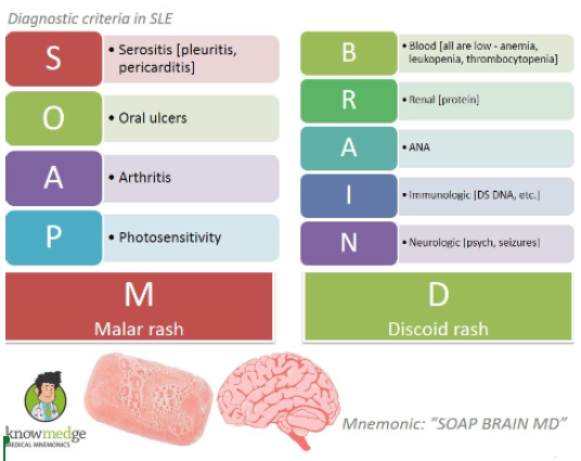
76
Skin Manifestations of lupus
Inflamed, red rash (butterfly/malar rash)
Discoid lesions
Discoid lesions
77
Renal Manifestations of lupus
Lupus Nephritis
78
CV manifestations of lupus
Pericarditis
Raynaud's phenomenon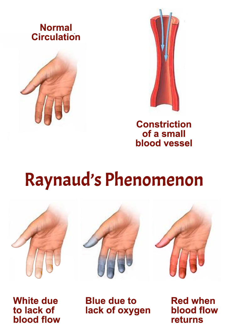
Raynaud's phenomenon
79
Pulmonary Manifestations of lupus
Pleural effusions
Pneumoniaa
Pneumoniaa
80
Neurologic Manifestations of lupus
CNS lupus
81
GI Manifestations of Lupus
Abdominal pain
82
Musculoskeletal Manifestations of Lupus
Joint Inflammation
Myositis
Myositis
83
SLE Therapeutic Management
1. Medications - steroids, NSAIDs, Hydroxychloroquine (Plaquenil)
2. Immunosuppresants - Methotrexate, Cyclophosphamide, Mycophenolate Mofetil, Azathioprine
3.Belimumab (Benlysta) - IV/SQ human monoclonal antibody (mAb)
Rituximab - off label use in pedi pts who have not responded to tx
4. Plasmapheresis - filter antibodies from plasma
5. Kidney transplant - for severe lupus nephritis
6. Calcium and Vitamin D - to prevent osteoporosis in vitamin D deficiency
2. Immunosuppresants - Methotrexate, Cyclophosphamide, Mycophenolate Mofetil, Azathioprine
3.Belimumab (Benlysta) - IV/SQ human monoclonal antibody (mAb)
Rituximab - off label use in pedi pts who have not responded to tx
4. Plasmapheresis - filter antibodies from plasma
5. Kidney transplant - for severe lupus nephritis
6. Calcium and Vitamin D - to prevent osteoporosis in vitamin D deficiency
84
SLE Nursing Management
Body image (think rashes)
Fear, anxiety, frustration - support groups, Lupus foundation
Chronic fatigue, weakness, anorexia, weight loss
Elective surgeries = need to increase steroids and antibiotics
Fear, anxiety, frustration - support groups, Lupus foundation
Chronic fatigue, weakness, anorexia, weight loss
Elective surgeries = need to increase steroids and antibiotics
85
SLE diet
Increase calcium, vitamin D (to help prevent osteoporosis)
Increase intake of Omega-3s (may help mitigate inflammation)
Increase consumption of lean meats and protein (to prevent muscle atrophy due to fatigue and not moving around much)
Lower consumption of fats, cholesterol, and sugars (Steroids can cause cholesterol, triglyceride, and blood sugar levels to increase, so it is important to limit it in diet)
Avoid red meats (contains saturated fats which can cause inflammation)
Avoid alcohol (esp. on steroids and methotrexate)
Avoid Saturated/Trans fats (increase inflammation flareups)
Increase intake of Omega-3s (may help mitigate inflammation)
Increase consumption of lean meats and protein (to prevent muscle atrophy due to fatigue and not moving around much)
Lower consumption of fats, cholesterol, and sugars (Steroids can cause cholesterol, triglyceride, and blood sugar levels to increase, so it is important to limit it in diet)
Avoid red meats (contains saturated fats which can cause inflammation)
Avoid alcohol (esp. on steroids and methotrexate)
Avoid Saturated/Trans fats (increase inflammation flareups)
86
SLE skin patient education
use mild soap and moisturizer
No drying substances
Limit exposure to UV and sunlight
For hair, use mild protein shampoos (biotin and collagen), no HARSH treatments
No drying substances
Limit exposure to UV and sunlight
For hair, use mild protein shampoos (biotin and collagen), no HARSH treatments
87
SLE medication patient education
Hydroxychloroquine - Eye exams before tx and then every 6 months
Immunosuppresants - increased risk of infection
Belimumab - risk for infection, no live vaccines for 30 days before treatment
Wear medical ID band for Lupus, *steroid dependence*
Immunosuppresants - increased risk of infection
Belimumab - risk for infection, no live vaccines for 30 days before treatment
Wear medical ID band for Lupus, *steroid dependence*
88
Systemic Sclerosis (Scleroderma)
Uncommon chronic, inflammatory, autoimmune connective tissue disease
Means hardening of the skin -> only one of the clinical manifestations
Children often only have skin involvement (localized/linear scleroderma)
Adults have skin AND organ involvement
Means hardening of the skin -> only one of the clinical manifestations
Children often only have skin involvement (localized/linear scleroderma)
Adults have skin AND organ involvement
89
Scleroderma is more common in _____
Women > men
25-55 years old
25-55 years old
90
Why is Scleroderma usually more fatal than other diseases such as SLE?
the disease process does not respond well to steroiods and immunosuppresants used in lupus
91
Scleroderma pathophysiology
Inflamed tissue becomes fibrotic then sclerotic (hardens)
92
Diffuse cutaneous Systemic Sclerosis (Scleroderma)
Edema over most of the body which is then subsequently replaced by tightening, hardening, and thickening of skin
Edema to upper extremities (1st symptom)
Edema to upper extremities (1st symptom)
93
Limited cutaneous Systemic Sclerosis (Scleroderma)
CREST Syndrome
Response to drug therapy can help differentiate from SLE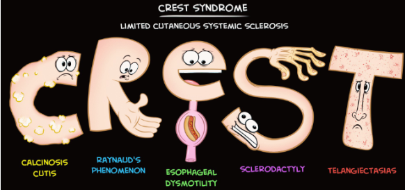
Response to drug therapy can help differentiate from SLE
94
Scleroderma Assessment findings
Arthralgia (joint pain)
Pitting edema of hands and fingers - may progress to entire upper/lower extremities and face (sausage-like fingers, skin is taut and shiny and free of wrinkles)
Loss of elasticity - ROM is markedly decreased
Pitting edema of hands and fingers - may progress to entire upper/lower extremities and face (sausage-like fingers, skin is taut and shiny and free of wrinkles)
Loss of elasticity - ROM is markedly decreased
95
Scleroderma GI manifestations
GERD - common in both types
Progresses to dysphagia
Reflux can lead to esophagititis and ulceration
Diminished peristalsis can have similar symptoms as partial bowel obstruction (Increase fiber + fluids, stool softeners)
Progresses to dysphagia
Reflux can lead to esophagititis and ulceration
Diminished peristalsis can have similar symptoms as partial bowel obstruction (Increase fiber + fluids, stool softeners)
96
Medications for GERD
PPIs (proton pump inhibitors such as -prazole)
97
Scleroderma CV manifestations
Raynaud's phenomenon in hands and feet (wear gloves and avoid extreme cold)
Severe cases -> digit necrosis -> can result in autoamputation of distal digits (basically they fall off by themselves
Myocardial fibrosis - ECG changes, Dysrhythmias, Chest pain
Severe cases -> digit necrosis -> can result in autoamputation of distal digits (basically they fall off by themselves
Myocardial fibrosis - ECG changes, Dysrhythmias, Chest pain
98
Scleroderma Lung Involvement manifestations
may go undetected until late in disease or even in the autopsy
Fibrosis of alveoli and interstitial tissues
Pulmonary arterial HTN (Blood vessel to the lung)
Fibrosis of alveoli and interstitial tissues
Pulmonary arterial HTN (Blood vessel to the lung)
99
Scleroderma Renal involvement manifestations
Often causes malignant HTN and death
100
Systemic Sclerosis (Scleroderma) goals of treatment
Use medications to force disease into remission - systemic steroids and immunosuppressants in large doses (often unsuccessful)
Goal is early ID of organ involvement and early treatment
Goal is early ID of organ involvement and early treatment
101
Scleroderma drug therapy
Bosentan (Tracleer) - improves pulmonary HTN
Tyrosine Kinase Inhibitors (TKIs) - slow down the disease process
NSAIDs - inflammation and pain
Tyrosine Kinase Inhibitors (TKIs) - slow down the disease process
NSAIDs - inflammation and pain
102
Bosentan patient teaching
Can cause liver toxicity and birth defects\
103
Tyrosine Kinase Inhibitors (TKIs)
TKIs are substances that block the action of tyrosine kinases which are enzymes that play a role in many cell functions. These functions include cell signaling, growth, and division.
nilotinib (Tasigna)
imatinib mesylate
nilotinib (Tasigna)
imatinib mesylate
104
Scleroderma nursing management
Skin protective measures
Reduce vasospasm and constriction
Swallow precautions
Joint protection (splints)
Energy conservation (rest, spread out ADLs)
Reduce vasospasm and constriction
Swallow precautions
Joint protection (splints)
Energy conservation (rest, spread out ADLs)
105
Scleroderma Skin-protective measur
Use mild soaps and lotions
Treat ulcers according to type and location
Bed cradle and foot board in severe cases (prevent the bedsheets from touching skin)
Treat ulcers according to type and location
Bed cradle and foot board in severe cases (prevent the bedsheets from touching skin)
106
How to reduce vasospasms and constriction in scleroderma?
Adjust room temperature
Wear gloves and socks
Avoid cigarette smokingi and extreme stress
Small, frequent meals
Wear gloves and socks
Avoid cigarette smokingi and extreme stress
Small, frequent meals
107
What should nurses do for patients with dysphagia due to scleroderma?
Follow swallow precautions:
1. Elevate HOB 60 degrees during AND 1-2 hours after meals
2. Limit spicy foods, caffeine, alcohol (can cause you to vomit)
3. Semisoft foods
4. Antacids or H2 antihistamines (to make acid less acidic)
5. Liquids should have a thick consistency to reduce risk of aspiration (avoid thin liquids like water)
1. Elevate HOB 60 degrees during AND 1-2 hours after meals
2. Limit spicy foods, caffeine, alcohol (can cause you to vomit)
3. Semisoft foods
4. Antacids or H2 antihistamines (to make acid less acidic)
5. Liquids should have a thick consistency to reduce risk of aspiration (avoid thin liquids like water)
108
Neoplasia
uncontrolled, abnormal growth of cells or tissues in the body
Can be either benign or malignant
Can be either benign or malignant
109
Benign neoplasia
Cells growing in the wrong place at the wrong time due to a small problem with cellular regulation
Harmless
Ex: moles, fibroids, polyps, endometriosis
Harmless
Ex: moles, fibroids, polyps, endometriosis
110
Malignant neoplasia
Cells are abnormal, serve no useful function, and are harmful to normal body tissues
111
anaplastic
A term used to describe cancer cells that divide rapidly and have little or no resemblance to normal cells.
112
Benign tumors characteristics
Cell division: continuous or inappropriate
Adherence: tight
Migratory: None
How does it grow: Expansion
Appearance: specific morphologic features
Adherence: tight
Migratory: None
How does it grow: Expansion
Appearance: specific morphologic features
113
Malignant tumors characteristics
Cell division: Rapid or continuous
Adherence: loose
Migratory: yes; metastasis
How does it grow: invades surrounding tissues
Appearance: anaplastic (no specific appearance of parent cells)
Adherence: loose
Migratory: yes; metastasis
How does it grow: invades surrounding tissues
Appearance: anaplastic (no specific appearance of parent cells)
114
Malignant transformation
Three stages of development
1. Initiation
2. Promotion
3. Progresison
1. Initiation
2. Promotion
3. Progresison
115
Initiation stage of malignant transformation
Irreversible event
Change in gene expression by anything that can damage cellular DNA (carcinogens)
If growth conditions are right, widespread metastatic disease can result from just one cancer cell
Change in gene expression by anything that can damage cellular DNA (carcinogens)
If growth conditions are right, widespread metastatic disease can result from just one cancer cell
116
Promotion stage of malignant transformation
enhanced growth of initiated cell by substances called promoters
117
Latency period
Time from initiation to overt tumor
Varies from months to years
Varies from months to years
118
Progression stage of malignant transformation
Continued change, becoming more malignant over time
Tumor develops own blood supply - vascular endothelial growth factor (VEGF) triggers growth of new capillaries into tumor
Tumor develops own blood supply - vascular endothelial growth factor (VEGF) triggers growth of new capillaries into tumor
119
Primary tumors
identified by tissue it derived from (parent tissue)
120
Metastatic/Secondary tumors
originates from some other part of the body
spread by blood and/or lymph
same cell, just in different location
spread by blood and/or lymph
same cell, just in different location
121
Common metastasis of tumors
Breast -> commonly spreads to bone and lung
Lung -> brain
Colorectal -> liver
Prostate -> bone (esp spine and legs)
Lung -> brain
Colorectal -> liver
Prostate -> bone (esp spine and legs)
122
Adeno tissue
epithelial glands
123
Glio tissue
glial cells in brain
124
Hemangio tissue
Blood vessel
125
Hepato tissue
liver
126
Lympho tissue
lymphoid tissue
127
Melano tissue
pigment producing cells
128
Meningio tissue
meninges
129
Neuro tissue
nerve tissue
130
Osteo tissue
bone
131
Renal tissue
kidney
132
Chemical carcinogens
tobacco
asbestos
asphalt fumes
benzene
gasoline
diesel exhaust
asbestos
asphalt fumes
benzene
gasoline
diesel exhaust
133
Physical carcinogens
UV radiation (sunlight, tanning beds)
Ionizing Radiation (X-rays, cosmic radiation)
Chronic irritation (GERD, skin cancer with burn scars, etc.)
Ionizing Radiation (X-rays, cosmic radiation)
Chronic irritation (GERD, skin cancer with burn scars, etc.)
134
Viral carcinogens
oncoviruses:
1. HPV (cervical)
2. Epstein-Barr (lymphoma)
3. Hep B & C (liver)
4. H. pylori (GI)
1. HPV (cervical)
2. Epstein-Barr (lymphoma)
3. Hep B & C (liver)
4. H. pylori (GI)
135
Cancer etiology: Personal factors
Immune function: cancer is increased in immunosuppressed (over 60 yo, organ transplant recipients, AIDs, etc.)
Genetics: Inherited cancer risks (breast, prostate, ovarian)
Down symdrome = increased risk for leukemia
Genetics: Inherited cancer risks (breast, prostate, ovarian)
Down symdrome = increased risk for leukemia
136
CAUTION warning signs of cancer
C: changes in bowel / bladder habits
A: a sore that does not heal
U: unusual bleeding or discharge
T: thickening or lump in breast or elsewhere
I: indigestion or difficulty swallowing
O: obvious change in wart or mole
N: nagging cough or hoarseness
A: a sore that does not heal
U: unusual bleeding or discharge
T: thickening or lump in breast or elsewhere
I: indigestion or difficulty swallowing
O: obvious change in wart or mole
N: nagging cough or hoarseness
137
Primary cancer prevention
Prevent the actual occurence of cancer. Most effective when there is a known cause
Avoid known/potential carcinogen
Modify associated risk factors
Remove "at-risk" tissues
Chemoprevention
Vaccination
Avoid known/potential carcinogen
Modify associated risk factors
Remove "at-risk" tissues
Chemoprevention
Vaccination
138
Chemoprevention drugs
Aspirin + Celebrex = colon cancer
Vitamin D + tamoxifen = breast cancer
Lycopene = prostate cancer
Vitamin D + tamoxifen = breast cancer
Lycopene = prostate cancer
139
Gardasil
HPV vaccination
140
Secondary Cancer Prevention
Screening to detect cancer early - Cure or control more likely
Participate in specific routine screenings - Mammogram, breast, prostate exam
Genetic screening
Participate in specific routine screenings - Mammogram, breast, prostate exam
Genetic screening
141
Tests/Diagnostics for Cancer
Blood tests (CEA, PSA)
Radiological studies
Specimens (biopsy)
Radiological studies
Specimens (biopsy)
142
Carcinoembryonic Antigen (CEA)
screening blood test that identifies a protein that is commonly found in some cancers
can be used to assess effectiveness of treatment
can be used to assess effectiveness of treatment
143
PSA (prostate specific antigen)
Lab test used to screen for prostate cancer
144
Imaging used to diagnose canceret
X-rays
CT scan
PET (positron emission tomography)
MRI
CT scan
PET (positron emission tomography)
MRI
145
Components of informed consent
(1) disclosure of information (procedure, reason for procedure, risk + benefits, etc.)
(2) competency of the patient (or surrogate) to make a decision
(3) voluntary nature of the decision
(2) competency of the patient (or surrogate) to make a decision
(3) voluntary nature of the decision
146
Staging and Grading of Cancer
Staging = How big cancer is + whether it has spread
Grading = how abnormal cancer cells and tissue look under a microscope
Grading = how abnormal cancer cells and tissue look under a microscope
147
Ploidy
description of cells by chromosome number & appearance
cancer cells can gain or lose whole chromosomes during the malignant transformation phase
remaining chromosomes may have structural abnormalities
cancer cells can gain or lose whole chromosomes during the malignant transformation phase
remaining chromosomes may have structural abnormalities
148
Types of cancer staging
Clinical staging: assesses clinical manifestations
Surgical staging: surgically assess tumor size, number, sites, and spread
Pathologic staging: most definitive type; by pathologic examination of tissues
Surgical staging: surgically assess tumor size, number, sites, and spread
Pathologic staging: most definitive type; by pathologic examination of tissues
149
Common side effects of cancer treatment
Reduced immunity and blood-producing functions (bone marrow suppression)
Altered GI structure and function (N/V, anorexia, altered taste, etc.) - high calorie and protein diet, vitamin C, increase fluids
Altered peripheral nerve function (chemo can cause neuropathy) - Gabapentin
Decreased gas exchange (inflammation from radiation)
Sterility
Psychosocial (fear of death, grieving, body image, etc.)
Altered GI structure and function (N/V, anorexia, altered taste, etc.) - high calorie and protein diet, vitamin C, increase fluids
Altered peripheral nerve function (chemo can cause neuropathy) - Gabapentin
Decreased gas exchange (inflammation from radiation)
Sterility
Psychosocial (fear of death, grieving, body image, etc.)
150
Which rays are used for radiation therapy?
Gamma rays are generally used because of its ability to deeply penetrate tissues
151
Exposure vs. Radiation Dose
Exposure = amount delivered to cells
Radiation dose = Amount absorbed by the tissues
Radiation dose = Amount absorbed by the tissues
152
Teletherapy
Radiation delivered from outside the body
Must be in exact position for each treatment
Patient is NOT radioactive
Must be in exact position for each treatment
Patient is NOT radioactive
153
Brachytherapy
Radiation source is within the patient
Patient is radioactive until radiation source is removed from the body
Patient is radioactive until radiation source is removed from the body
154
Types of Brachytherapy
Sealed or solid: implanted within or near tumor cells (seeds, needles, rods)
Unsealed: suspended in body fluids (given PO, IV, instilled in body cavities)
Unsealed: suspended in body fluids (given PO, IV, instilled in body cavities)
155
Rules for brachytherapy
Patient will need to be in isolation while emitting radiation
Staff must be cautious about how physically close they are as well as amount of time in close proximity to patient
Staff required to wear dosimeter film badge
Everything in patient's room is radioactive (yes, even their toothbrush)
Pregnant or nursing nurses can NOT take care of these patients
Staff must be cautious about how physically close they are as well as amount of time in close proximity to patient
Staff required to wear dosimeter film badge
Everything in patient's room is radioactive (yes, even their toothbrush)
Pregnant or nursing nurses can NOT take care of these patients
156
Inverse square law
Intensity of radiation decreases with the distance from the radiation source
157
Brachytherapy Patient Teaching
Visitors are typically discouraged
Children should NOT be visiting the patient
Visitors should be limited to 30 minutes
Nurses may only be in the room for 30 minutes of a 12 hour shift
Children should NOT be visiting the patient
Visitors should be limited to 30 minutes
Nurses may only be in the room for 30 minutes of a 12 hour shift
158
Radiation therapy symptoms
Effects are seen only on or in tissues within the radiation field or path:
1. Skin changes
2. Hair Loss
3. Taste changes
4. Fatigue
5. Bone Marrow Suppression (Neutropenic, low platelet, anemic)
6. Increased risk for development of second malignancies (can basically cause other cancers)
1. Skin changes
2. Hair Loss
3. Taste changes
4. Fatigue
5. Bone Marrow Suppression (Neutropenic, low platelet, anemic)
6. Increased risk for development of second malignancies (can basically cause other cancers)
159
Care of patient with sealed implants of radioactive sources
Private room with proper signage
Keep door closed
Portable lead shields, lead aprons
Dosimeter film badge
If pregnant or trying to conceive, DO NOT CARE FOR PT
Limit visitors to 30 min/day, stay 6 ft away at all times
Do NOT remove anything from the room (including dressings and bed linens)
Keep door closed
Portable lead shields, lead aprons
Dosimeter film badge
If pregnant or trying to conceive, DO NOT CARE FOR PT
Limit visitors to 30 min/day, stay 6 ft away at all times
Do NOT remove anything from the room (including dressings and bed linens)
160
Chemotherapy
Chemical agents that act systemically to selectively kill cancer cells by damaging the DNA of the cell to prevent reproduction
Normal cells can still be damaged
Administered systemically or locally
Normal cells can still be damaged
Administered systemically or locally
161
Chemotherapy routes
IV
PO
Topical
Intraperitoneal (within abdominal cavity)
Intra-arterial
Intrathecal (fluid-filled space between the thin layers of tissue that cover the brain and spinal cord)
Intraventricular (spaces in the brain that contain the cerebral spinal fluid)
Intravesicular (in the bladder)
PO
Topical
Intraperitoneal (within abdominal cavity)
Intra-arterial
Intrathecal (fluid-filled space between the thin layers of tissue that cover the brain and spinal cord)
Intraventricular (spaces in the brain that contain the cerebral spinal fluid)
Intravesicular (in the bladder)
162
antimetabolites
Impair cell division
163
antimitotic
Prevents cancer cells from dividing or divides only once
164
Antitumor
interrupts DNA or RNA synthesis
165
Topoisomerase inhibitors
cause cell death by disrupting enzyme essential for DNA synthesis and cell division
166
Nurse certification to administer chemotherapy
Don't need to be board certified, just need to be trained within a facility or hospital system
167
Extravasation
The leakage of blood, lymph, or other fluid, such as an anticancer drug, from a blood vessel or tube into the tissue around it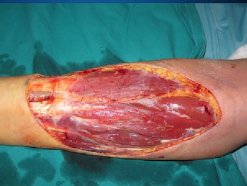
168
Extravasation nursing interventions
Most important nursing intervention is prevention
Ensure patency before administration of medication (aspirate for blood)
Use an implanted port or central line
Cold (vasoconstriction)/warm (vasodilation) compresses
Injection of antidote
Ensure patency before administration of medication (aspirate for blood)
Use an implanted port or central line
Cold (vasoconstriction)/warm (vasodilation) compresses
Injection of antidote
169
What to do with oral anticancer
DO NOT CRUSH, SPLIT, BREAK, OR CHEW
Adhere to medication schedule
Proper disposal
Adhere to medication schedule
Proper disposal
170
Chemoprotectants/Cytoprotectants
Amifostine (Ethyol) - reduce kidney damage by cisplatin
Mesna (mesnex) - prevent hemorrhagic cystitis from ifosfamide and cyclophosphamide
Mesna (mesnex) - prevent hemorrhagic cystitis from ifosfamide and cyclophosphamide
171
Myelosuppression
bone marrow suppression of leukocytes, erythrocytes, platelets
172
Nadir
time when bone marrow activity and WBCs are at lowest levels
173
Myelosuppression nursing care
Private room w/ reverse isolation (anyone sick cannot come in) or neutropenic precautions
Good handwashing technique
Room and bathroom cleaned daily
Good hygeine
Dedicated equipment stays in room
Single use food items, individually wrapped items
Avoid indwelling foley catheters
Monitor vital signs and IV sites q.4H
Inspect mouth, skin, and mucous membranes q.8H
Basically just want to minimize risk for infection
Good handwashing technique
Room and bathroom cleaned daily
Good hygeine
Dedicated equipment stays in room
Single use food items, individually wrapped items
Avoid indwelling foley catheters
Monitor vital signs and IV sites q.4H
Inspect mouth, skin, and mucous membranes q.8H
Basically just want to minimize risk for infection
174
What should a person with low WBC count report to PCP?
temp above 100°F
Cough
UTI
abscess
discharge
Cough
UTI
abscess
discharge
175
Thrombocytopenia Nursing Care
Goal of care to reduce risk of bleeding
Wear shoes when ambulating
No enemas, caution with suppositories
Use electric razor
Use soft-bristle toothbrush
Fall precautions
Apply ice to areas of trauma
*Observe IV sites q.4H for bleeding*
Don't pick nose
Wear shoes when ambulating
No enemas, caution with suppositories
Use electric razor
Use soft-bristle toothbrush
Fall precautions
Apply ice to areas of trauma
*Observe IV sites q.4H for bleeding*
Don't pick nose
176
Chemotherapy Induced Nausea and Vomiting
Anticipatory - before receiving chemo
Acute - within first 24 hours after chemo
Delayed - after first 24 hours
Breakthrough - occurs intermittently during therapy
Acute - within first 24 hours after chemo
Delayed - after first 24 hours
Breakthrough - occurs intermittently during therapy
177
Antiemetics drugs
Zofran (ondansetron) - side effects include bradycardia, hypotension, vertigo, and headache
Reglan (metoclopramide) - side effect is drowsiness
Emend (aprepitant) - can decrease the effectiveness of oral contraceptives
Reglan (metoclopramide) - side effect is drowsiness
Emend (aprepitant) - can decrease the effectiveness of oral contraceptives
178
Emend (aprepitant) and Coumadin (warfarin) drug interaction
Aprepitant is a known inducer of CYP2C9, the main warfarin-metabolizing enzyme
Co-administration of these two drugs may result in reduction of the anticoagulation activity of warfarin.
Check INR before and after 3 days of treatment if taking warfarin alongside aprepitant
Co-administration of these two drugs may result in reduction of the anticoagulation activity of warfarin.
Check INR before and after 3 days of treatment if taking warfarin alongside aprepitant
179
Mucositis Nursing care
mucous membranes = entire GI tract
Oral hygeine, mouth rinsing
Change PO meds to IV
IV hydration
Parenteral, enteral nutrition if needed
Oral hygeine, mouth rinsing
Change PO meds to IV
IV hydration
Parenteral, enteral nutrition if needed
180
Stomatitis (oral cavity) Patient teaching
Examine mouth q.4H for fissures, blisters, sores, drainage, etc.
Brush with soft-bristle toothbrush or sponge-tipped applicators q.8H
Avoid mouthwash with alcohol
Take pain meds PRN
Avoid alcohol, tobacco, spicy, salty, acidic, dry, rough, or hard food
Brush with soft-bristle toothbrush or sponge-tipped applicators q.8H
Avoid mouthwash with alcohol
Take pain meds PRN
Avoid alcohol, tobacco, spicy, salty, acidic, dry, rough, or hard food
181
Alopecia patient teaching
it is temporary - regrowth starts about 1 month after completion of chemo
Hair may grow back different color, texture, and thickness
Cold caps during chemo
Wigs, hats, and scarves during chemo
Protect bare scalp from injury
Recommend getting wigs prior to treatment
Hair may grow back different color, texture, and thickness
Cold caps during chemo
Wigs, hats, and scarves during chemo
Protect bare scalp from injury
Recommend getting wigs prior to treatment
182
Chemo brain
Changes in cognitive function
Difficulty with concentration, memory loss, learning new info
Can last months to years
Avoid alcohol, recreational drug use, activities that increase risk for head injury
Difficulty with concentration, memory loss, learning new info
Can last months to years
Avoid alcohol, recreational drug use, activities that increase risk for head injury
183
Chemotherapy induced Peripheral Neuropathy
Altered sensation in hands and feet - may or may not resolve
Impaired gaibat and balance, orthostatic hypotension, erectile dysfunction, pain, taste alteration, severe constipation.
Impaired gaibat and balance, orthostatic hypotension, erectile dysfunction, pain, taste alteration, severe constipation.
184
Chemo-induced Peripheral Neuropathy Patient Teaching
Promote safety - protect areas with altered sensation
Avoid shoes that would cause rubbing
Assess feet daily for signs of injury
High fiber diet, 2-3 liters of fluids (non-alcoholic) per day UNLESS contraindicated
Avoid shoes that would cause rubbing
Assess feet daily for signs of injury
High fiber diet, 2-3 liters of fluids (non-alcoholic) per day UNLESS contraindicated
185
Interleukins
Regulate inflammation and immunity
Interferons - slow tumor cell division, inhibit expression of oncogenes, stimulate growth and activation of NK cells
Interferons - slow tumor cell division, inhibit expression of oncogenes, stimulate growth and activation of NK cells
186
Biological Response Modifiers as supportive cancer therapy
"Growth Factors"
Treat chemotherapy induced bone marrow suppression
Neupogen = neutrophils
Epogen = erythrocytes
Neumega = platelets
Treat chemotherapy induced bone marrow suppression
Neupogen = neutrophils
Epogen = erythrocytes
Neumega = platelets
187
Photodynamic Therapy
use of certain types of lasers
Agent given IV to sensitize cells to light, cancer cells will eliminate agent more slowly than normal cells so after 48-72 hours, most of the drug is concentrated in cancer cells
A laser light is focused on the tumor and activates a reaction that causes irreversible damage and death to cells retaining the sensitizing agent.
Agent given IV to sensitize cells to light, cancer cells will eliminate agent more slowly than normal cells so after 48-72 hours, most of the drug is concentrated in cancer cells
A laser light is focused on the tumor and activates a reaction that causes irreversible damage and death to cells retaining the sensitizing agent.
188
Spinal cord compression
Caused by cord compression and its nerve roots
Associated with a vertebral tumor which extends into epidural space
Vertebral bone metatasis leads to bone collapse/displacement
May lead to permanent neurological impairment
Associated with a vertebral tumor which extends into epidural space
Vertebral bone metatasis leads to bone collapse/displacement
May lead to permanent neurological impairment
189
Cancers that can metastasize to bone
Lung
Breast
Prostate
Lymphoma
Multiple myeloma
Breast
Prostate
Lymphoma
Multiple myeloma
190
Spinal cord compression clinical manifestations
inflammation
back/neck pain
pain exacerbated by movement
Neuro impairment (both motor and sensory)
Bowel and bladder dysfunction
back/neck pain
pain exacerbated by movement
Neuro impairment (both motor and sensory)
Bowel and bladder dysfunction
191
Spinal Cord Compression Diagnostics
X-rays
CT scan
Bone scan
MRI Gold Standard
CT scan
Bone scan
MRI Gold Standard
192
Spinal Cord Compression Medical Management
Corticosteroids
Pain management
XRT
Vertebral Augmentation (Minimally invasive)
Debulking tumors
Vertebroplasty
Kyphoplasty
Pain management
XRT
Vertebral Augmentation (Minimally invasive)
Debulking tumors
Vertebroplasty
Kyphoplasty
193
Superior Vena Cava Syndrome
Compression or invasion of the superior vena cava by:
-Tumor
-Enlarged lymph nodes
-Intraluminal thrombus obstructing the venous circulation
-Drainage of head, neck arms, thorax
if left untreated -> can lead to cerebral anoxia
-Tumor
-Enlarged lymph nodes
-Intraluminal thrombus obstructing the venous circulation
-Drainage of head, neck arms, thorax
if left untreated -> can lead to cerebral anoxia
194
Superior Vena Cava Syndrome Clinical Manifestations
Impaired venous drainage
Edema arms, neck, thorax with decreased sensations
Engorged/distended jugular temporal, arm veins
Increased ICP
Edema arms, neck, thorax with decreased sensations
Engorged/distended jugular temporal, arm veins
Increased ICP
195
Superior Vena Cava Syndrome Diagnostics
X-Ray
CT Thorax
MRI Thorax
Venogram if transuminal thrombus suspected
CT Thorax
MRI Thorax
Venogram if transuminal thrombus suspected
196
Medical Management of Superior Vena Cava Syndrome
XRT (radiation therapy)
Chemotherapy
Percutaneous stents
Anticoagulants and Thrombolytics
Diuretics
Oxygen
Chemotherapy
Percutaneous stents
Anticoagulants and Thrombolytics
Diuretics
Oxygen
197
Tumor Lysis syndrome
Potentially fatal
Occurs most commonly after the start of chemotherapy
Refers to the constellation of metabolic disturbances:
1. Hyperuricemia (high uric acid)
2. Hyperkalemia
3. Hyperphosphatemia
4. Hypocalcemia
5. Acute kidney injury (AKI)
Occurs most commonly after the start of chemotherapy
Refers to the constellation of metabolic disturbances:
1. Hyperuricemia (high uric acid)
2. Hyperkalemia
3. Hyperphosphatemia
4. Hypocalcemia
5. Acute kidney injury (AKI)
198
Tumor Lysis Syndrome Clinical Manifestations
Prescence of urinary symptoms
Symptoms of hyperkalemia (Weakness, paralysis, cardiac dysrhythmias)
Symptoms of hypocalcemia (Anorexia, vomiting, cramps, SZ, spasms, AMS, tetany)
Abdominal pain/distention
Sudden death
Symptoms of hyperkalemia (Weakness, paralysis, cardiac dysrhythmias)
Symptoms of hypocalcemia (Anorexia, vomiting, cramps, SZ, spasms, AMS, tetany)
Abdominal pain/distention
Sudden death
199
Tumor Lysis Syndrome diagnostics
Electrolyte panel (CMP)
Urinalysis
Urinalysis
200
Medical Management of Tumor Lysis Syndrome
Agressive fluid hydration
Allopurinol
Diuretics
Correct electrolyte imbalance
Dialysis
Allopurinol
Diuretics
Correct electrolyte imbalance
Dialysis
201
How to correct hyperkalemia
Sodium polystrene
Insulin + sodium bicarbonate
Insulin + sodium bicarbonate
202
How to correct hyperphosphate
Aluminum hydroxide
203
Oncologic Emergencies: Hypercalcemia
Potentially fatal metabolic abnormality
Ocurs when Ca is released by the bones is more than the kidneys can excrete or the bones can reabsorb
May result from growth factors of cancer cells
when serum Ca is >10.4 mg/dL
Ocurs when Ca is released by the bones is more than the kidneys can excrete or the bones can reabsorb
May result from growth factors of cancer cells
when serum Ca is >10.4 mg/dL
204
Hypercalcemia Clinical Manifestations
Fatigue
Weakness
Confusions
Nausea
Constipation
Hyporeflexia
Dysrhythmias
Ileus (hypoactive)
Weakness
Confusions
Nausea
Constipation
Hyporeflexia
Dysrhythmias
Ileus (hypoactive)
205
Hypercalcemia Medical Management
Identify clients at risk
Oral/IV hydration
Diuretics
Oral/IV hydration
Diuretics
206
Prostate Cancer
One of the slowest growing and predictable
Can have 100% cure rate if found early
Metastasizes to nearby lymph nodes, bones, lungs and liver
Most are androgen-sensitive
Can have 100% cure rate if found early
Metastasizes to nearby lymph nodes, bones, lungs and liver
Most are androgen-sensitive
207
Prostate Cancer Risk Ractors
Advancing Age
1st degree relative
Race (African Americans)
Diet
Vasectomy
Environmental toxins
1st degree relative
Race (African Americans)
Diet
Vasectomy
Environmental toxins
208
Prostate Cancer: Urination Assessment
Symptoms include:
- Dysuria
- Painful ejaculations
- Blood in semen and urine
- Dysuria
- Painful ejaculations
- Blood in semen and urine
209
Prostate Cancer Screening
Prostate-specific antigen testing (PSA); normal is < 2.5ng/ml
Age 50 or family history - maybe earlier
Check before DRE(digital rectal exam) because can cause an abnormal elevation of results
Early prostate cancer antigen (EPCA-2) – sensitive and specific
Age 50 or family history - maybe earlier
Check before DRE(digital rectal exam) because can cause an abnormal elevation of results
Early prostate cancer antigen (EPCA-2) – sensitive and specific
210
Digital Rectal Exam (DRE)
Exactly what it sounds like
Check for prostate cancer
Hard and stony with irregularities or indurations
Check for prostate cancer
Hard and stony with irregularities or indurations
211
Transrectal ultrasound biopsy
Type of biopsy that uses ultrasound probe to check for prostate cancer
212
Purpose of using imaging for prostate cancer screening
To rule out metastasis
213
Asymptomatic Prostate Cancer Interventions
Check PSA levels, aggravated symptoms, or if tumor is growing
214
Prostate Cancer Surgical Interventions
TURP (Transurethal resection of the prostate)
Laparoscopic Radical Prostatectomy
Open surgery
Bilateral orchiectomy (removal of both testicles)
*Lithotomy Position*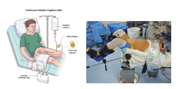
Laparoscopic Radical Prostatectomy
Open surgery
Bilateral orchiectomy (removal of both testicles)
*Lithotomy Position*
215
Prostate Cancer Nonsurgical Interventions
Radiation (external or internal)
Chemotherapy (Cisplatin)
Chemotherapy (Cisplatin)
216
Prostate Cancer Hormone Therapy
Luteinizing hormone releasing hormone agonists or anti-androgens
Lupron - Can cause medical castration
Lupron - Can cause medical castration
217
Lung Cancer
Third leading cause of new cancer
Leading cause of cancer-related death
Metastasis to lymph nodes, bone, liver, brain, adrenal glands
Leading cause of cancer-related death
Metastasis to lymph nodes, bone, liver, brain, adrenal glands
218
Lung Cancer risk factors
Major risk factor is smoking
Exposure to second hand smoke puts individuals at greater risk than non-smokers
Chronic inhalation of carcinogens
COPD and TB can increase risk of developing lung cancer
Exposure to second hand smoke puts individuals at greater risk than non-smokers
Chronic inhalation of carcinogens
COPD and TB can increase risk of developing lung cancer
219
Lung Cancer complications
Poor oxygenation (tumor can obstruct bronchioles, cause compression of other parts of the lung)
Paraneoplastic Syndrome - tumors can secrete hormones
Paraneoplastic Syndrome - tumors can secrete hormones
220
Paraneoplastic Syndrome Hormones
ADH (antidiuretic hormone) - can cause SIADH (syndrome of inappropriate secretion of antidiuretic hormone)
ACTH (Adrenocorticotropic hormone) - Cushings
FSH (follicle stimulating hormone) - gynecomastia (moobs)
Parathyroid hormone - increased serum calcium
Ectopic insulin - hypoglycemia
ACTH (Adrenocorticotropic hormone) - Cushings
FSH (follicle stimulating hormone) - gynecomastia (moobs)
Parathyroid hormone - increased serum calcium
Ectopic insulin - hypoglycemia
221
Lung Cancer Warning Signs
Hoarseness
Change in respiratory pattern
Persistent cough or change in cough
Blood-tinged sputum
Rust colored or purulent sputum
Frank hemoptysis
Chest pain or tightness
Shoulder, arm, chest wall pain
Recurring episodes of pleural effusion, pneumonia, bronchitis
Dyspnea
Fever associated with one or two other signs
Wheezing
Weight loss
Clubbing of the fingers
Change in respiratory pattern
Persistent cough or change in cough
Blood-tinged sputum
Rust colored or purulent sputum
Frank hemoptysis
Chest pain or tightness
Shoulder, arm, chest wall pain
Recurring episodes of pleural effusion, pneumonia, bronchitis
Dyspnea
Fever associated with one or two other signs
Wheezing
Weight loss
Clubbing of the fingers
222
Lung Cancer Assessment
Ask pt about warning symptoms (how long? when did it start?)
Pack year hx
Physical assessment
Pain
Dyspnea
Breath sounds
Heart sounds (may be muffled by tumor)
Fatigue
Dysphagia
N/V
Superior Vena Cava Syndrome
Pack year hx
Physical assessment
Pain
Dyspnea
Breath sounds
Heart sounds (may be muffled by tumor)
Fatigue
Dysphagia
N/V
Superior Vena Cava Syndrome
223
Lung Cancer Diagnostics
Cytologic Testing – early morning sputum, thoracentesis fluid
CXR
Lung CT
MRI
Thoracoscopy / bronchoscopy
Needle Biopsy
PET Scan
CXR
Lung CT
MRI
Thoracoscopy / bronchoscopy
Needle Biopsy
PET Scan
224
Lung Cancer treatment
Chemotherapy
Targeted Therapy
Radiation
Photodynamic therapy
Surgical Intervention
Targeted Therapy
Radiation
Photodynamic therapy
Surgical Intervention
225
Lung Cancer Surgical Interventions
*Side-lying jacknife position*
Removal of tumor only
Lung segment
Lobectomy (lobe removal)
Pneumonectomy (remove entire lung)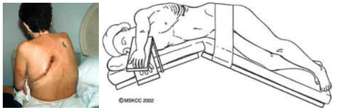
Removal of tumor only
Lung segment
Lobectomy (lobe removal)
Pneumonectomy (remove entire lung)
226
Lung Cancer Palliative measures
Oxygen therapy
Drug therapy - Bronchodilators, corticosteroids, mucolytics
Radiation therapy
Thoracentesis - removal of fluid for lung expansion
Dyspnea management - Oxygen, positioning (high fowlers)
Pain Management - morphine
Hospice care
Drug therapy - Bronchodilators, corticosteroids, mucolytics
Radiation therapy
Thoracentesis - removal of fluid for lung expansion
Dyspnea management - Oxygen, positioning (high fowlers)
Pain Management - morphine
Hospice care
227
Colorectal Cancer
Third most common cause of cancer death
Not common before 40 but is increasing 2/3 of colon cancer in descending colon / rectum
Spread by blood/lymph
Metastasize to other abdominal organs, brain, bone
Not common before 40 but is increasing 2/3 of colon cancer in descending colon / rectum
Spread by blood/lymph
Metastasize to other abdominal organs, brain, bone
228
Colon Cancer risk factors
Older than 50
Genetic/Family history of cancer
Disease of the colon
Crohn’s/Ulcerative Colitis
*Infectious Agents - H-Pylori, Streptococcus bovis, HPV*
Red meat, Processed foods, High Fat Diet
Long-term smoking
Heavy alcohol use
Obesity, physical inactivity
Increased bile acid secretions
*Lack of fiber in diet*
Genetic/Family history of cancer
Disease of the colon
Crohn’s/Ulcerative Colitis
*Infectious Agents - H-Pylori, Streptococcus bovis, HPV*
Red meat, Processed foods, High Fat Diet
Long-term smoking
Heavy alcohol use
Obesity, physical inactivity
Increased bile acid secretions
*Lack of fiber in diet*
229
Colon Cancer Screening
Screening at 50 years of age:
- Earlier and more frequently depending on family history
- Fecal occult blood test (FOCT) yearly
- Colonoscopy every 10 years OR double-contract barium enema every 5 years OR sigmoidoscopy every 5 years
- Earlier and more frequently depending on family history
- Fecal occult blood test (FOCT) yearly
- Colonoscopy every 10 years OR double-contract barium enema every 5 years OR sigmoidoscopy every 5 years
230
Colon Cancer Prevention
Diet: low fat, high fiber
Smoking: smoking cessation
Exercise: Aerobic exercise at least 3x weekly for at least 30 minutes
Smoking: smoking cessation
Exercise: Aerobic exercise at least 3x weekly for at least 30 minutes
231
Early and Late manifestations of colon cancer
Early:
- Bleeding (probably not hemorrhoids, but GI bleeding)
Late:
- Fatigue
- Weight loss
- Pain
- Bleeding (probably not hemorrhoids, but GI bleeding)
Late:
- Fatigue
- Weight loss
- Pain
232
Colon Cancer Diagnostics
Labs:
- CEA
- Fecal occult blood
- CBCs
- Liver function tests (LFTs) used to rule out metastasis
- CEA
- Fecal occult blood
- CBCs
- Liver function tests (LFTs) used to rule out metastasis
233
Colon Cancer Treatment
Radiation
Chemotherapy
- Vascular endothelial growth factor (VEGF) inhibitors
- Epidermal growth factor receptor inhibitor (EGFRI)
Surgical Management
- Colon resection
- Colectomy
- Abdominoperineal (AP) resection
Chemotherapy
- Vascular endothelial growth factor (VEGF) inhibitors
- Epidermal growth factor receptor inhibitor (EGFRI)
Surgical Management
- Colon resection
- Colectomy
- Abdominoperineal (AP) resection
234
Colon Cancer Surgery teaching
Pre-Op Education
- Bowel prep (laxatives, NPO after midnight)
- Post surgical care (pain control, PACU)
- Wound Care (may take 6-8 months to heal)
- Colostomy care/education (make sure it isn't purple, keep it clean)
- Bowel prep (laxatives, NPO after midnight)
- Post surgical care (pain control, PACU)
- Wound Care (may take 6-8 months to heal)
- Colostomy care/education (make sure it isn't purple, keep it clean)
235
Breast Cancer
Inherited Gene Mutation - BRCA1 or BRCA2 genes
Second most common diagnosed cancer in women after skin cancer
small % of cases are under 40 years of age
1% of all cases are male
Even with no family hx, risk for breast cancer increases with age
Second most common diagnosed cancer in women after skin cancer
small % of cases are under 40 years of age
1% of all cases are male
Even with no family hx, risk for breast cancer increases with age
236
Primary prevention of breast cancer
Surgical
Chemoprevention: Vitamin D + Tamoxifen
Chemoprevention: Vitamin D + Tamoxifen
237
Types of breast cancer
Non-Invasive – ductal and lobular carcinoma in situ: have not invaded the tissues outside of the ducts and milk-producing glands
Invasive – *most common* - infiltrating ductal carcinoma
Inflammatory breast cancer (IBC) – rare and highly aggressive
Invasive – *most common* - infiltrating ductal carcinoma
Inflammatory breast cancer (IBC) – rare and highly aggressive
238
Breast Cancer Risk Factors
High Risk: female, age >65, genetic factors, previous hx of breast CA, breast density, atypical hyperplasia
Moderate: family hx, ionizing radiation exposure, high postmenopausal bone density
Low risk: Reproductive hx – nulliparity or 1st child after age 30, early menarche or late menopause or both, recent oral contraceptive use, recent HRT, obesity
Other risk factors – alcohol, high socioeconomic status, Jewish heritage → BRCA1 and BRCA2 genetic mutations
Moderate: family hx, ionizing radiation exposure, high postmenopausal bone density
Low risk: Reproductive hx – nulliparity or 1st child after age 30, early menarche or late menopause or both, recent oral contraceptive use, recent HRT, obesity
Other risk factors – alcohol, high socioeconomic status, Jewish heritage → BRCA1 and BRCA2 genetic mutations
239
Breast Self-Examination
1 week after menstrual period
Decreased swelling and tenderness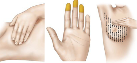
Decreased swelling and tenderness
240
Modifiable Risk Factors of Breast Cancer
Obesity
Alcohol consumption
Alcohol consumption
241
Breast Cancer assessment findings
Irregular, poorly defined mass
Dimpling (advanced disease)
Peau d'orange (orange peel skin) - late stage breast cancer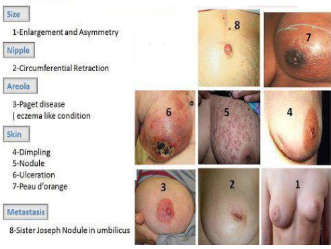
Dimpling (advanced disease)
Peau d'orange (orange peel skin) - late stage breast cancer
242
Inflammatory Breast Cancer assessment findings
Swelling, skin redness, pain
Seldom presents as a palpable lump
*May not show up on mammogram*
Seldom presents as a palpable lump
*May not show up on mammogram*
243
Breast Cancer in Males assessment findings
Hard, painless, sub-areolar mass
Gynecomastia (moobs)
Nipple discharge, retraction, erosion, or ulceration
Gynecomastia (moobs)
Nipple discharge, retraction, erosion, or ulceration
244
Breast Cancer Imaging
Mammogram – controversy about when to begin & how often – follow MD’s recommendation
Tomosynthesis
Ultrasound
CT/MRI
Tomosynthesis
Ultrasound
CT/MRI
245
Needle Biopsy
Breast tissue
Lymph nodes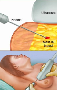
Lymph nodes
246
Breast Cancer labs
Liver enzymes - to rule out liver involvement
Calcium
Alkaline phosphatase
Calcium
Alkaline phosphatase
247
Breast Cancer Surgical Interventions
Breast-conserving - Lumpectomy, Partial Mastectomy
Simple Mastectomy – entire breast removed & some lymph nodes
Modified Radical Mastectomy - entire breast removed & some lymph nodes as well as some chest wall muscle
Simple Mastectomy – entire breast removed & some lymph nodes
Modified Radical Mastectomy - entire breast removed & some lymph nodes as well as some chest wall muscle
248
Neoadjuvant therapy
Chemo before surgery to shrink tumor
249
Adjuvant Chemo Therapy
Chemo after surgery
250
Breast Cancer Treatment
Targeted Therapy Medication
- Trastuzumab (Herceptin) – 1st targeted drug for breast cancer
Hormonal Therapy
- Estrogen receptor (ER) positive
- Progesterone receptor (PR) positive
Selective estrogen receptor modulators (SERMs)
- Tamoxifen
Aromatase inhibitors (AIs) – postmenopausal women
- Usually follows treatment with tamoxifen
- Loss of bone density
Complementary and Alternative Therapies
- Stem cell transplantation possible for those at high risk of reoccurrence
- Trastuzumab (Herceptin) – 1st targeted drug for breast cancer
Hormonal Therapy
- Estrogen receptor (ER) positive
- Progesterone receptor (PR) positive
Selective estrogen receptor modulators (SERMs)
- Tamoxifen
Aromatase inhibitors (AIs) – postmenopausal women
- Usually follows treatment with tamoxifen
- Loss of bone density
Complementary and Alternative Therapies
- Stem cell transplantation possible for those at high risk of reoccurrence
251
Breast Cancer: Preoperative Care
Focus is preop teaching and psychological preparation
Include spouse or partner
Review type of procedure planned and possible post-op therapies
How to care for drains
Mobility restriction - Nothing over 10 pounds, no IV or BP cuff on side of operation
Include spouse or partner
Review type of procedure planned and possible post-op therapies
How to care for drains
Mobility restriction - Nothing over 10 pounds, no IV or BP cuff on side of operation
252
SIRs Requirement
Two or more of the following:
- Febrile
- Tachycardia
- Tachypnea
- Alteration in WBC count (over / under)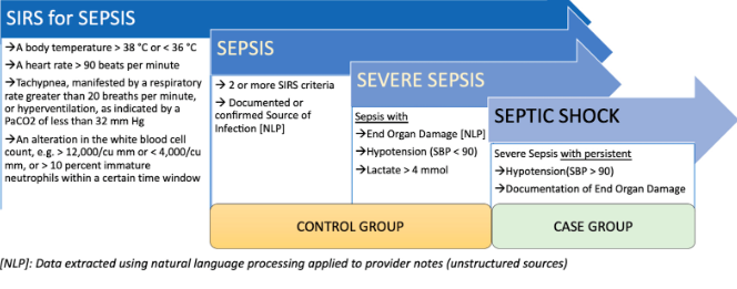
- Febrile
- Tachycardia
- Tachypnea
- Alteration in WBC count (over / under)
253
JP drain teaching
Drainage should be serosanguineous
If it gets clogged, notify the SURGEON STAT
If it gets clogged, notify the SURGEON STAT
254
Breast Cancer Post-Op teaching
Monitor for signs of infection
Monitor Pain, Vital Signs, Dressing
Drains care
ROM exercises - do not try to do full ROM immediately
Monitor Pain, Vital Signs, Dressing
Drains care
ROM exercises - do not try to do full ROM immediately
255
Post-Mastectomy Reconstruction
Breast implant -May need tissue expanders first to create a pocket for implant
Myocutaneous flap - Flap of skin, fat, and muscle transferred from donor site to the operative site
Myocutaneous flap - Flap of skin, fat, and muscle transferred from donor site to the operative site
256
Cervical Cancer
Third most common cancer of female genital system
Usually in 30’s-40’s
Human Papilloma Virus (HPV)
-HPV vaccine Gardasil/Cervarix
Usually in 30’s-40’s
Human Papilloma Virus (HPV)
-HPV vaccine Gardasil/Cervarix
257
Cervical Cancer Risk Factors
Unprotected sexual intercourse
Low socioeconomic status
Family history of cervical cancer (mother or sisters)
Sexual intercourse with uncircumcised men
Female offspring of mothers who took diethylstilbestrol (DES)
Infections with genital herpes or chronic chlamydia
Multiple sex partners
Cigarette smoking
Immunocompromised state
HIV infection
Oral contraceptive use
Moderate dysplasia on Pap smear within past 5 years
HPV infection
Low socioeconomic status
Family history of cervical cancer (mother or sisters)
Sexual intercourse with uncircumcised men
Female offspring of mothers who took diethylstilbestrol (DES)
Infections with genital herpes or chronic chlamydia
Multiple sex partners
Cigarette smoking
Immunocompromised state
HIV infection
Oral contraceptive use
Moderate dysplasia on Pap smear within past 5 years
HPV infection
258
Cervical cancer Assessment findings
Preinvasive cancer is asymptomatic
Classic symptom: Painless vaginal bleeding
Classic symptom: Painless vaginal bleeding
259
Late Symptoms of Cervical Cancer
Pain
Watery, blood-tinged vaginal discharge that becomes dark and foul-smelling
Leg pain along sciatic nerve
Flank pain – hydronephrosis (kidney swelling)
Unexplained weight loss
Dysuria
Pelvic pain
Hematuria
Watery, blood-tinged vaginal discharge that becomes dark and foul-smelling
Leg pain along sciatic nerve
Flank pain – hydronephrosis (kidney swelling)
Unexplained weight loss
Dysuria
Pelvic pain
Hematuria
260
Cervical Cancer Diagnostics
PAP smear
Colposcopy (examine the cervix)
Cervical biopsy
Endocervical scraping of endo-cervix wall - small amount of bleeding expected for up to 2 weeks
Colposcopy (examine the cervix)
Cervical biopsy
Endocervical scraping of endo-cervix wall - small amount of bleeding expected for up to 2 weeks
261
Loop Electrosurgical Exision Procedure (LEEP)
Local anesthetic in cervix
Thin loop wire electrode uses electrical current to cut away affected tissue
Both diagnostic and treatment
Thin loop wire electrode uses electrical current to cut away affected tissue
Both diagnostic and treatment
262
Laser Therapy for cervical cancer
Laser beam vaporizes abnormal tissue
263
Cryotherapy for cervical cancer
freezing of the cancer cells which will cause subsequent necrosis & tissue sloughing
264
Conization
removal of cone shaped section of cervical tissue & sent to lab for examination 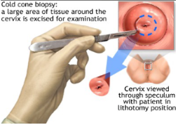
265
Radical trachelectomy
Cervix and upper part of vagina removed. Uterus remains intact.
266
Hysterectomy
Removal of the uterus
laparoscopic, vaginal, open depending on extent of disease
laparoscopic, vaginal, open depending on extent of disease
267
Nonsurgical Treatment of Cervical Cancer
Radiation: external beam, brachytherapy
Chemo: cisplatin, paclitaxel, carboplatin, 5 - fluorouracil (5 - FU)
5-FU is the drug of choice for cervical cancer chemo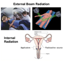
Chemo: cisplatin, paclitaxel, carboplatin, 5 - fluorouracil (5 - FU)
5-FU is the drug of choice for cervical cancer chemo