Lab 12: Ear
Study these flashcardsFree · spaced repetition · iPhone, Android, Mac & web
All cards
1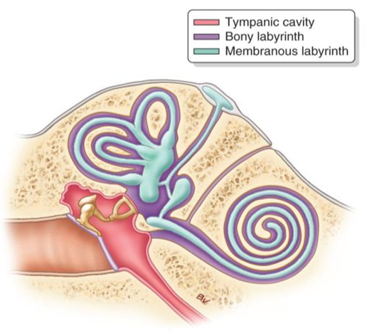
Image occlusion: name the hidden labels
2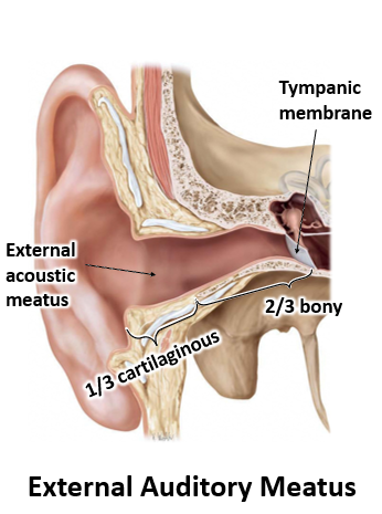
Image occlusion: name the hidden labels
3
External acoustic (auditory) meatus
Extends from the auricle to the tympanic membrane. Lateral 1/3 is cartilaginous, medial 2/3 is bony. Somatic sensory innervation is carried by CN X ("vagus behind tragus") and the auriculotemporal nerve (CN V3).
4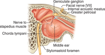
Image occlusion: name the hidden labels
5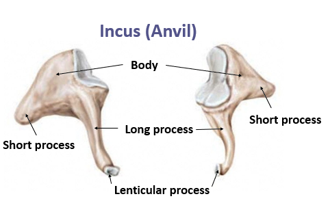
Image occlusion: name the hidden labels
6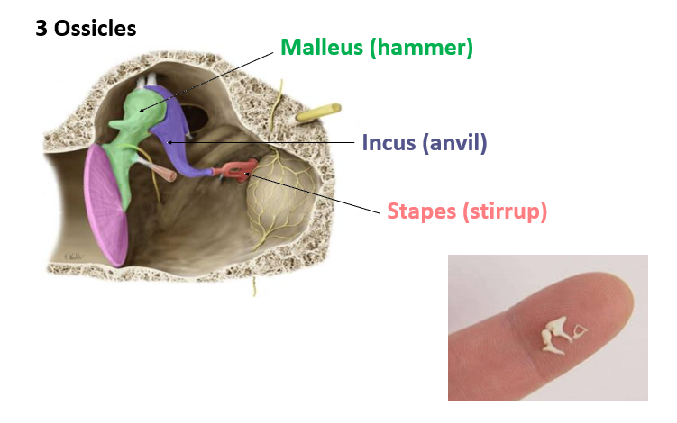
Image occlusion: name the hidden labels
7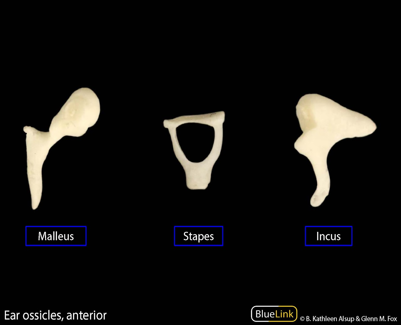
Image occlusion: name the hidden labels
8
Incus
The incus is connected to the malleus by a ball-and-socket joint at the articular facet. The lenticular process articulates with the head of the stapes.
9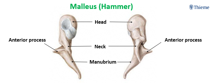
Image occlusion: name the hidden labels
10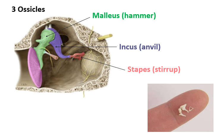
Image occlusion: name the hidden labels
11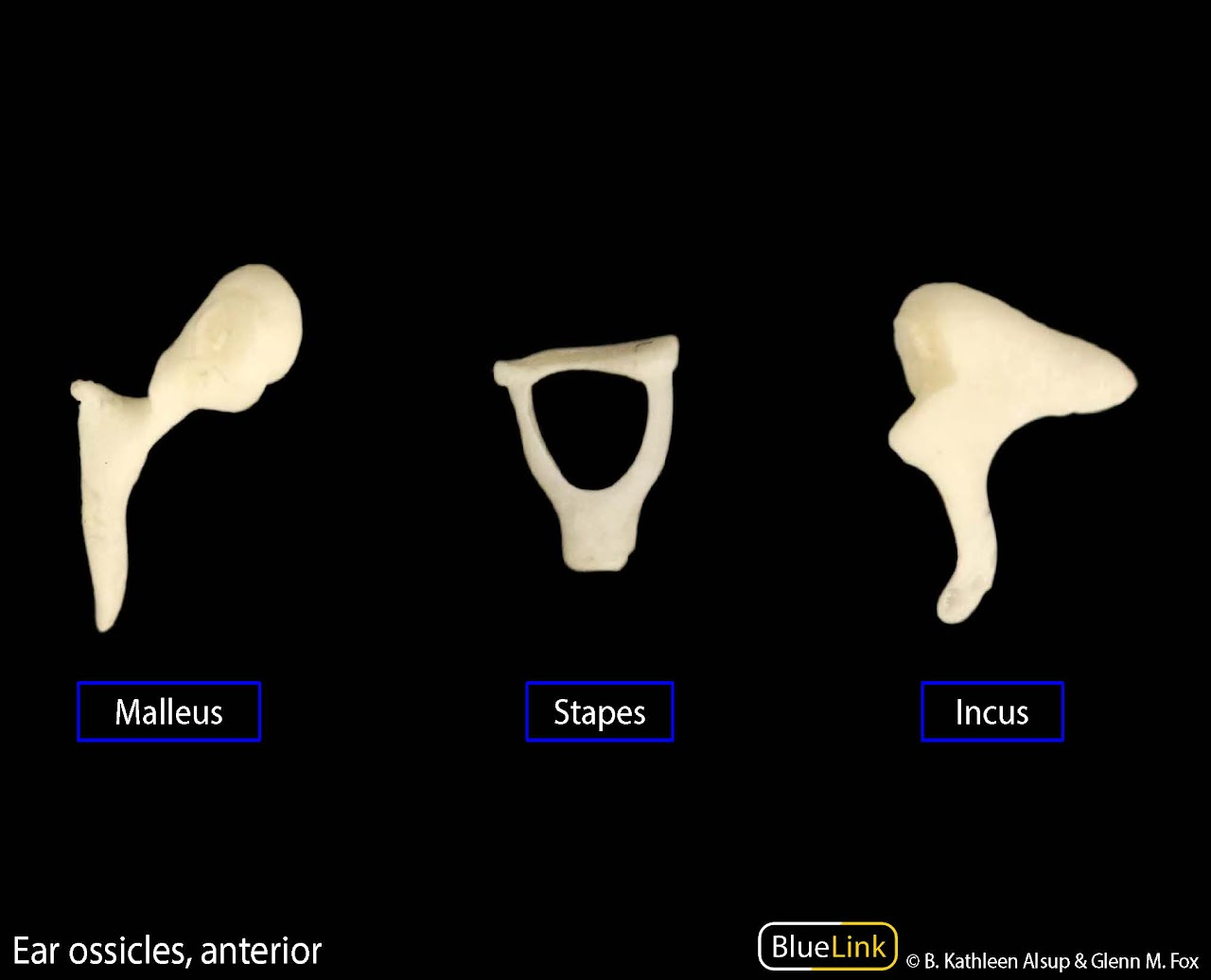
Image occlusion: name the hidden labels
12
Malleus
The malleus is the largest and heaviest of the three ossicles. The manubrium is attached to the medial surface of the tympanic membrane.
13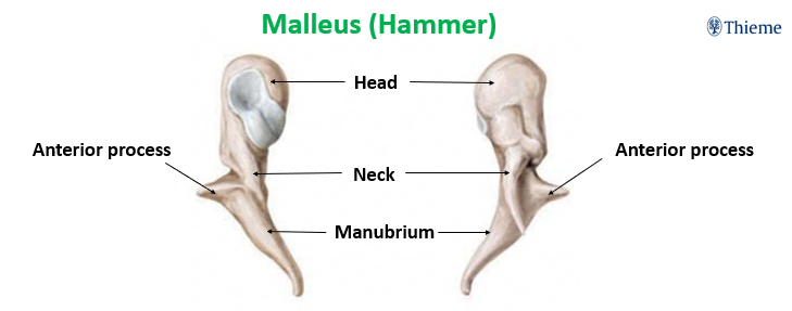
Image occlusion: name the hidden labels
14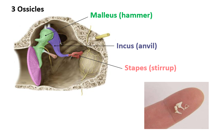
Image occlusion: name the hidden labels
15
Image occlusion: name the hidden labels
16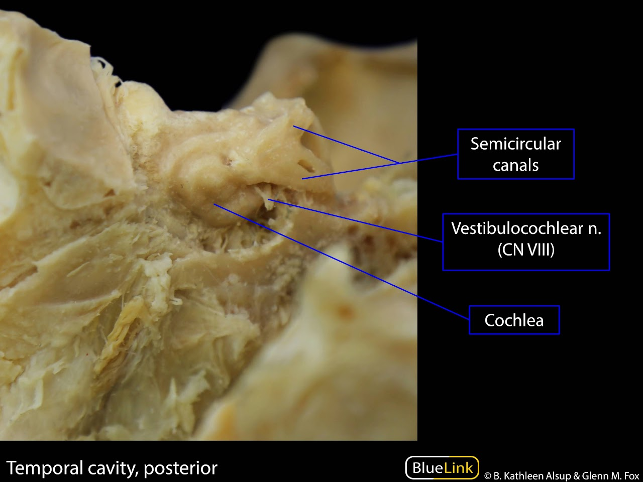
Image occlusion: name the hidden labels
17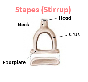
Image occlusion: name the hidden labels
18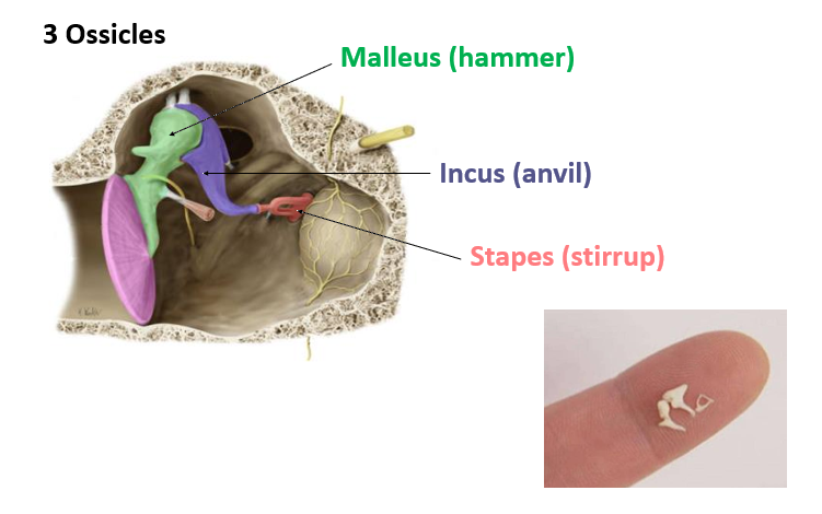
Image occlusion: name the hidden labels
19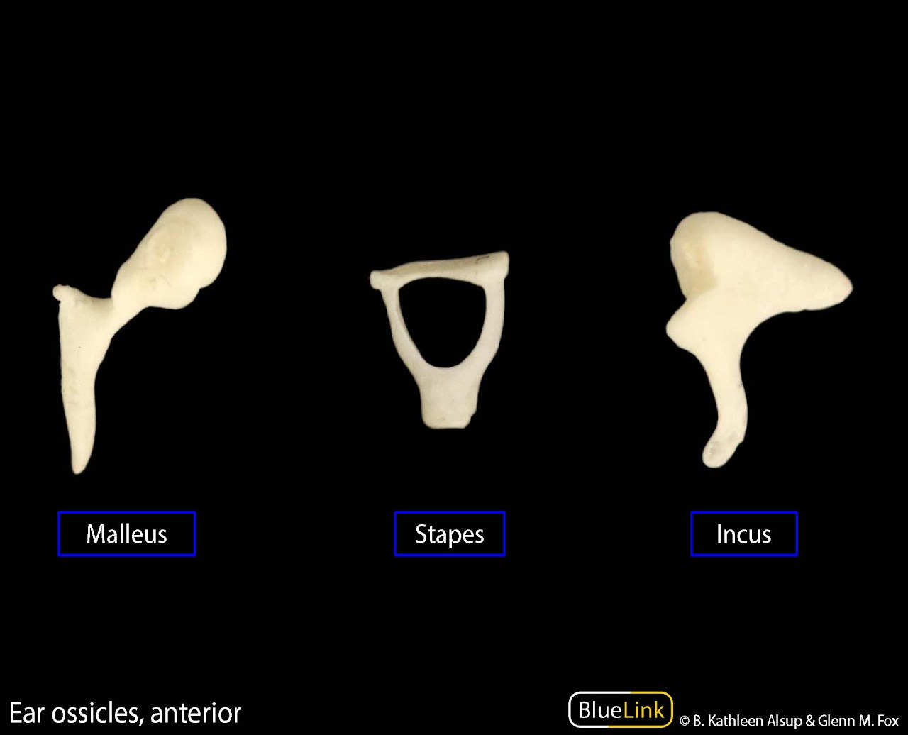
Image occlusion: name the hidden labels
20
Image occlusion: name the hidden labels
21
Stapes
The stapes is the smallest bone in the body. Each crus has a channel on its inner surface to reduce mass. The footplate has both osseous and cartilaginous components. The footplate is fixed within the oval window of the cochlea.
22
Tegmen tympani
A thin plate of the petrous part of the temporal bone that separates the cranial cavity and middle ear
23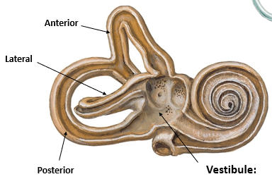
Image occlusion: name the hidden labels
24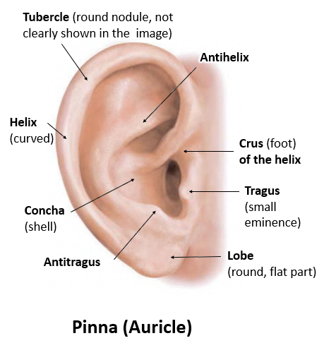
Image occlusion: name the hidden labels
25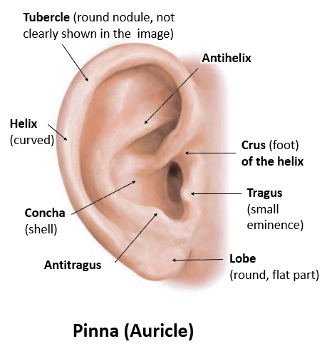
Image occlusion: name the hidden labels
26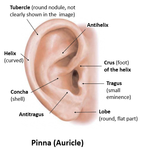
Image occlusion: name the hidden labels
27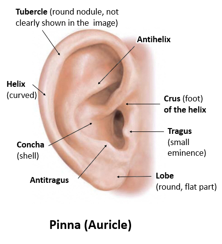
Image occlusion: name the hidden labels
28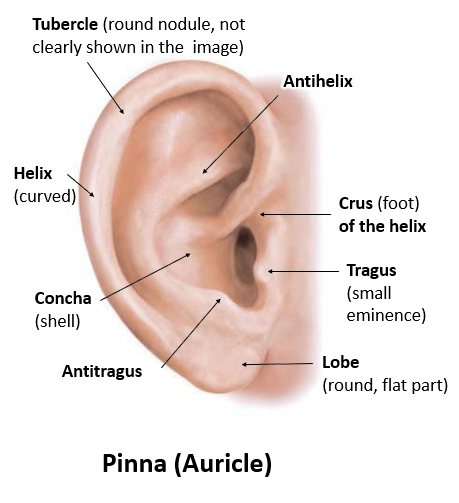
Image occlusion: name the hidden labels
29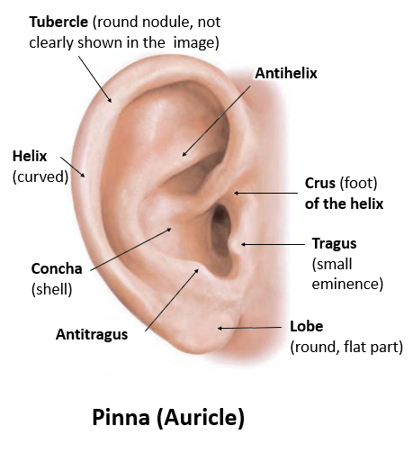
Image occlusion: name the hidden labels
30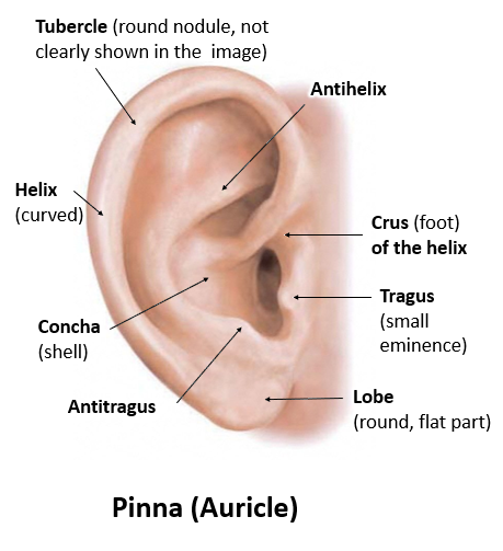
Image occlusion: name the hidden labels
31
Image occlusion: name the hidden labels
32
Image occlusion: name the hidden labels
33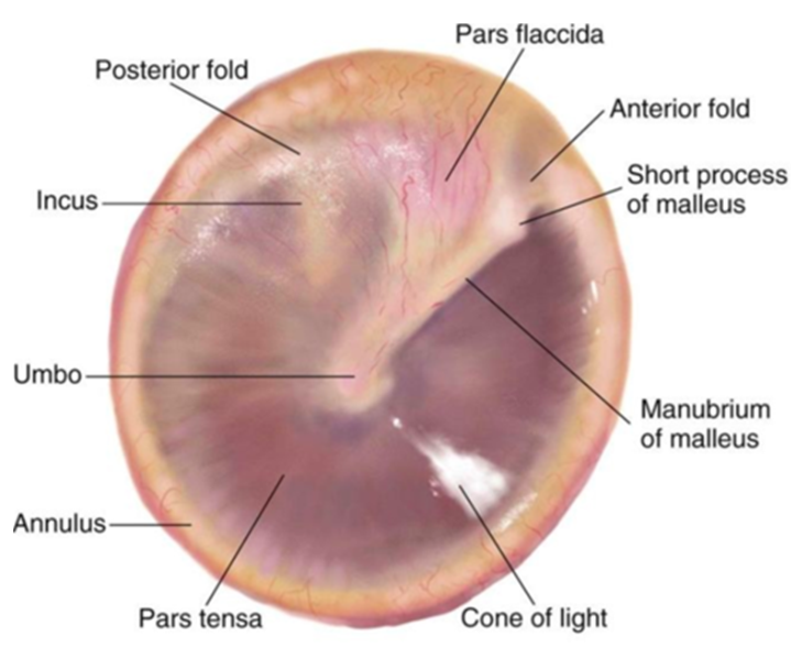
Image occlusion: name the hidden labels
34
Cone of light
A visible phenomenon which occurs upon examination of the tympanic membrane with an otoscope, which causes a cone-shaped reflection of light to appear in the anterior inferior quadrant
35
Image occlusion: name the hidden labels
36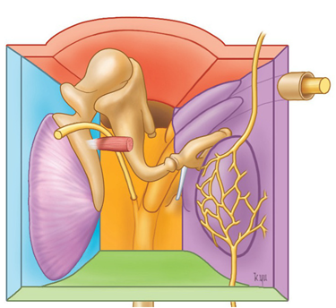
Image occlusion: name the hidden labels
37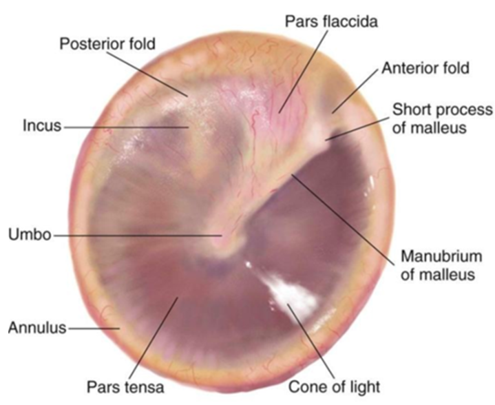
Image occlusion: name the hidden labels
38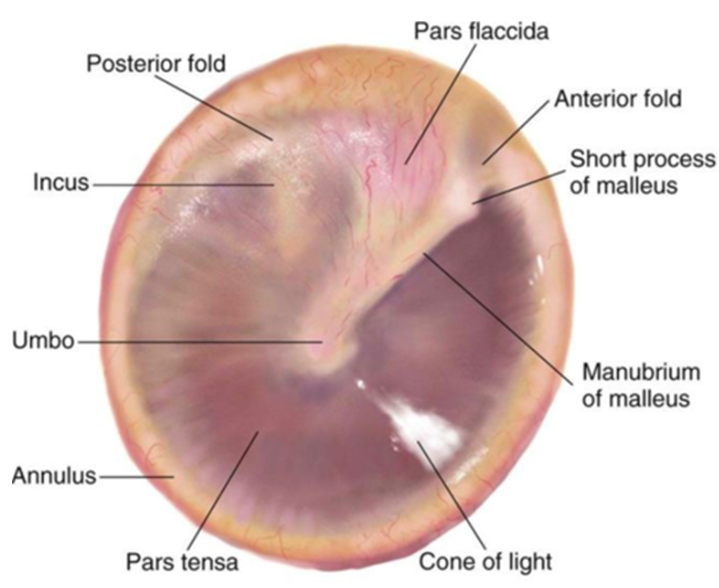
Image occlusion: name the hidden labels
39
Image occlusion: name the hidden labels
40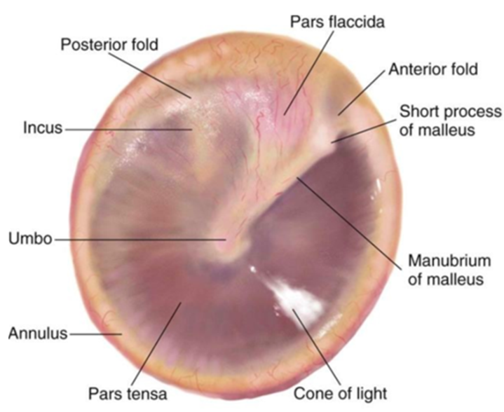
Image occlusion: name the hidden labels
41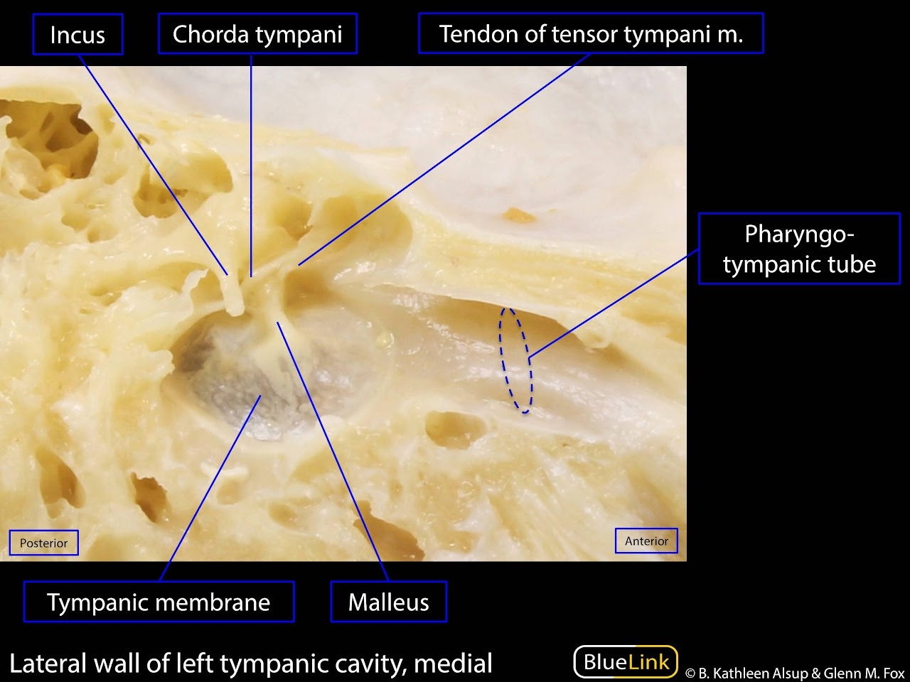
Image occlusion: name the hidden labels
42
Tympanic membrane
The "ear drum." It separates the external from the middle ear. It functions as a sound pressure receptor and transformer. It's external surface is innervated (sensory) by the vagus (CN X) & the auriculotemporal nerve (CN V3). It's inner surface is innervated (sensory) by the glossopharyngeal nerve (CN IX).
43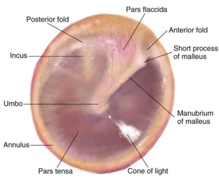
Image occlusion: name the hidden labels
44
Image occlusion: name the hidden labels
45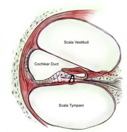
Image occlusion: name the hidden labels
46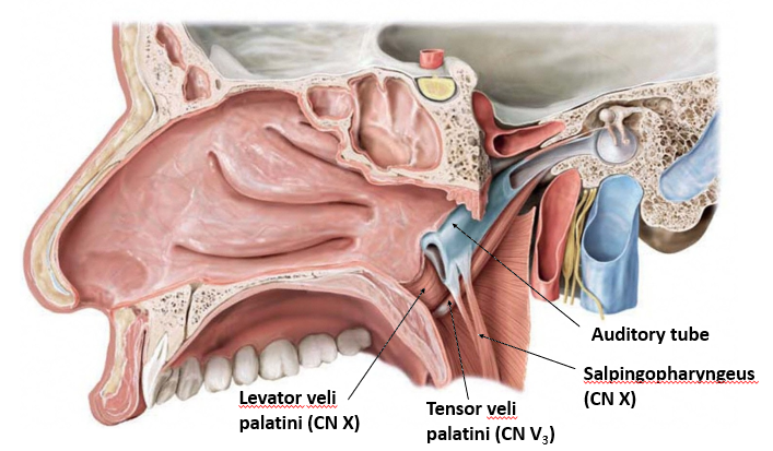
Image occlusion: name the hidden labels
47
Pharyngotympanic (Auditory, Eustachian) tube
Pharyngotympanic (Auditory, Eustachian) tube
Extends from the anterior wall of the middle ear to the lateral wall of the nasopharynx, approximately at the level of the inferior nasal concha. It equalizes pressure in middle ear and allows drainage of fluid. Salpingopharyngeus (CN X: pharyngeal plexus), levator veli palatini (CN X), tensor veli palatini (CN V3, MAST MATT) are the muscles that open the tube.
48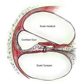
Image occlusion: name the hidden labels
49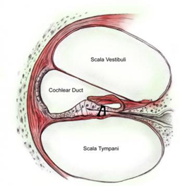
Image occlusion: name the hidden labels
50
Image occlusion: name the hidden labels
51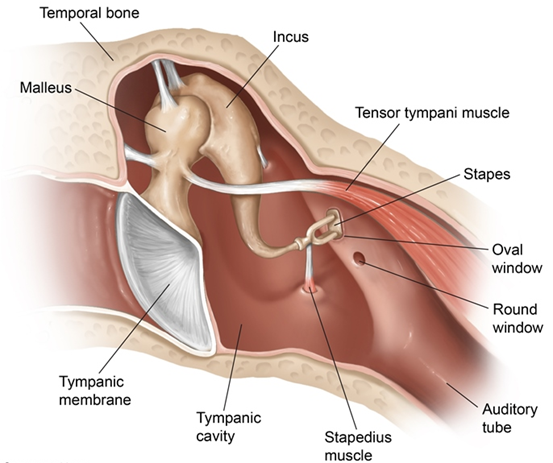
Image occlusion: name the hidden labels
52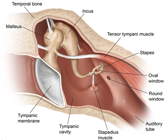
Image occlusion: name the hidden labels