Chap 1,2,3,4
Study these flashcardsFree · spaced repetition · iPhone, Android, Mac & web
All cards
1
Electrons
- Electrons are not always shared equally and are closer to one atom than the other.
- Polar vs. Nonpolar
- Dipole moment
- Polar vs. Nonpolar
- Dipole moment
2
Polar vs. Nonpolar
- Polar molecules contain O,N,S,P
- Nonpolar molecules contain C,H
- Nonpolar molecules contain C,H
3
Dipole Moment
- measure of seperation of electrical charge
- has a magnitude and direction
- has a magnitude and direction
4
The Inner Life of the Cell
5
Eukaryotic Cells
- Different eukaryotic cells have the same organelles
- differ in number, shape and distribution of organelles
- differ in number, shape and distribution of organelles
6
Eukaryotic cell cytoplasm
- Eukaryotic cell cytoplasm is a crowded compartment
- Cytoplasm = organelles + cytosol
- Cytoplasm = organelles + cytosol
7
a simpler system
- Much of what we know about cell biology comes from cultured cells
- Cells from Henrietta Lacks (HeLa) have been cultured since 1951
- Cells from Henrietta Lacks (HeLa) have been cultured since 1951
8
Bonds
- Atoms are held together by bonds
- Covalent and Noncovalent bonds
- Covalent and Noncovalent bonds
9
Covalent Bonds
- Sharing of electrons
- Polar vs Nonpolar
- Strong (stronger than thermal energies of random collisions)
- keep organic molecules together
- broken using enzymes
- Energy is stored in bonds and breaking bonds can release energy for useful work
- need a "just right" attraction ex. If molecules are too close repelling will take place
- Polar vs Nonpolar
- Strong (stronger than thermal energies of random collisions)
- keep organic molecules together
- broken using enzymes
- Energy is stored in bonds and breaking bonds can release energy for useful work
- need a "just right" attraction ex. If molecules are too close repelling will take place
10
Noncovalent bonds
- relatively weak
- not sharing of electrons, but attractive forces between atoms
- imp. for folding macromolecules into proper shape (e.g., enzymes)
- imp. for holding molecules together
- electrostatic attraction
- not sharing of electrons, but attractive forces between atoms
- imp. for folding macromolecules into proper shape (e.g., enzymes)
- imp. for holding molecules together
- electrostatic attraction
11
electrostatic attraction
- attractions between molecules with polar, covalent bonds can form complementary charges
- relatively weak and easily break and reform
- although weak, many together can be strong (think of Velcro®)
- relatively weak and easily break and reform
- although weak, many together can be strong (think of Velcro®)
12
Four types of noncovalent bonds bring molecules together:
1. Ionic bonds*
2. Hydrogen bonds
3. van der Waals attractions*
4. Hydrophobic force*
2. Hydrogen bonds
3. van der Waals attractions*
4. Hydrophobic force*
13
Hydrogen Bonds
- Between bonded electronegative atom and bonded H atom
H is “sandwiched” between two electron-attracting atoms
- H bonds form between most polar molecules and are especially important in the behavior of water.
- Partial charges between molecules attract each other and from hydrogen bonds.
- Hydrogen bonds stabilize adjacent molecules to form arrays or crystals, e.g., water, or stabilize 3 dimensional configurations of large molecules, e.g., proteins.
H is “sandwiched” between two electron-attracting atoms
- H bonds form between most polar molecules and are especially important in the behavior of water.
- Partial charges between molecules attract each other and from hydrogen bonds.
- Hydrogen bonds stabilize adjacent molecules to form arrays or crystals, e.g., water, or stabilize 3 dimensional configurations of large molecules, e.g., proteins.
14
Importance of synthesis by polymerization of small molecules
- most cellular structures (e.g., ribosomes, chromosomes, membranes) are made up by linear molecules (macromolecules)
15
Synthesis of macromolecules
monomers and polymers
- Lipids are macromolecules, but not polymers
- Condenstation vs Hydrolysis
- Lipids are macromolecules, but not polymers
- Condenstation vs Hydrolysis
16
Condenstation vs. Hydrolysis
17
Macromolecules
1. Carbohydrates (monosaccharides)
2. Lipids
3. Nucleic acids (nucleotides)
4. Proteins (amino acids)
2. Lipids
3. Nucleic acids (nucleotides)
4. Proteins (amino acids)
18
Polymers
- Carbohydrates (monosaccharides)
- Nucleic acids (nucleotides)
- Proteins (amino acids)
- Nucleic acids (nucleotides)
- Proteins (amino acids)
19
creation of macrocellular assemblies
20
Carbohydrates
Forms:
- monosaccharides (monomer)
- oligosaccharides (short polymer; 3–10 sugars)
- polysaccharides (long polymer)
Functions:
- chemical energy stored in covalent bonds is primary source of cell energy
- sugar polymers are the most abundant structural components on earth
- monosaccharides (monomer)
- oligosaccharides (short polymer; 3–10 sugars)
- polysaccharides (long polymer)
Functions:
- chemical energy stored in covalent bonds is primary source of cell energy
- sugar polymers are the most abundant structural components on earth
21
Structure of monosaccharides (simple sugars)
Glucose is a key molecule in the metabolism of plants and animals
22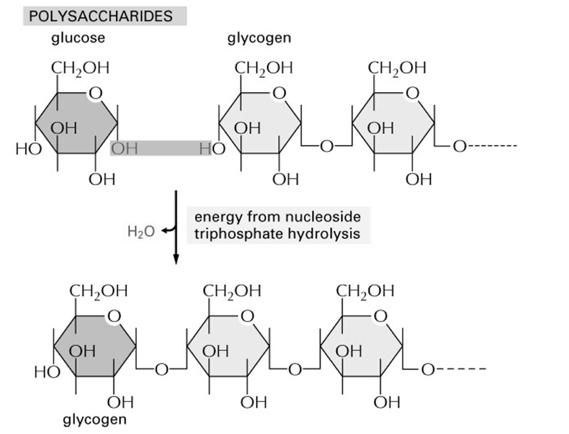
sythesis of polysachrides
23
Disaccharides
- 2 sugar units
- ex. Lactose (milk sugar) glucose + galactose
- ex. Lactose (milk sugar) glucose + galactose
24
Glycosidic bond
in between disaccharides
- covalent bond
- covalent bond
25
Polysaccharides
- many sugar units
- imp. for energy storage and cell structure
- storange vs structural polysacride
- imp. for energy storage and cell structure
- storange vs structural polysacride
26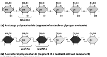
storange vs structural polysacride
27
Lipids
Forms: (diverse structures; all mostly hydrophobic)
-fats
-steroids (sterols)
-phospholipids
Functions:
-energy storage (2X as much as carbohydrates).
-membrane structure
-specific biological functions
-fats
-steroids (sterols)
-phospholipids
Functions:
-energy storage (2X as much as carbohydrates).
-membrane structure
-specific biological functions
28
Fats
- CH2 (carboxyl) - non poplar molecules
- storage form of energy - carbons are broken off
- Glycerol + Fatty acid = triacylglycerol
- 3 fatty acids esterified (combined to an alchol or alchol combined to acid) to glycerol
- Storage form of fat in plants and animals
- storage form of energy - carbons are broken off
- Glycerol + Fatty acid = triacylglycerol
- 3 fatty acids esterified (combined to an alchol or alchol combined to acid) to glycerol
- Storage form of fat in plants and animals
29
Fatty acid
- saturated vs. unsaturated
- amphipathic (polar/nonpolar) one end hydrophilic the other hydrophobic
- size (usually even & 14 to 20 carbons)
- one hydrogen (double bond) - forms a kink
- amphipathic (polar/nonpolar) one end hydrophilic the other hydrophobic
- size (usually even & 14 to 20 carbons)
- one hydrogen (double bond) - forms a kink
30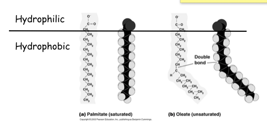
saturated vs unsaturated fatty acid
31
Fats (more specific to their bonds)
- double bonds make kinks
- the more double bonds the more liquid at RT
- animal fats tend to be saturated
- plants fats tend to be unsaturated and liquid (oil)
- hydrogenate vegetable fat to make solid margarine
- Storage form in plants & animals
- the more double bonds the more liquid at RT
- animal fats tend to be saturated
- plants fats tend to be unsaturated and liquid (oil)
- hydrogenate vegetable fat to make solid margarine
- Storage form in plants & animals
32
straight vs double bonds
33
Fats and Temperature
- lower temperature it will freeze
- temp goes up will eventually become liquid
- temp goes up will eventually become liquid
34
Phospholipids
- glycerol + 2 fatty acids + phosphate group bound to a polar group
- amphipathic and make up the backbone of cell membranes
- some are kink and some are straight - changes the fluid of the membrane
- third carbon is bonded to a phosphate and something else (in this case a choline group)
- This phosphate group is very hydrophilic (has a very hydrophilic and very hydrophobic side) - important in the formation of the plasma membrane
- amphipathic and make up the backbone of cell membranes
- some are kink and some are straight - changes the fluid of the membrane
- third carbon is bonded to a phosphate and something else (in this case a choline group)
- This phosphate group is very hydrophilic (has a very hydrophilic and very hydrophobic side) - important in the formation of the plasma membrane
35
Steroids (sterols)
- characteristic 4 ring structure
- cholesterol:
** membrane component
** hormone precursor (non perscription hormones)
- cholesterol:
** membrane component
** hormone precursor (non perscription hormones)
36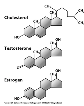
Types of steroids
37
Proteins (amino acids)
- experiance condensation rxns
DNA - transmission of genetic info
RNA - structural and catalytic job
Forms:
- nucleotides (monomers)
- nucleic acids (polymers)--RNA and DNA
Functions:
- storage and transmission of genetic info
- structure
- catalytic
DNA - transmission of genetic info
RNA - structural and catalytic job
Forms:
- nucleotides (monomers)
- nucleic acids (polymers)--RNA and DNA
Functions:
- storage and transmission of genetic info
- structure
- catalytic
38
Nucleotides
- nucleic acid monomers (Building Blocks to make polynucleotide)
- Bond breaks by the phosphate which is the energy source for forming the nucleotide
- 2 types of sugars: Deoxyribose & Ribose
- #2 prime Carbon - OH group different in DNA (DEOXY) H (store house information - 2 daughter cells exact same copy of genes **needs to be stable versus RNA (OH) (does not have to be as stable)
- RNA does not use U; looks very similar to thymine but NO methyl group
- Purines and Pyrimidnes
- Bond breaks by the phosphate which is the energy source for forming the nucleotide
- 2 types of sugars: Deoxyribose & Ribose
- #2 prime Carbon - OH group different in DNA (DEOXY) H (store house information - 2 daughter cells exact same copy of genes **needs to be stable versus RNA (OH) (does not have to be as stable)
- RNA does not use U; looks very similar to thymine but NO methyl group
- Purines and Pyrimidnes
39
Purines and Pyrimidnes
Purines: have two rings
Pyrimidines: single ring
Pyrimidines: single ring
40
Nucleidic Acids Hydrolysis Reaction
41
DNA fucntions
- template for protein production
- template for daughter cell genome
- Two chains of DNA molecules can H-bond to form DNA double-helix
- some regions code for polypeptides
- template for protein production and daughter cells
- the nucleotides could hydrogen bond to another nucleotide of the strands
- template for daughter cell genome
- Two chains of DNA molecules can H-bond to form DNA double-helix
- some regions code for polypeptides
- template for protein production and daughter cells
- the nucleotides could hydrogen bond to another nucleotide of the strands
42
RNAs
- Although RNAs generally consist of single strands, they can fold back on themselves to form double-stranded segments
- There are double-stranded RNA viruses
- RNA - does not have a double helix like DNA - more complex structure — in some region sit folds back on themselves (the structure is important to its function!!) - mutation will lead to a different shape —— bad FUNCTION
- RNA come together in the cytoplasm to make a protein
- There are double-stranded RNA viruses
- RNA - does not have a double helix like DNA - more complex structure — in some region sit folds back on themselves (the structure is important to its function!!) - mutation will lead to a different shape —— bad FUNCTION
- RNA come together in the cytoplasm to make a protein
43
RNA functions
RNA functions
1. genetic information and transmission - (virus)
2. catalytic enzymes (activation energy not broken down as part of the rxn) -- ribozymes (RNA ENZYMES) — example. peptransferace ** a ribosome RNA NOT A PROTEIN
3. structure -- ribosome structure
1. genetic information and transmission - (virus)
2. catalytic enzymes (activation energy not broken down as part of the rxn) -- ribozymes (RNA ENZYMES) — example. peptransferace ** a ribosome RNA NOT A PROTEIN
3. structure -- ribosome structure
44
Protein
Forms:
- polymers of 20 different AA (amino acids) in genome
- unique arrangement of AA gives protein unique shapes allowing selective interactions
- Typical cell may have 10,000 different proteins
Functions:
- Molecular tools and machines that perform nearly all the cell’s activities
- polymers of 20 different AA (amino acids) in genome
- unique arrangement of AA gives protein unique shapes allowing selective interactions
- Typical cell may have 10,000 different proteins
Functions:
- Molecular tools and machines that perform nearly all the cell’s activities
45
synthesis protein polymer
- Reaction occurs on ribosomes in the cell
- protein + amino acid --> protein (w energy from nucleoside triphospahate hydrolysis)
- protein + amino acid --> protein (w energy from nucleoside triphospahate hydrolysis)
46
Four levels of protein structure
- average = 100 amino acids long
- shape and structure of polypeptide at different levels
1. Primary Structure
2. Secondary Structure
3. Tertiary Structure
4. Quaternary Structure
- shape and structure of polypeptide at different levels
1. Primary Structure
2. Secondary Structure
3. Tertiary Structure
4. Quaternary Structure
47
Primary Structure of a protein
e.g., Arg-Gly-Asp-Ser (RGDS)
- precise order of AA determined by genes
- sequence determines protein shape
- held together by peptide bonds
- Changes in amino acid sequence caused by changes (mutations) in DNA
- Average protein in human genome contains ~100 aa
- Order of amino acids: (the sequence of the polypeptide) - in the DNA there is a series of nucleotides and the genes determine the order - changes the shape which changes the function
- Held together by peptide covalent bonds
- MUTATION = CHANGE = WRONG AMINO ACIDS ** change in the primary structure
- precise order of AA determined by genes
- sequence determines protein shape
- held together by peptide bonds
- Changes in amino acid sequence caused by changes (mutations) in DNA
- Average protein in human genome contains ~100 aa
- Order of amino acids: (the sequence of the polypeptide) - in the DNA there is a series of nucleotides and the genes determine the order - changes the shape which changes the function
- Held together by peptide covalent bonds
- MUTATION = CHANGE = WRONG AMINO ACIDS ** change in the primary structure
48
Amino Acid Side Chain
- Once part of a polypeptide, an individual amino acid is called a residue
- Side chains (aka R groups) give each AA its unique properties
- Some side chains are nonpolar and some are neg or pos uncharged polar
- R chains could be hydrophilic or hydrophobic
- DO NOT have to know abbreviations
- hydrophobic (center of protein - away from water)
- If charged, could bond to water - they are polar
- If not charged, but polar bonds they will have hydrogen bonds
- Non polar - on the INSIDE of the protein
- Lots of hydrophll or phobic in a row will change the hold of the protein
- Side chains (aka R groups) give each AA its unique properties
- Some side chains are nonpolar and some are neg or pos uncharged polar
- R chains could be hydrophilic or hydrophobic
- DO NOT have to know abbreviations
- hydrophobic (center of protein - away from water)
- If charged, could bond to water - they are polar
- If not charged, but polar bonds they will have hydrogen bonds
- Non polar - on the INSIDE of the protein
- Lots of hydrophll or phobic in a row will change the hold of the protein
49
Side Chain and peptide bonds
- side chains are not part of peptide bonds
- even though the backbone amino and carboxyl are polar, the H- bond with each other and not with
- water, so the properties of the R-groups determines the hydrophobicity of the polypeptide
- even though the backbone amino and carboxyl are polar, the H- bond with each other and not with
- water, so the properties of the R-groups determines the hydrophobicity of the polypeptide
50
Secondary Structure
- describes conformation (spatial organization) of polypeptide chain portions
- formed between N-H and C=O of polypeptide backbone and doesn’t involve R-groups. Thus can be formed by many AA sequences (the identities of the R-groups, however, puts constraints on which type of conformations can form)
- preferred ones provide maximum possible number of H–bonds between neighboring AA
- there are two common conformations found in many proteins -- alpha(α) helix and beta (Ω) pleated-sheet
- formed between N-H and C=O of polypeptide backbone and doesn’t involve R-groups. Thus can be formed by many AA sequences (the identities of the R-groups, however, puts constraints on which type of conformations can form)
- preferred ones provide maximum possible number of H–bonds between neighboring AA
- there are two common conformations found in many proteins -- alpha(α) helix and beta (Ω) pleated-sheet
51
α-helix
- cylindrical, twisting spiral
- H-bonds between AA of same molecule
- peptide bonds of
every 4th aa close
- H-bonds parallel to cylinder axis
- R-groups to outside
- α-helices can cross lipid bilayer
e.g., wool protein; can be stretched
- H-bonds between AA of same molecule
- peptide bonds of
every 4th aa close
- H-bonds parallel to cylinder axis
- R-groups to outside
- α-helices can cross lipid bilayer
e.g., wool protein; can be stretched
52
α-helix structural components **
- hydrogen bonds keep it together
- hydrogen bonding (hydrogen in positively charged)
- hydrogen bonds are parallel t the axis of the helix
- 3.6 residues
- could be embedded in hydrophobic lipid bilayer (takes 22 amino acids to make through)
- hydrogen bonding (hydrogen in positively charged)
- hydrogen bonds are parallel t the axis of the helix
- 3.6 residues
- could be embedded in hydrophobic lipid bilayer (takes 22 amino acids to make through)
53
what will a - helices sometimes take part in?
- Sometimes 2-3 α-helices wrap around each other
- Intertwined
α helices can
form a coiled- coil
- Heptad (7 aa repeat)
~ every 4th aa is hydrophobic and so a stripe of hydrophobic on inner surface
- attract together bc they are repelled from water — more energetically stable
- Intertwined
α helices can
form a coiled- coil
- Heptad (7 aa repeat)
~ every 4th aa is hydrophobic and so a stripe of hydrophobic on inner surface
- attract together bc they are repelled from water — more energetically stable
54
B -pleated sheet
- several polypeptide segments lie side-by-side; adopt folded or pleated conformation
- H bonds perpendicular to molecule axis
- In some regions there could be hydrogen bonding between CO & O2
- If it could be strengthen - it would not, because you are breaking covalent bonds ex. why silk wont stretch
e.g., silk protein; can’t be stretched
- H bonds perpendicular to molecule axis
- In some regions there could be hydrogen bonding between CO & O2
- If it could be strengthen - it would not, because you are breaking covalent bonds ex. why silk wont stretch
e.g., silk protein; can’t be stretched
55
example of B - pleated sheet
- In the average polypeptide, 60% of the chain is α-helix or �- sheet
- Chain portions not organized into α-helix or
�-pleated sheet may
consist of hinges, turns, loops
- often most flexible portions of chain & sites of greatest biological activity
- Chain portions not organized into α-helix or
�-pleated sheet may
consist of hinges, turns, loops
- often most flexible portions of chain & sites of greatest biological activity
56
Tertiary Structure
- Makes up the conformation of the entire protein
- noncovalent interactions between R groups in same chain
- Fibrous proteins (highly elongated shape - more stretched out) - long strands or flattened sheets that resist pulling
- Globular proteins (shaped like a ball) – most proteins in the cell; compact shape; chains folded & twisted into
- complex shapes
- noncovalent interactions between R groups in same chain
- Fibrous proteins (highly elongated shape - more stretched out) - long strands or flattened sheets that resist pulling
- Globular proteins (shaped like a ball) – most proteins in the cell; compact shape; chains folded & twisted into
- complex shapes
57
Tertiary Structure (more specific to the bonds)
- Noncovalent interactions between R groups in same polypeptide chain (VDW and hydrogen bonds)
Disulfide (—SS—) bridge:
- cysteine (—SH) group often covalently linked to another cysteine
- helps stabilize protein structure - once it is formed these covalent bonds are added to help STABILIZE
- oxidation and reduction
- 1 bond that is covalent: disulfide
ex. hair perm: breaks down disulfide to put hair in a certain shape
Disulfide (—SS—) bridge:
- cysteine (—SH) group often covalently linked to another cysteine
- helps stabilize protein structure - once it is formed these covalent bonds are added to help STABILIZE
- oxidation and reduction
- 1 bond that is covalent: disulfide
ex. hair perm: breaks down disulfide to put hair in a certain shape
58
Protein Folding within Tertiary Structure
- Protein folding is inherent in the molecule
- may be sped up by accessory proteins
a. Denaturation and rentuaration of rbonuclease
b. Synthesis and self-assembly of ribonuclease
ex. take cow protein - you could change shape and ph to make it stretched out — back to same — back to original conditions
- may be sped up by accessory proteins
a. Denaturation and rentuaration of rbonuclease
b. Synthesis and self-assembly of ribonuclease
ex. take cow protein - you could change shape and ph to make it stretched out — back to same — back to original conditions
59
Unfolded polypeptide (Tertiary Structure)
60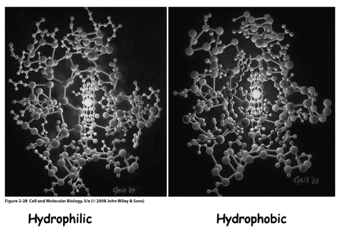
Cytochrome C
61
Quaternary Structure
- the linking of polypeptide chains to form multisubunit functional protein (e.g., dimer, trimer or tetramer)
- intermolecular R group interactions
subunits synthesized separately
- may be linked by disulfide bonds, but more often noncovalent bonds
- Molecular chaperones
Note: quaternary structure DOES NOT mean four subunits
- intermolecular R group interactions
subunits synthesized separately
- may be linked by disulfide bonds, but more often noncovalent bonds
- Molecular chaperones
Note: quaternary structure DOES NOT mean four subunits
62
Quaternary Structure (more specific)
- Many proteins do not have the quaternary structure - a dimer
- alpha and beta come together to create a dimer, it functions as a dimer. Meaning, when the alpha and beta are separate they do not have a function; when they are together they do!
- Contains 2,3,4,5 subunits quit DOES NOT MEAN 4; our example just shows that
- R group interaction between polypeptides
- disulfide bonds (covalent interactions holding together)
- alpha and beta come together to create a dimer, it functions as a dimer. Meaning, when the alpha and beta are separate they do not have a function; when they are together they do!
- Contains 2,3,4,5 subunits quit DOES NOT MEAN 4; our example just shows that
- R group interaction between polypeptides
- disulfide bonds (covalent interactions holding together)
63
Molecular chaperones
- not all proteins assume proper tertiary or quaternary structure by self-assembly
- interactions with other proteins may inhibit correct folding
- chaperones help unfolded/misfolded proteins
- When proteins are being synthesized. a lot of times they have things helping them out: chaperones
- Endoplasmic Retic: most of the proteins within the ER or cytoplasm help the protein fold correctly (need ATP for this interaction); shield them from the cytoplasm
- Temperature effects protein folding
- too many misfiled proteins - cell will stop producing protein - increasing chaperone proteins so that proteins can finally be folded in the correct way
- interactions with other proteins may inhibit correct folding
- chaperones help unfolded/misfolded proteins
- When proteins are being synthesized. a lot of times they have things helping them out: chaperones
- Endoplasmic Retic: most of the proteins within the ER or cytoplasm help the protein fold correctly (need ATP for this interaction); shield them from the cytoplasm
- Temperature effects protein folding
- too many misfiled proteins - cell will stop producing protein - increasing chaperone proteins so that proteins can finally be folded in the correct way
64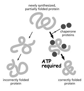
Molecular chaperones = The Process
65
Protein vs. Polypeptide
- Polypeptides --chains of AA
- Proteins -- one or more polypeptides folded into a specific conformation; a functional molecule
- enzyme is a protein, but to unfold the protein and not in its functional form it is called a polypeptide
- Proteins -- one or more polypeptides folded into a specific conformation; a functional molecule
- enzyme is a protein, but to unfold the protein and not in its functional form it is called a polypeptide
66
Protein domains
- (level of organization distinct from primary to quaternary; sometimes called “supersecondary structure”)
- proteins often composed of 2 or more distinct modules (domains) that fold independently of one another (structurally or functionally)
- often represent parts that function in semi-independent manner
- part of a protein sequence that can function, evolve and exist independently of the rest of the protein chain
- usually 25-500 AA in length
- proteins often composed of 2 or more distinct modules (domains) that fold independently of one another (structurally or functionally)
- often represent parts that function in semi-independent manner
- part of a protein sequence that can function, evolve and exist independently of the rest of the protein chain
- usually 25-500 AA in length
67
Protein Domains images
68
cutting up single polypeptide..
- It would still have its same shape
Domains: its own little region folding on its own
- protein molecule could be made up of two differnt domains
Domains: its own little region folding on its own
- protein molecule could be made up of two differnt domains
69
Proteins are built of structural units called..
- DOMAINS
- part of a protein sequence that can function, evolve and exist independently of the rest of the protein chain
- usually 25-500 AA in length
- multi-domain proteins probably evolved by the fusion of genes that once encoded separate proteins
- part of a protein sequence that can function, evolve and exist independently of the rest of the protein chain
- usually 25-500 AA in length
- multi-domain proteins probably evolved by the fusion of genes that once encoded separate proteins
70
HOW PROTEINS ARE CONTROLLED
- Many times the activity of proteins is controlled by altering the shape of the protein
- Most proteins are allosteric; can adopt 2 or
more conformations, which is regulated by something binding to its non- active site
- If a cell could change the shape of the protein - it could change its function
allosteric: changing the shape in order to bind to the enzyme
- Most proteins are allosteric; can adopt 2 or
more conformations, which is regulated by something binding to its non- active site
- If a cell could change the shape of the protein - it could change its function
allosteric: changing the shape in order to bind to the enzyme
71
ADP
regulates the activity of this hypothetical enzyme
- ADP can only bind to closed conformation
- Closed protein now more likely to bind substrat
- ADP can only bind to closed conformation
- Closed protein now more likely to bind substrat
72
Protein phosphorylation
- can regulate protein activity
- cell signaling - surface of the cell causes shape change to the protein of the cell
- cell signaling - surface of the cell causes shape change to the protein of the cell
73
Protein phosphorylation (the process)
- This shows a polypeptide folded in its tertiary shape — it is going left to right (showing a SHAPE CHANGE) — adding a phosphate covalently to change the shape of the polypeptide protein
Kinase: are specific; need specific phosphate in a certain way to change the shape of the polypeptide
- Phophtase: an enzyme that takes off phosphate to revert back to original condition (shown in the right image)
Changing the sequence is CHANGING the shape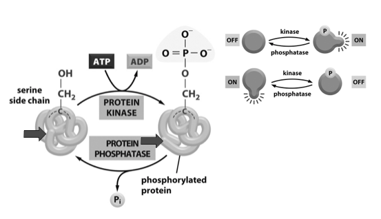
Kinase: are specific; need specific phosphate in a certain way to change the shape of the polypeptide
- Phophtase: an enzyme that takes off phosphate to revert back to original condition (shown in the right image)
Changing the sequence is CHANGING the shape
74
GTP - binding proteins
Many GTP-binding proteins can be switched on/off by a gain/loss of a phosphate group
- different between GTP and GDP - this is not a phosphorylation, this is a binding site that activates and inactivates
example of this is: vision (photons binding and G-proteins changes shape and ions temporarily — vision is very quick) — rapid change by changing the charge
- Large conformational change protein synthesis enlongation factor releases tRNA when GTP to GDP
- different between GTP and GDP - this is not a phosphorylation, this is a binding site that activates and inactivates
example of this is: vision (photons binding and G-proteins changes shape and ions temporarily — vision is very quick) — rapid change by changing the charge
- Large conformational change protein synthesis enlongation factor releases tRNA when GTP to GDP
75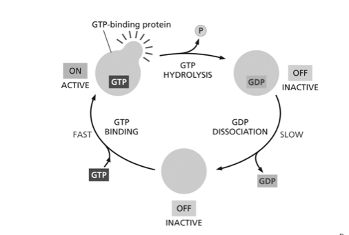
GTP - binding proteins (the process)
76
Large conformational change protein synthesis
- Large conformational change protein synthesis enlongation factor releases tRNA when GTP to GDP
- During Protein Synthesis: G Protein binds to GTP and GDP to make different shape and activity
- When GTP to GDP tRNA is release with the change of shape, meaning 2-3 phosphates could make a large difference in the shape of the protein
- Elongations: 20 amino acids added per second >> a fast process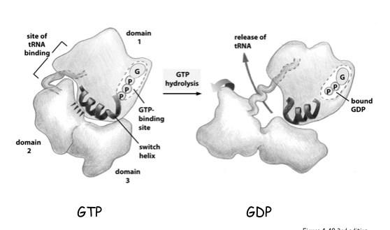
- During Protein Synthesis: G Protein binds to GTP and GDP to make different shape and activity
- When GTP to GDP tRNA is release with the change of shape, meaning 2-3 phosphates could make a large difference in the shape of the protein
- Elongations: 20 amino acids added per second >> a fast process
77
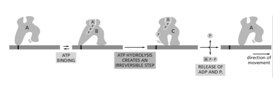
Cellular motors
- Cellular motors can use conformation change with ATP hydrolysis to move along a track