Ophthalmology and Eye Injuries
Diese Karteikarten lernenKostenlos · Spaced Repetition · iPhone, Android, Mac & Web
Die ersten 300 von 794 Karten
1
What is the likely diagnosis in a patient with recent cataract surgery that presents with fever, decreased vision, and pain over the left eye for 12 hours? Physical examination reveals a swollen eyelid and exudates in the anterior chamber.
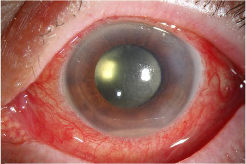
What is the likely diagnosis in a patient with recent cataract surgery that presents with fever, decreased vision, and pain over the left eye for 12 hours? Physical examination reveals a swollen eyelid and exudates in the anterior chamber.
Post-operative endopthalmitis
Post-operative endopthalmitis
2
What is the likely diagnosis in a patient that presents with "floating spots" and blurred vision in the right eye? Several weeks ago, the patient had a penetrating injury to his left eye, causing vision loss
What is the likely diagnosis in a patient that presents with "floating spots" and blurred vision in the right eye? Several weeks ago, the patient had a penetrating injury to his left eye, causing vision loss
Sympathetic ophthalmia ("spared eye injury")
Sympathetic ophthalmia ("spared eye injury")
3
If a patient's ptosis is due to myasthenia gravis, application of a(n) over the eyelids will result in improvement in ptosis
If a patient's ptosis is due to myasthenia gravis, application of a(n) ice pack over the eyelids will result in improvement in ptosis
4
Corneal vascularization is a symptom of which vitamin deficiency?
Vitamin B2 (Riboflavin)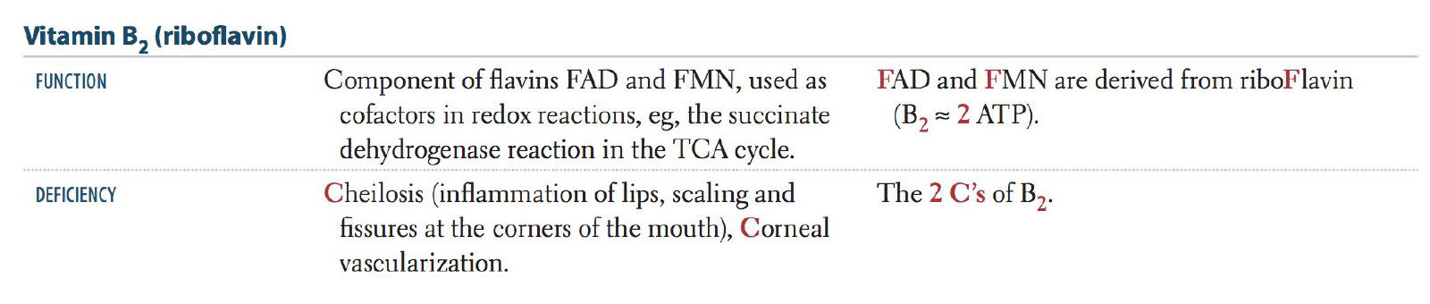
5
What is the likely diagnosis in a child that presents with pain with eye movements as well as fever, periorbital edema, ophthalmoplegia, and proptosis?
What is the likely diagnosis in a child that presents with pain with eye movements as well as fever, periorbital edema, ophthalmoplegia, and proptosis?
Orbital cellulitis
Orbital cellulitis
6
What is the ocular finding in patients with Vitamin B2 (Riboflavin) deficiency?
Corneal vascularization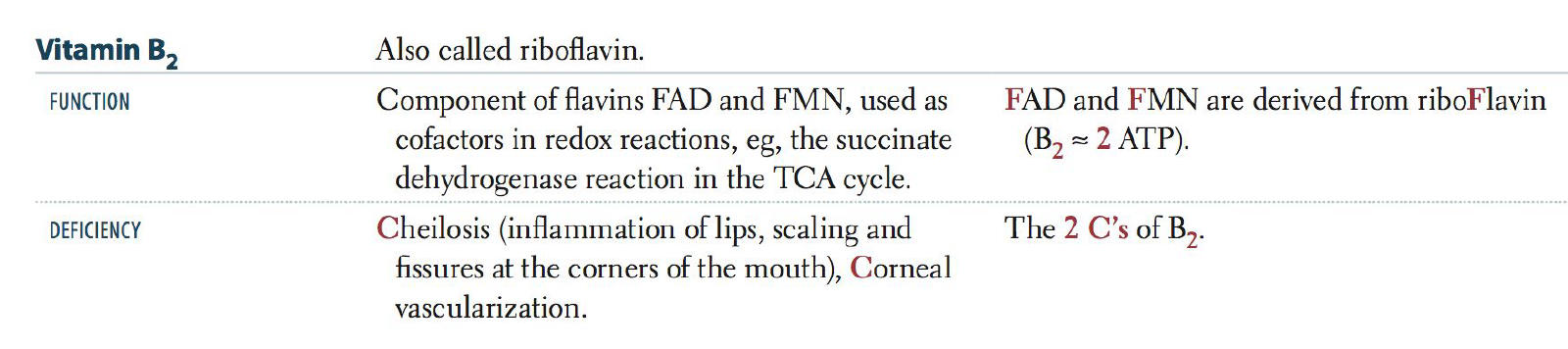
7
Pupils not reactive to light after an eye trauma: Dx?
1) Retina- Retinal detachment
2) Optic nerve- Retrobulbar hematoma
Tx for retrobulbar hematoma: Lateral canthotomy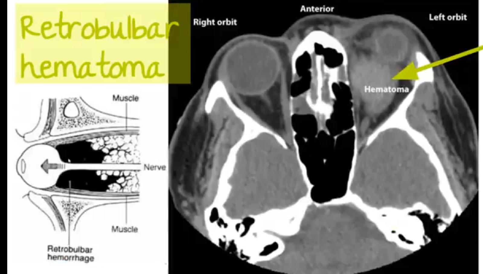
2) Optic nerve- Retrobulbar hematoma
Tx for retrobulbar hematoma: Lateral canthotomy
8
Dilated pupils few days after eye trauma in an otherwise healthy person?
Traumatic iritis.
(nothing to worry about)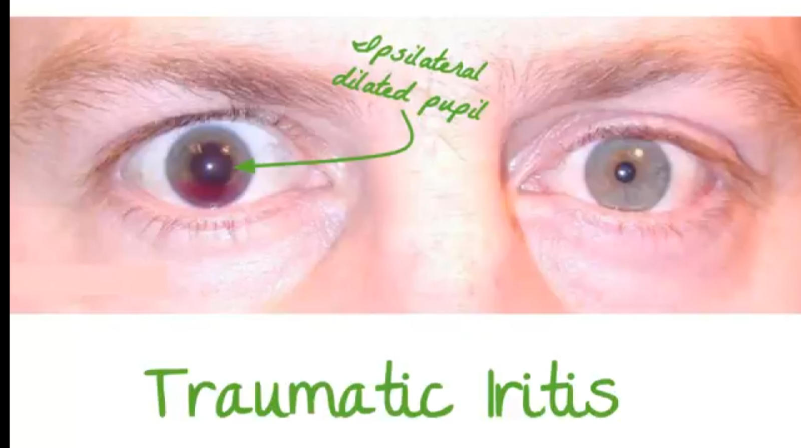
(nothing to worry about)
9
Tear drop pupil after an eye trauma?
Signifies foreign body penetrated cornea and damaged that part of iris.
Concerning for open globe injury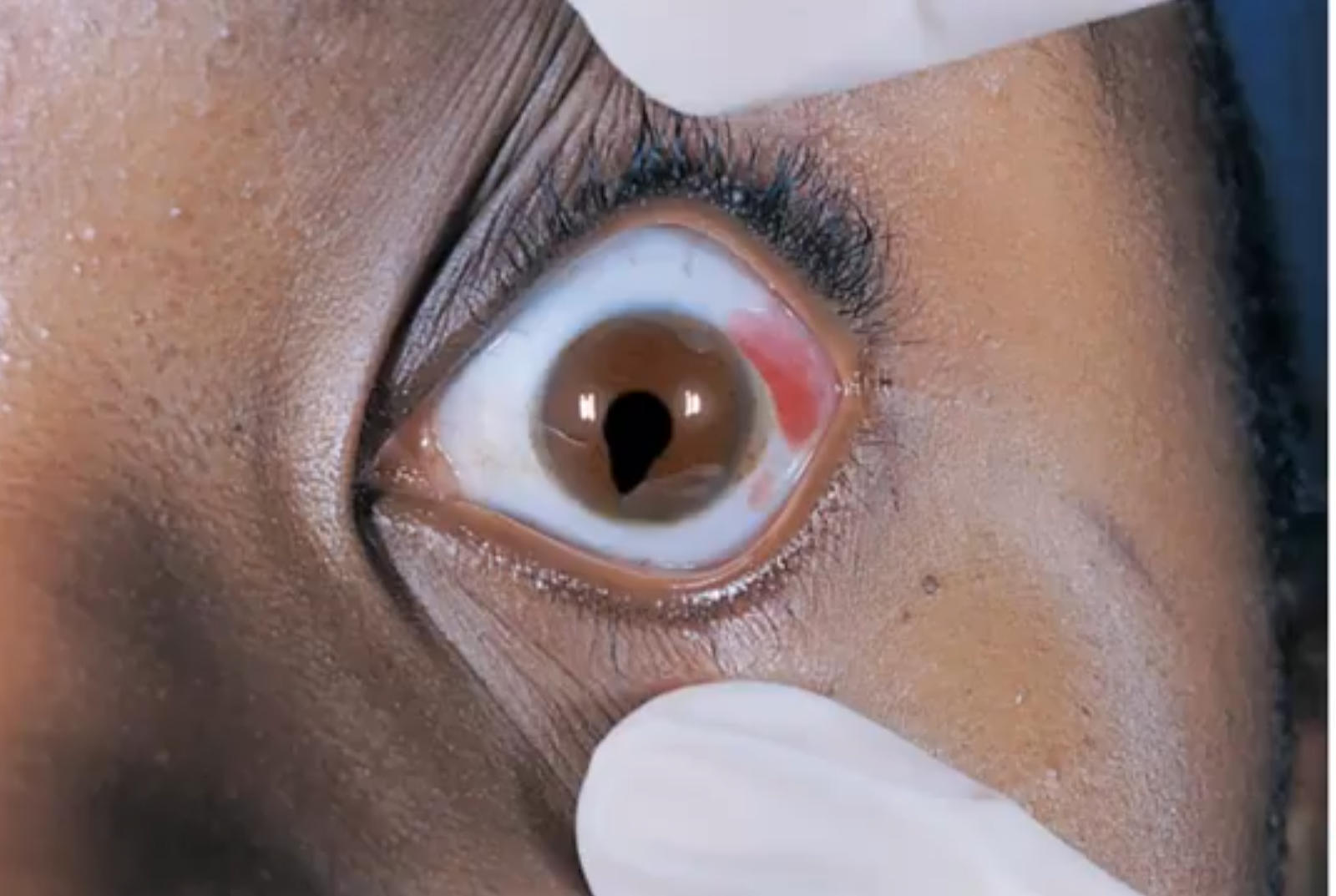
Concerning for open globe injury
10
Eye movements not free after an eye trauma?
Orbital blow out fracture
(usually floor of eye trapping inferior rectus)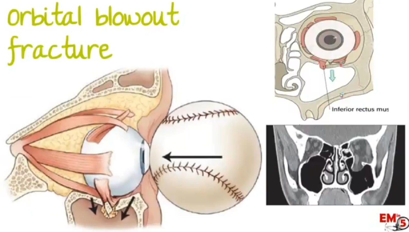
(usually floor of eye trapping inferior rectus)
11
Lid laceration after eye trauma- considerations?
If no subcutaneous fat coming out of wound: repair in ED
If it is medial and involved tear drainage system, call optha
If subcutaneous fat comes out OR full thickness wound: concerning for penetration into orbit-call ophtha
Do CT scan to look for open globe injury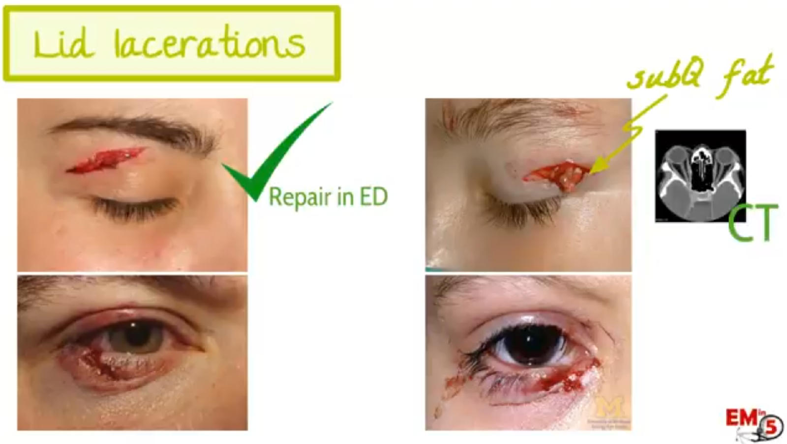
If it is medial and involved tear drainage system, call optha
If subcutaneous fat comes out OR full thickness wound: concerning for penetration into orbit-call ophtha
Do CT scan to look for open globe injury
12
Diagnosis and emergency management of eye injuries?
Symptoms of orbital compartment syndrome: Periorbital swelling with proptosis, tight eyelids, or tight orbit.
Open globe injury signs:
1) obvious perforation are penetrating foreign body
2) teardrop pupil
3) ↑/↓ Anterior chamber depth
4) vitreous extrusion
5) prolapse of intraocular structures
6) corneal or scleral tenting at puncture site
7) full thickness lid laceration or orbital fat protrusion.
NOTE: decreased visual acquity and rAPD is also seen in orbital compartment syndrome and open globe injury, however we rule them out first in the algorithm using their specific manifestations.
Traumatic optic neuropathy:
Due to trauma to brow/temporal skull, forces are transmitted to the optic canal, and the shearing forces can contuse the optic nerve travelling within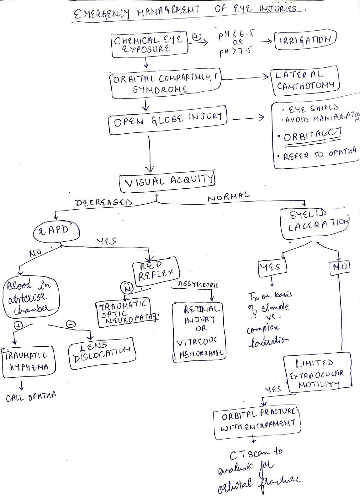
Open globe injury signs:
1) obvious perforation are penetrating foreign body
2) teardrop pupil
3) ↑/↓ Anterior chamber depth
4) vitreous extrusion
5) prolapse of intraocular structures
6) corneal or scleral tenting at puncture site
7) full thickness lid laceration or orbital fat protrusion.
NOTE: decreased visual acquity and rAPD is also seen in orbital compartment syndrome and open globe injury, however we rule them out first in the algorithm using their specific manifestations.
Traumatic optic neuropathy:
Due to trauma to brow/temporal skull, forces are transmitted to the optic canal, and the shearing forces can contuse the optic nerve travelling within
13
9 y/o boy is brought to the ED after a bicycling accident. Examination shows ecchymosis over the right superior orbit. Visual acquity is 20/20 in left eye. Using the right eye,the patient is only able to perceive light. There is rAPD in the right eye. Extraocular movements are normal, and the red reflex is symmetrical bilaterally. Fluorescein testing is unremarkable. Dx?
Traumatic optic nerve injury
Blow to the brow or temple can get transmitted to the walls of optic canal where the optic nerve can get bruised due to shearing forces.(indirect mechanism)
Direct injury to the optic nerve results from lacerating trauma to the orbit (less common)
Symptoms of orbital compartment syndrome: Periorbital swelling with proptosis, tight eyelids, or tight orbit.
Open globe injury signs:
1) obvious perforation are penetrating foreign body
2) teardrop pupil
3) ↑/↓ Anterior chamber depth
4) vitreous extrusion
5) prolapse of intraocular structures
6) corneal or scleral tenting at puncture site
7) full thickness lid laceration or orbital fat protrusion.
NOTE: decreased visual acquity and rAPD is also seen in orbital compartment syndrome and open globe injury, however we rule them out first in the algorithm using their specific manifestations.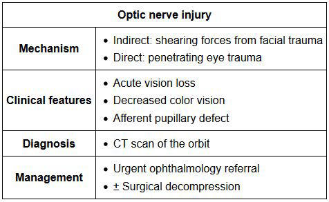
Blow to the brow or temple can get transmitted to the walls of optic canal where the optic nerve can get bruised due to shearing forces.(indirect mechanism)
Direct injury to the optic nerve results from lacerating trauma to the orbit (less common)
Symptoms of orbital compartment syndrome: Periorbital swelling with proptosis, tight eyelids, or tight orbit.
Open globe injury signs:
1) obvious perforation are penetrating foreign body
2) teardrop pupil
3) ↑/↓ Anterior chamber depth
4) vitreous extrusion
5) prolapse of intraocular structures
6) corneal or scleral tenting at puncture site
7) full thickness lid laceration or orbital fat protrusion.
NOTE: decreased visual acquity and rAPD is also seen in orbital compartment syndrome and open globe injury, however we rule them out first in the algorithm using their specific manifestations.
14
is associated with loss of Eyelash or Eyebrow
Madarosis is associated with loss of Eyelash or Eyebrow
15
Madarosis is associated with
Madarosis is associated with loss of Eyelash or Eyebrow
16
A patient presents with left sided head tilt, which on straightening leads to right hypotropia, which increases on dextroversion and right head tilt. Which muscle is involved?
A patient presents with left sided head tilt, which on straightening leads to right hypotropia, which increases on dextroversion and right head tilt. Which muscle is involved?
Right Superior Rectus
Right Superior Rectus
17
MusclePrimary actionSecondary actionTertiary action
Lateral Rectus Medial RectusAdductionnone
none
Superior RectusElevationIntorsionAdductionInferior RectusDepressionExtorsionAdductionSuperior ObliqueIntorsionDepressionAbduction Inferior ObliqueExtorsionElevationAbduction
Lateral Rectus Medial RectusAdductionnone
none
Superior RectusElevationIntorsionAdductionInferior RectusDepressionExtorsionAdductionSuperior ObliqueIntorsionDepressionAbduction Inferior ObliqueExtorsionElevationAbduction
MusclePrimary actionSecondary actionTertiary action
Lateral RectusAbductionnonenoneMedial RectusAdductionnone
none
Superior RectusElevationIntorsionAdductionInferior RectusDepressionExtorsionAdductionSuperior ObliqueIntorsionDepressionAbduction Inferior ObliqueExtorsionElevationAbduction
Lateral RectusAbductionnonenoneMedial RectusAdductionnone
none
Superior RectusElevationIntorsionAdductionInferior RectusDepressionExtorsionAdductionSuperior ObliqueIntorsionDepressionAbduction Inferior ObliqueExtorsionElevationAbduction
18
MusclePrimary actionSecondary actionTertiary action
Lateral RectusAbductionnonenoneMedial Rectus
Superior RectusElevationIntorsionAdductionInferior RectusDepressionExtorsionAdductionSuperior ObliqueIntorsionDepressionAbduction Inferior ObliqueExtorsionElevationAbduction
Lateral RectusAbductionnonenoneMedial Rectus
Superior RectusElevationIntorsionAdductionInferior RectusDepressionExtorsionAdductionSuperior ObliqueIntorsionDepressionAbduction Inferior ObliqueExtorsionElevationAbduction
MusclePrimary actionSecondary actionTertiary action
Lateral RectusAbductionnonenoneMedial RectusAdductionnone
none
Superior RectusElevationIntorsionAdductionInferior RectusDepressionExtorsionAdductionSuperior ObliqueIntorsionDepressionAbduction Inferior ObliqueExtorsionElevationAbduction
Lateral RectusAbductionnonenoneMedial RectusAdductionnone
none
Superior RectusElevationIntorsionAdductionInferior RectusDepressionExtorsionAdductionSuperior ObliqueIntorsionDepressionAbduction Inferior ObliqueExtorsionElevationAbduction
19
MusclePrimary actionSecondary actionTertiary action
Lateral RectusAbductionnonenoneMedial RectusAdductionnone
none
Superior Rectus Inferior RectusDepressionExtorsionAdductionSuperior ObliqueIntorsionDepressionAbduction Inferior ObliqueExtorsionElevationAbduction
Lateral RectusAbductionnonenoneMedial RectusAdductionnone
none
Superior Rectus Inferior RectusDepressionExtorsionAdductionSuperior ObliqueIntorsionDepressionAbduction Inferior ObliqueExtorsionElevationAbduction
MusclePrimary actionSecondary actionTertiary action
Lateral RectusAbductionnonenoneMedial RectusAdductionnone
none
Superior RectusElevationIntorsionAdductionInferior RectusDepressionExtorsionAdductionSuperior ObliqueIntorsionDepressionAbduction Inferior ObliqueExtorsionElevationAbduction
Lateral RectusAbductionnonenoneMedial RectusAdductionnone
none
Superior RectusElevationIntorsionAdductionInferior RectusDepressionExtorsionAdductionSuperior ObliqueIntorsionDepressionAbduction Inferior ObliqueExtorsionElevationAbduction
20
MusclePrimary actionSecondary actionTertiary action
Lateral RectusAbductionnonenoneMedial RectusAdductionnone
none
Superior RectusElevationIntorsionAdductionInferior Rectus Superior ObliqueIntorsionDepressionAbduction Inferior ObliqueExtorsionElevationAbduction
Lateral RectusAbductionnonenoneMedial RectusAdductionnone
none
Superior RectusElevationIntorsionAdductionInferior Rectus Superior ObliqueIntorsionDepressionAbduction Inferior ObliqueExtorsionElevationAbduction
MusclePrimary actionSecondary actionTertiary action
Lateral RectusAbductionnonenoneMedial RectusAdductionnone
none
Superior RectusElevationIntorsionAdductionInferior RectusDepressionExtorsionAdductionSuperior ObliqueIntorsionDepressionAbduction Inferior ObliqueExtorsionElevationAbduction
Lateral RectusAbductionnonenoneMedial RectusAdductionnone
none
Superior RectusElevationIntorsionAdductionInferior RectusDepressionExtorsionAdductionSuperior ObliqueIntorsionDepressionAbduction Inferior ObliqueExtorsionElevationAbduction
21
MusclePrimary actionSecondary actionTertiary action
Lateral RectusAbductionnonenoneMedial RectusAdductionnone
none
Superior RectusElevationIntorsionAdductionInferior RectusDepressionExtorsionAdductionSuperior Oblique Inferior ObliqueExtorsionElevationAbduction
Lateral RectusAbductionnonenoneMedial RectusAdductionnone
none
Superior RectusElevationIntorsionAdductionInferior RectusDepressionExtorsionAdductionSuperior Oblique Inferior ObliqueExtorsionElevationAbduction
MusclePrimary actionSecondary actionTertiary action
Lateral RectusAbductionnonenoneMedial RectusAdductionnone
none
Superior RectusElevationIntorsionAdductionInferior RectusDepressionExtorsionAdductionSuperior ObliqueIntorsionDepressionAbduction Inferior ObliqueExtorsionElevationAbduction
Lateral RectusAbductionnonenoneMedial RectusAdductionnone
none
Superior RectusElevationIntorsionAdductionInferior RectusDepressionExtorsionAdductionSuperior ObliqueIntorsionDepressionAbduction Inferior ObliqueExtorsionElevationAbduction
22
MusclePrimary actionSecondary actionTertiary action
Lateral RectusAbductionnonenoneMedial RectusAdductionnone
none
Superior RectusElevationIntorsionAdductionInferior RectusDepressionExtorsionAdductionSuperior ObliqueIntorsionDepressionAbduction Inferior Oblique
Lateral RectusAbductionnonenoneMedial RectusAdductionnone
none
Superior RectusElevationIntorsionAdductionInferior RectusDepressionExtorsionAdductionSuperior ObliqueIntorsionDepressionAbduction Inferior Oblique
MusclePrimary actionSecondary actionTertiary action
Lateral RectusAbductionnonenoneMedial RectusAdductionnone
none
Superior RectusElevationIntorsionAdductionInferior RectusDepressionExtorsionAdductionSuperior ObliqueIntorsionDepressionAbduction Inferior ObliqueExtorsionElevationAbduction
Lateral RectusAbductionnonenoneMedial RectusAdductionnone
none
Superior RectusElevationIntorsionAdductionInferior RectusDepressionExtorsionAdductionSuperior ObliqueIntorsionDepressionAbduction Inferior ObliqueExtorsionElevationAbduction
23
test is used to distinguish between colored halos of cataract and glaucoma?
Fincham test is used to distinguish between colored halos of cataract and glaucoma?
24
In Fincham test, if the halos break when the slit moves, then diagnosis of is made.
In Fincham test, if the halos break when the slit moves, then diagnosis of cataract is made.
25
In Fincham test, if the halos remain intact when the slit moves, then diagnosis of is made.
In Fincham test, if the halos remain intact when the slit moves, then diagnosis of glaucoma is made.
26
Halos in glaucoma are due to
Halos in glaucoma are due to corneal edema
27
This eye finding is called 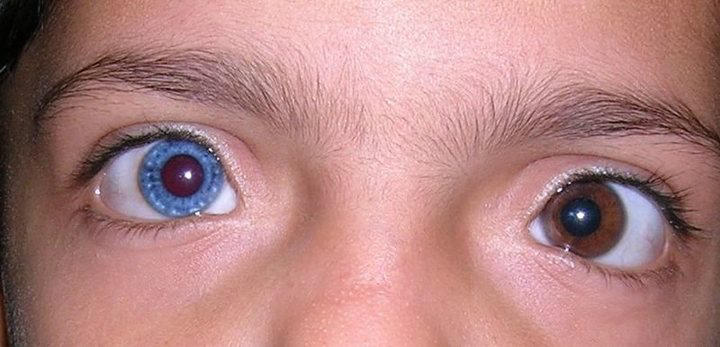
This eye finding is called heterochromia iridum
28
This type of cataract is seen in .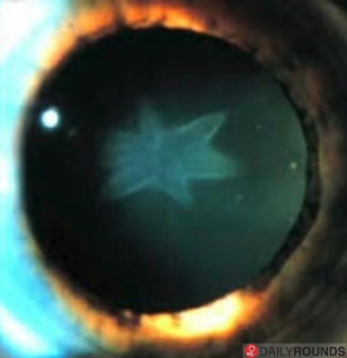
This type of cataract is seen in Blunt trauma.
29
This type of cataract is seen in and is called cataract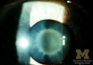
This type of cataract is seen in Wilson Disease and is called sunflower cataract
30
This type of cataract is seen in and is called cataract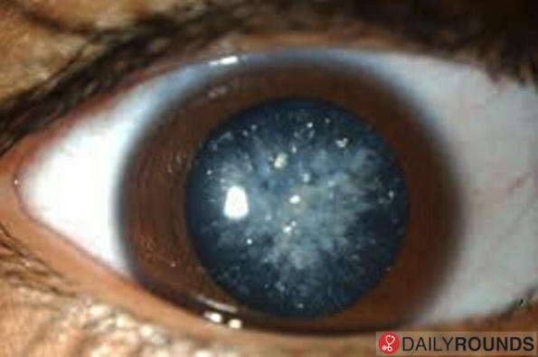
This type of cataract is seen in Diabetes and is called snowflake cataract
31
This type of cataract is seen in and is called cataract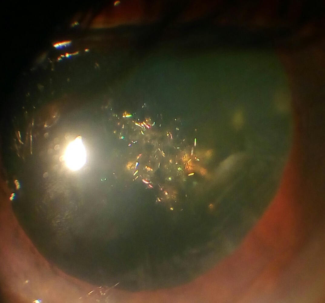
This type of cataract is seen in Myotonic dystrophy and is called Christmas tree cataract
32
In congenital Rubella, there is cataract.
In congenital Rubella, there is nuclear cataract.
33
cararact is seen in atopic dermatitis.It is also called shield cataract
Anterior subcapsular cararact is seen in atopic dermatitis.It is also called shield cataract
34
Anterior subcapsular cararact is seen in atopic dermatitis.It is also called cataract
Anterior subcapsular cararact is seen in atopic dermatitis.It is also called shield cataract
35
Systemic steroids cause cataract.
Systemic steroids cause posterior subcapsular cataract.
36
Complicated cataract has appearance. It has polychromatic lusture. Location of the cataract is posterior subcapsular
Complicated cataract has bread crum appearance. It has polychromatic lusture. Location of the cataract is posterior subcapsular
37
Complicated cataract has bread crum appearance. It has lusture. Location of the cataract is posterior subcapsular
Complicated cataract has bread crum appearance. It has polychromatic lusture. Location of the cataract is posterior subcapsular
38
Complicated cataract has bread crum appearance. It has polychromatic lusture. Location of the cataract is
Complicated cataract has bread crum appearance. It has polychromatic lusture. Location of the cataract is posterior subcapsular
39
Morgagnian cataract is hypermature cataract
Morgagnian cataract is hypermature cortical cataract
40
Sclerotic cataract is hypermature cataract.
Sclerotic cataract is hypermature nuclear cataract.
41
cataract is seen in galactosemia
Oil droplet cataract is seen in galactosemia
42
Oil droplet cataract is seen in
Oil droplet cataract is seen in galactosemia
43
Chlorpromazine causes shaped cataract
Chlorpromazine causes stellate shaped cataract
44
Which structure in eye is most sensitive to radiation?
Which structure in eye is most sensitive to radiation?
Eye lens
Eye lens
45
Which cataract has hemeralopia?
Which cataract has hemeralopia?
Nuclear cataract
Nuclear cataract
46
Cortical cataract causes
Cortical cataract causes night blindness
47
Which cataract causes maximum visual handicap?
Which cataract causes maximum visual handicap?
Posterior subcapsular cataract.
Posterior subcapsular cataract.
48
According to WHO, what is the definition of legally blind?
According to WHO, what is the definition of legally blind?
<3/60 vision in better eye with best possible correction
<3/60 vision in better eye with best possible correction
49
2 MC infectious causes of blindness
1)
2) Oncocercosis(River Blindness)
1)
2) Oncocercosis(River Blindness)
2 MC infectious causes of blindness
1) Trachoma
2) Oncocercosis(River Blindness)
1) Trachoma
2) Oncocercosis(River Blindness)
50
2 MC infectious causes of blindness
1) Trachoma
2)
1) Trachoma
2)
2 MC infectious causes of blindness
1) Trachoma
2) Oncocercosis(River Blindness)
1) Trachoma
2) Oncocercosis(River Blindness)
51
Pigment deposition in anterior vitreous (tobacco dusting) is seen in and is called Schaffer sign
Pigment deposition in anterior vitreous (tobacco dusting) is seen in fresh rhegmatogenous retinal detachment and is called Schaffer sign
52
Pigment deposition in anterior vitreous (tobacco dusting) is seen in fresh rhegmatogenous retinal detachment and is called sign
Pigment deposition in anterior vitreous (tobacco dusting) is seen in fresh rhegmatogenous retinal detachment and is called Schaffer sign
53
Most important clinical features of Retinal Detachment?
Photopsia(flashes of light due to retinal break)
Floaters(due to associated vitreous hemorrhage)
Photopsia(flashes of light due to retinal break)
Floaters(due to associated vitreous hemorrhage)
Most important clinical features of Retinal Detachment?
Black curtain/veil in front of eye
Photopsia(flashes of light due to retinal break)
Floaters(due to associated vitreous hemorrhage)
Black curtain/veil in front of eye
Photopsia(flashes of light due to retinal break)
Floaters(due to associated vitreous hemorrhage)
54
Most important clinical features of Retinal Detachment?
Black curtain/veil in front of eye
Floaters(due to associated vitreous hemorrhage)
Black curtain/veil in front of eye
Floaters(due to associated vitreous hemorrhage)
Most important clinical features of Retinal Detachment?
Black curtain/veil in front of eye
Photopsia(flashes of light due to retinal break)
Floaters(due to associated vitreous hemorrhage)
Black curtain/veil in front of eye
Photopsia(flashes of light due to retinal break)
Floaters(due to associated vitreous hemorrhage)
55
Most important clinical features of Retinal Detachment?
Black curtain/veil in front of eye
Photopsia(flashes of light due to retinal break)
Black curtain/veil in front of eye
Photopsia(flashes of light due to retinal break)
Most important clinical features of Retinal Detachment?
Black curtain/veil in front of eye
Photopsia(flashes of light due to retinal break)
Floaters(due to associated vitreous hemorrhage)
Black curtain/veil in front of eye
Photopsia(flashes of light due to retinal break)
Floaters(due to associated vitreous hemorrhage)
56
This sign seen in is called Munson Sign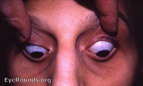
This sign seen in keratoconus is called Munson Sign
57
This sign seen in keratoconus is called Sign
This sign seen in keratoconus is called Munson Sign
58
This sign in is called Vogt Striae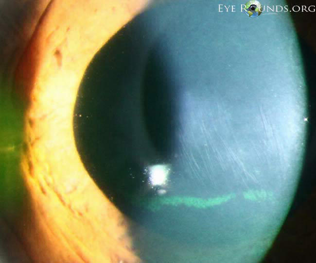
This sign in keratoconus is called Vogt Striae
59
This sign in keratoconus is called
This sign in keratoconus is called Vogt Striae
60
A residual stromal bed thickness of is generally recognised as a minimum requirement before LASIK surgery.
A residual stromal bed thickness of 250 micrometer is generally recognised as a minimum requirement before LASIK surgery.
61
If LASIK surgery is performed in a person with residual stromal thickness <250μm, it predisposes to
If LASIK surgery is performed in a person with residual stromal thickness <250μm, it predisposes to keratectasia
62
What is the minimum age at which LASIK surgery can be performed?
What is the minimum age at which LASIK surgery can be performed?
21yrs
21yrs
63
Sudden onset vision loss in right eye. Pain while moving eyeballs. Fundus normal.Dx?
Sudden onset vision loss in right eye. Pain while moving eyeballs. Fundus normal.Dx?
Optic neuritis
Optic neuritis
64
Depth of anterior chamber with age.
Depth of anterior chamber decreases with age.
65
Congenital glaucoma has a triad of clinical features:
Photophobia
Blepharospasm
Photophobia
Blepharospasm
Congenital glaucoma has a triad of clinical features:
Lacrimation
Photophobia
Blepharospasm
Lacrimation
Photophobia
Blepharospasm
66
Congenital glaucoma has a triad of clinical features:
Lacrimation
Blepharospasm
Lacrimation
Blepharospasm
Congenital glaucoma has a triad of clinical features:
Lacrimation
Photophobia
Blepharospasm
Lacrimation
Photophobia
Blepharospasm
67
Congenital glaucoma has a triad of clinical features:
Lacrimation
Photophobia
Lacrimation
Photophobia
Congenital glaucoma has a triad of clinical features:
Lacrimation
Photophobia
Blepharospasm
Lacrimation
Photophobia
Blepharospasm
68
Congenital glaucoma is defined when glaucoma is seen within years of life.
Congenital glaucoma is defined when glaucoma is seen within 3 years of life.
69
Buphthalmos is seen in
Buphthalmos is seen in congenital glaucoma
70
are horizontal/circumfrential breaks in Descement membrane seen in congenital glaucoma.
Haab's striae are horizontal/circumfrential breaks in Descement membrane seen in congenital glaucoma.
71
Haab's striae are horizontal/circumfrential breaks in membrane seen in congenital glaucoma.
Haab's striae are horizontal/circumfrential breaks in Descement membrane seen in congenital glaucoma.
72
Haab's striae are horizontal/circumfrential breaks in Descement membrane seen in .
Haab's striae are horizontal/circumfrential breaks in Descement membrane seen in congenital glaucoma.
73
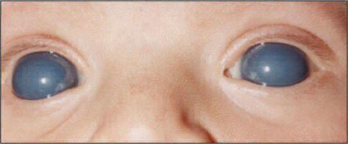
Large, hazy corneas, photophobia, lacrimation in a newborn few days after birth. Diagnosis?
`
`
Large, hazy corneas, photophobia, lacrimation in a newborn few days after birth. Diagnosis?
`Congenital glaucoma
`Congenital glaucoma
74
Reason for hazy cornea in congenital glaucoma?
Reason for hazy cornea in congenital glaucoma?
Corneal edema
Corneal edema
75
MCC of congenital glaucoma is presence of membrane
MCC of congenital glaucoma is presence of Burkan membrane
76
Treatment of congenital glaucoma in a patient with clear cornea?
Treatment of congenital glaucoma in a patient with clear cornea?
Goniotomy
Goniotomy
77
Treatment of congenital glaucoma in a patient with hazy cornea?
Treatment of congenital glaucoma in a patient with hazy cornea?
Trabeculotomy + Trabeculotomy(Trab + Trab)
Trabeculotomy + Trabeculotomy(Trab + Trab)
78
Is buphthalmos seen in glaucoma cases arising after age 3?
Is buphthalmos seen in glaucoma cases arising after age 3?
No, eye ball is not elastic enough to grow due to pressure after age 3
No, eye ball is not elastic enough to grow due to pressure after age 3
79
Expected pupil condition in a patient of acute angle closure glaucoma?
Expected pupil condition in a patient of acute angle closure glaucoma?
Verically oval mid dilated pupil which is non reactive to light
Verically oval mid dilated pupil which is non reactive to light
80
In a patient with past h/o acute angle closure glaucoma, triad can be seen.
In a patient with past h/o acute angle closure glaucoma, vogt triad can be seen.
81
Vogt triad components:
Peripheral anterior synechiae
Glaukomaflecken
Peripheral anterior synechiae
Glaukomaflecken
Vogt triad components:
Iris atrophy
Peripheral anterior synechiae
Glaukomaflecken
Iris atrophy
Peripheral anterior synechiae
Glaukomaflecken
82
Vogt triad components:
Iris atrophy
Glaukomaflecken
Iris atrophy
Glaukomaflecken
Vogt triad components:
Iris atrophy
Peripheral anterior synechiae
Glaukomaflecken
Iris atrophy
Peripheral anterior synechiae
Glaukomaflecken
83
Vogt triad components:
Iris atrophy
Peripheral anterior synechiae
Iris atrophy
Peripheral anterior synechiae
Vogt triad components:
Iris atrophy
Peripheral anterior synechiae
Glaukomaflecken
Iris atrophy
Peripheral anterior synechiae
Glaukomaflecken
84
Frequent change of presbyopic glasses should clue you to a diagnosis of .
Frequent change of presbyopic glasses should clue you to a diagnosis of open angle glaucoma.
85
Frequent change of distant vision glasses in an aged person?
Frequent change of distant vision glasses in an aged person?
Nuclear cataract
Nuclear cataract
86
Frequent change of glasses in young is seen in what pathologies of eye?
Pathological myopia
Pathological myopia
Frequent change of glasses in young is seen in what pathologies of eye?
Keratoconus
Pathological myopia
Keratoconus
Pathological myopia
87
Frequent change of glasses in young is seen in what pathologies of eye?
Keratoconus
Keratoconus
Frequent change of glasses in young is seen in what pathologies of eye?
Keratoconus
Pathological myopia
Keratoconus
Pathological myopia
88
Normal visual field angles:
Superior: degrees
Nasal: 60 degrees
Inferior: 70 degrees
Temporal: 100 degrees
Superior: degrees
Nasal: 60 degrees
Inferior: 70 degrees
Temporal: 100 degrees
Normal visual field angles:
Superior: 50 degrees
Nasal: 60 degrees
Inferior: 70 degrees
Temporal: 100 degrees
Superior: 50 degrees
Nasal: 60 degrees
Inferior: 70 degrees
Temporal: 100 degrees
89
Normal visual field angles:
Superior: 50 degrees
Nasal: degrees
Inferior: 70 degrees
Temporal: 100 degrees
Superior: 50 degrees
Nasal: degrees
Inferior: 70 degrees
Temporal: 100 degrees
Normal visual field angles:
Superior: 50 degrees
Nasal: 60 degrees
Inferior: 70 degrees
Temporal: 100 degrees
Superior: 50 degrees
Nasal: 60 degrees
Inferior: 70 degrees
Temporal: 100 degrees
90
Normal visual field angles:
Superior: 50 degrees
Nasal: 60 degrees
Inferior: degrees
Temporal: 100 degrees
Superior: 50 degrees
Nasal: 60 degrees
Inferior: degrees
Temporal: 100 degrees
Normal visual field angles:
Superior: 50 degrees
Nasal: 60 degrees
Inferior: 70 degrees
Temporal: 100 degrees
Superior: 50 degrees
Nasal: 60 degrees
Inferior: 70 degrees
Temporal: 100 degrees
91
Normal visual field angles:
Superior: 50 degrees
Nasal: 60 degrees
Inferior: 70 degrees
Temporal: degrees
Superior: 50 degrees
Nasal: 60 degrees
Inferior: 70 degrees
Temporal: degrees
Normal visual field angles:
Superior: 50 degrees
Nasal: 60 degrees
Inferior: 70 degrees
Temporal: 100 degrees
Superior: 50 degrees
Nasal: 60 degrees
Inferior: 70 degrees
Temporal: 100 degrees
92
Physiological blind spot is located in visual field
Physiological blind spot is located in temporal visual field
93
This fundus exam is of eye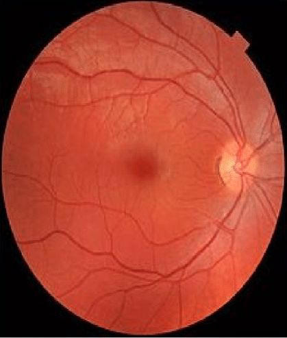
This fundus exam is of right eye
94
This fundus exam is of eye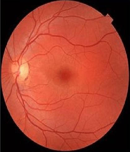
This fundus exam is of left eye
95
Glaucoma visual field changes:
Paracentral scotoma(scotoma in Bjerrum area)
Siedel scotoma
Arcuate scotoma aka Bjerrum scotoma
Double arcuate scotoma showing Ronne nasal step
Tubular vision
Paracentral scotoma(scotoma in Bjerrum area)
Siedel scotoma
Arcuate scotoma aka Bjerrum scotoma
Double arcuate scotoma showing Ronne nasal step
Tubular vision
Glaucoma visual field changes:
Isopter contraction with baring of blind spot
Paracentral scotoma(scotoma in Bjerrum area)
Siedel scotoma
Arcuate scotoma aka Bjerrum scotoma
Double arcuate scotoma showing Ronne nasal step
Tubular vision
Isopter contraction with baring of blind spot
Paracentral scotoma(scotoma in Bjerrum area)
Siedel scotoma
Arcuate scotoma aka Bjerrum scotoma
Double arcuate scotoma showing Ronne nasal step
Tubular vision
96
Glaucoma visual field changes:
Isopter contraction with baring of blind spot
Siedel scotoma
Arcuate scotoma aka Bjerrum scotoma
Double arcuate scotoma showing Ronne nasal step
Tubular vision
Isopter contraction with baring of blind spot
Siedel scotoma
Arcuate scotoma aka Bjerrum scotoma
Double arcuate scotoma showing Ronne nasal step
Tubular vision
Glaucoma visual field changes:
Isopter contraction with baring of blind spot
Paracentral scotoma(scotoma in Bjerrum area)
Siedel scotoma
Arcuate scotoma aka Bjerrum scotoma
Double arcuate scotoma showing Ronne nasal step
Tubular vision
Isopter contraction with baring of blind spot
Paracentral scotoma(scotoma in Bjerrum area)
Siedel scotoma
Arcuate scotoma aka Bjerrum scotoma
Double arcuate scotoma showing Ronne nasal step
Tubular vision
97
Glaucoma visual field changes:
Isopter contraction with baring of blind spot
Paracentral scotoma(scotoma in Bjerrum area)
Arcuate scotoma aka Bjerrum scotoma
Double arcuate scotoma showing Ronne nasal step
Tubular vision
Isopter contraction with baring of blind spot
Paracentral scotoma(scotoma in Bjerrum area)
Arcuate scotoma aka Bjerrum scotoma
Double arcuate scotoma showing Ronne nasal step
Tubular vision
Glaucoma visual field changes:
Isopter contraction with baring of blind spot
Paracentral scotoma(scotoma in Bjerrum area)
Siedel scotoma
Arcuate scotoma aka Bjerrum scotoma
Double arcuate scotoma showing Ronne nasal step
Tubular vision
Isopter contraction with baring of blind spot
Paracentral scotoma(scotoma in Bjerrum area)
Siedel scotoma
Arcuate scotoma aka Bjerrum scotoma
Double arcuate scotoma showing Ronne nasal step
Tubular vision
98
Glaucoma visual field changes:
Isopter contraction with baring of blind spot
Paracentral scotoma(scotoma in Bjerrum area)
Siedel scotoma
Double arcuate scotoma showing Ronne nasal step
Tubular vision
Isopter contraction with baring of blind spot
Paracentral scotoma(scotoma in Bjerrum area)
Siedel scotoma
Double arcuate scotoma showing Ronne nasal step
Tubular vision
Glaucoma visual field changes:
Isopter contraction with baring of blind spot
Paracentral scotoma(scotoma in Bjerrum area)
Siedel scotoma
Arcuate scotoma aka Bjerrum scotoma
Double arcuate scotoma showing Ronne nasal step
Tubular vision
Isopter contraction with baring of blind spot
Paracentral scotoma(scotoma in Bjerrum area)
Siedel scotoma
Arcuate scotoma aka Bjerrum scotoma
Double arcuate scotoma showing Ronne nasal step
Tubular vision
99
Glaucoma visual field changes:
Isopter contraction with baring of blind spot
Paracentral scotoma(scotoma in Bjerrum area)
Siedel scotoma
Arcuate scotoma aka Bjerrum scotoma
Tubular vision
Isopter contraction with baring of blind spot
Paracentral scotoma(scotoma in Bjerrum area)
Siedel scotoma
Arcuate scotoma aka Bjerrum scotoma
Tubular vision
Glaucoma visual field changes:
Isopter contraction with baring of blind spot
Paracentral scotoma(scotoma in Bjerrum area)
Siedel scotoma
Arcuate scotoma aka Bjerrum scotoma
Double arcuate scotoma showing Ronne nasal step
Tubular vision
Isopter contraction with baring of blind spot
Paracentral scotoma(scotoma in Bjerrum area)
Siedel scotoma
Arcuate scotoma aka Bjerrum scotoma
Double arcuate scotoma showing Ronne nasal step
Tubular vision
100
Bjerrum scotoma is a finding of glaucoma.
Bjerrum scotoma is a late finding of glaucoma.
101
Glaucoma scotomas are scotomas.
Glaucoma scotomas are negative scotomas.
102
Glaucoma scotoma is measured by perimeter
Glaucoma scotoma is measured by Humphrey's perimeter
103
Normal cup disc ratio is upto
Normal cup disc ratio is upto 0.6
104
What is ISNT rule?
What is ISNT rule?
Normally, Inferior neuroretinal rim is broadest f\/b superior>nasal>temporal
Normally, Inferior neuroretinal rim is broadest f\/b superior>nasal>temporal
105
Cupping in glaucoma occurs
Cupping in glaucoma occurs vertically
106
Horizontal cupping of optic disc is seen in
Horizontal cupping of optic disc is seen in normal individuals
107
Glaucoma is bilaterally
Glaucoma is bilaterally asymmetrical
108
Laminar dot sign is seen in
Laminar dot sign is seen in glaucoma
109
Which of the fundus signs are seen in this patient?
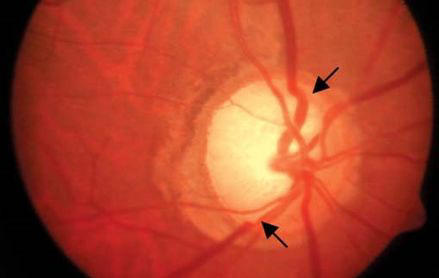
Which of the fundus signs are seen in this patient?
Bayonetting of vessels
Bayonetting of vessels
110
The last visual field to be affected in glaucoma is
The last visual field to be affected in glaucoma is temporal
111
Initial drug to choose in patients of open angle glaucoma?
Initial drug to choose in patients of open angle glaucoma?
Topical prostaglandins
Topical prostaglandins
112
If a patient with open angle glaucoma cannot afford a prostaglandin analog, what drug should be considered?
If a patient with open angle glaucoma cannot afford a prostaglandin analog, what drug should be considered?
Topical ßblocker(eg timolol)
Topical ßblocker(eg timolol)
113
MOA of prostaglandins in glaucoma?
MOA of prostaglandins in glaucoma?
↑uveoscleral outflow
↑uveoscleral outflow
114
Drugs to be given in angle closure glaucoma?
Drugs to be given in angle closure glaucoma?
Mannitol
Acetazolamide
Glycerol
Mannitol
Acetazolamide
Glycerol
115
From iris to cornea, the structures seen in gonioscopy are
Scleral Spur
Trabecular meshwork
Schwalbe line
Scleral Spur
Trabecular meshwork
Schwalbe line
From iris to cornea, the structures seen in gonioscopy are
Ciliary body band
Scleral Spur
Trabecular meshwork
Schwalbe line
Ciliary body band
Scleral Spur
Trabecular meshwork
Schwalbe line
116
From iris to cornea, the structures seen in gonioscopy are
Ciliary body band
Trabecular meshwork
Schwalbe line
Ciliary body band
Trabecular meshwork
Schwalbe line
From iris to cornea, the structures seen in gonioscopy are
Ciliary body band
Scleral Spur
Trabecular meshwork
Schwalbe line
Ciliary body band
Scleral Spur
Trabecular meshwork
Schwalbe line
117
From iris to cornea, the structures seen in gonioscopy are
Ciliary body band
Scleral Spur
Schwalbe line
Ciliary body band
Scleral Spur
Schwalbe line
From iris to cornea, the structures seen in gonioscopy are
Ciliary body band
Scleral Spur
Trabecular meshwork
Schwalbe line
Ciliary body band
Scleral Spur
Trabecular meshwork
Schwalbe line
118
From iris to cornea, the structures seen in gonioscopy are
Ciliary body band
Scleral Spur
Trabecular meshwork
Ciliary body band
Scleral Spur
Trabecular meshwork
From iris to cornea, the structures seen in gonioscopy are
Ciliary body band
Scleral Spur
Trabecular meshwork
Schwalbe line
Ciliary body band
Scleral Spur
Trabecular meshwork
Schwalbe line
119
According to the Shaffers grading, what is seen in grade 1 of anterior chamber angle?
According to the Shaffers grading, what is seen in grade 1 of anterior chamber angle?
Only schwalbe line
Only schwalbe line
120
According to the Shaffers grading, what is seen in grade 2 of anterior chamber angle?
According to the Shaffers grading, what is seen in grade 2 of anterior chamber angle?
schwalbe line + Trabecular meshwork
schwalbe line + Trabecular meshwork
121
According to the Shaffers grading, what is seen in grade 3 of anterior chamber angle?
According to the Shaffers grading, what is seen in grade 3 of anterior chamber angle?
schwalbe line + Trabecular meshwork + Scleral spur
schwalbe line + Trabecular meshwork + Scleral spur
122
According to the Shaffers grading, what is seen in grade 4 of anterior chamber angle?
According to the Shaffers grading, what is seen in grade 4 of anterior chamber angle?
schwalbe line + Trabecular meshwork + Scleral spur + ciliary body
schwalbe line + Trabecular meshwork + Scleral spur + ciliary body
123
Treatment of choice for angle closure glaucoma?
Treatment of choice for angle closure glaucoma?
Laser iridotomy
Laser iridotomy
124
is lens opacity due to recent angle closure glaucoma.
Glaukomflecken is lens opacity due to recent angle closure glaucoma.
125
Glaukomflecken is due to recent angle closure glaucoma.
Glaukomflecken is lens opacity due to recent angle closure glaucoma.
126
Glaukomflecken is lens opacity due to recent .
Glaukomflecken is lens opacity due to recent angle closure glaucoma.
127
Ciliary block glaucoma was formerly called glaucoma.
Ciliary block glaucoma was formerly called Malignant glaucoma.
128
Why is malignant glaucoma also called ciliary block glaucoma?
Why is malignant glaucoma also called ciliary block glaucoma?
Due to malrotation of the ciliary body, which leads to secretion of aqueous into vitreous humor.
Due to malrotation of the ciliary body, which leads to secretion of aqueous into vitreous humor.
129
Posterior aqeuous mis-direction syndrome can be seen after .
Posterior aqeuous mis-direction syndrome can be seen after intraocular surgery.
130
Is pupillary block seen in ciliary block glaucoma?
Is pupillary block seen in ciliary block glaucoma?
NO
NO
131
Ciliary block glaucoma is characterized by
Anterior chamber
Elevated IOP
Absence of pupilary block
Anterior chamber
Elevated IOP
Absence of pupilary block
Ciliary block glaucoma is characterized by
Shallow Anterior chamber
Elevated IOP
Absence of pupilary block
Shallow Anterior chamber
Elevated IOP
Absence of pupilary block
132
Ciliary block glaucoma is characterized by
Shallow Anterior chamber
Elevated IOP
of pupilary block
Shallow Anterior chamber
Elevated IOP
of pupilary block
Ciliary block glaucoma is characterized by
Shallow Anterior chamber
Elevated IOP
Absence of pupilary block
Shallow Anterior chamber
Elevated IOP
Absence of pupilary block
133
Treatment of malignant glaucoma?
Treatment of malignant glaucoma?
Atropine
Atropine
134
Krukenberg spindle is seen in glaucoma.
Krukenberg spindle is seen in pigment dispersion glaucoma.
135
This defect in iris is seen in glaucoma.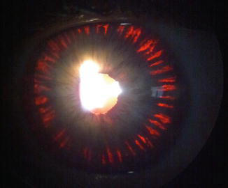
This defect in iris is seen in pigmentary glaucoma.
136
Sampaolesi line is seen in glaucoma
Sampaolesi line is seen in pseudoexfoliation glaucoma
137
The most reliable tonometer is tonometer.
The most reliable tonometer is Goldmann tonometer.
138
Goldmann tonometer is a type of tonometer.
Goldmann tonometer is a type of applanation tonometer.
139
Schiotz tonometer is a type of tonometer
Schiotz tonometer is a type of indentation tonometer
140
tonometer is non contact applanation tonometer
Pulsair tonometer is non contact applanation tonometer
141
Which tonometer reading is dependent on scleral rigidity?
Which tonometer reading is dependent on scleral rigidity?
Indentation(eg Schiotz tonometer)
Indentation(eg Schiotz tonometer)
142
100 day glaucoma is a neovascular glaucoma seen in
100 day glaucoma is a neovascular glaucoma seen in Central Retinal vein occlusion(CRVO)
143
MCC of secondary open-angle glaucoma:
MCC of secondary open-angle glaucoma:
Pseudo exfoliation syndrome
Pseudo exfoliation syndrome
144
Which area of retina has the highest concentration of the rods?
Which area of retina has the highest concentration of the rods?
Mid-periphery of retina
Mid-periphery of retina
145
Stages of ROPDescription of fundus
Stage 1 Stage 2Intraretinal ridge formationStage 3NeovascularizationStage 4Subtotal retinal detachmentStage 5Total retinal detachment
Stage 1 Stage 2Intraretinal ridge formationStage 3NeovascularizationStage 4Subtotal retinal detachmentStage 5Total retinal detachment
Stages of ROPDescription of fundus
Stage 1Demarcation lineStage 2Intraretinal ridge formationStage 3NeovascularizationStage 4Subtotal retinal detachmentStage 5Total retinal detachment
Stage 1Demarcation lineStage 2Intraretinal ridge formationStage 3NeovascularizationStage 4Subtotal retinal detachmentStage 5Total retinal detachment
146
Stages of ROPDescription of fundus
Stage 1Demarcation lineStage 2 Stage 3NeovascularizationStage 4Subtotal retinal detachmentStage 5Total retinal detachment
Stage 1Demarcation lineStage 2 Stage 3NeovascularizationStage 4Subtotal retinal detachmentStage 5Total retinal detachment
Stages of ROPDescription of fundus
Stage 1Demarcation lineStage 2Intraretinal ridge formationStage 3NeovascularizationStage 4Subtotal retinal detachmentStage 5Total retinal detachment
Stage 1Demarcation lineStage 2Intraretinal ridge formationStage 3NeovascularizationStage 4Subtotal retinal detachmentStage 5Total retinal detachment
147
Stages of ROPDescription of fundus
Stage 1Demarcation lineStage 2Intraretinal ridge formationStage 3 Stage 4Subtotal retinal detachmentStage 5Total retinal detachment
Stage 1Demarcation lineStage 2Intraretinal ridge formationStage 3 Stage 4Subtotal retinal detachmentStage 5Total retinal detachment
Stages of ROPDescription of fundus
Stage 1Demarcation lineStage 2Intraretinal ridge formationStage 3NeovascularizationStage 4Subtotal retinal detachmentStage 5Total retinal detachment
Stage 1Demarcation lineStage 2Intraretinal ridge formationStage 3NeovascularizationStage 4Subtotal retinal detachmentStage 5Total retinal detachment
148
Stages of ROPDescription of fundus
Stage 1Demarcation lineStage 2Intraretinal ridge formationStage 3NeovascularizationStage 4 Stage 5Total retinal detachment
Stage 1Demarcation lineStage 2Intraretinal ridge formationStage 3NeovascularizationStage 4 Stage 5Total retinal detachment
Stages of ROPDescription of fundus
Stage 1Demarcation lineStage 2Intraretinal ridge formationStage 3NeovascularizationStage 4Subtotal retinal detachmentStage 5Total retinal detachment
Stage 1Demarcation lineStage 2Intraretinal ridge formationStage 3NeovascularizationStage 4Subtotal retinal detachmentStage 5Total retinal detachment
149
Stages of ROPDescription of fundus
Stage 1Demarcation lineStage 2Intraretinal ridge formationStage 3NeovascularizationStage 4Subtotal retinal detachmentStage 5
Stage 1Demarcation lineStage 2Intraretinal ridge formationStage 3NeovascularizationStage 4Subtotal retinal detachmentStage 5
Stages of ROPDescription of fundus
Stage 1Demarcation lineStage 2Intraretinal ridge formationStage 3NeovascularizationStage 4Subtotal retinal detachmentStage 5Total retinal detachment
Stage 1Demarcation lineStage 2Intraretinal ridge formationStage 3NeovascularizationStage 4Subtotal retinal detachmentStage 5Total retinal detachment
150
Which stage of retinopathy is one of the criteria of threshold retinopathy of prematurity which is eligible for treatment?
Which stage of retinopathy is one of the criteria of threshold retinopathy of prematurity which is eligible for treatment?
Stage 3
Stage 3
151
Involvement of what zones of the retina is one of the criteria for threshold retinopathy of prematurity?
Involvement of what zones of the retina is one of the criteria for threshold retinopathy of prematurity?
Zones 1 and zone 2
Zones 1 and zone 2
152
One of the criteria of threshold of retinopathy of prematurity is plus disease which is characterized by .
One of the criteria of threshold of retinopathy of prematurity is plus disease which is characterized by dilation and tortuosity of blood vessels.
153
All 4 of the following are the criteria of threshold retinopathy of prematurity:
Zone: Zone of retina
Stage: Stage 3
Extent: 5 continuous clock hours or 8 non contiguous clock hours
Plus disease
Zone: Zone of retina
Stage: Stage 3
Extent: 5 continuous clock hours or 8 non contiguous clock hours
Plus disease
All 4 of the following are the criteria of threshold retinopathy of prematurity:
Zone: Zone 1 & 2 of retina
Stage: Stage 3
Extent: 5 continuous clock hours or 8 non contiguous clock hours
Plus disease
Zone: Zone 1 & 2 of retina
Stage: Stage 3
Extent: 5 continuous clock hours or 8 non contiguous clock hours
Plus disease
154
All 4 of the following are the criteria of threshold retinopathy of prematurity:
Zone: Zone 1 & 2 of retina
Stage: Stage
Extent: 5 continuous clock hours or 8 non contiguous clock hours
Plus disease
Zone: Zone 1 & 2 of retina
Stage: Stage
Extent: 5 continuous clock hours or 8 non contiguous clock hours
Plus disease
All 4 of the following are the criteria of threshold retinopathy of prematurity:
Zone: Zone 1 & 2 of retina
Stage: Stage 3
Extent: 5 continuous clock hours or 8 non contiguous clock hours
Plus disease
Zone: Zone 1 & 2 of retina
Stage: Stage 3
Extent: 5 continuous clock hours or 8 non contiguous clock hours
Plus disease
155
All 4 of the following are the criteria of threshold retinopathy of prematurity:
Zone: Zone 1 & 2 of retina
Stage: Stage 3
Extent: continuous clock hours or non contiguous clock hours
Plus disease
Zone: Zone 1 & 2 of retina
Stage: Stage 3
Extent: continuous clock hours or non contiguous clock hours
Plus disease
All 4 of the following are the criteria of threshold retinopathy of prematurity:
Zone: Zone 1 & 2 of retina
Stage: Stage 3
Extent: 5 continuous clock hours or 8 non contiguous clock hours
Plus disease
Zone: Zone 1 & 2 of retina
Stage: Stage 3
Extent: 5 continuous clock hours or 8 non contiguous clock hours
Plus disease
156
All 4 of the following are the criteria of threshold retinopathy of prematurity:
Zone: Zone 1 & 2 of retina
Stage: Stage 3
Extent: 5 continuous clock hours or 8 non contiguous clock hours
disease
Zone: Zone 1 & 2 of retina
Stage: Stage 3
Extent: 5 continuous clock hours or 8 non contiguous clock hours
disease
All 4 of the following are the criteria of threshold retinopathy of prematurity:
Zone: Zone 1 & 2 of retina
Stage: Stage 3
Extent: 5 continuous clock hours or 8 non contiguous clock hours
Plus disease
Zone: Zone 1 & 2 of retina
Stage: Stage 3
Extent: 5 continuous clock hours or 8 non contiguous clock hours
Plus disease
157
Screening of retinopathy of prematurity should be performed at after birth in children born 28-34wks.
If the gestational age is <28wks, screening is done at 2wks
If the gestational age is <28wks, screening is done at 2wks
Screening of retinopathy of prematurity should be performed at 4 wks after birth in children born 28-34wks.
If the gestational age is <28wks, screening is done at 2wks
If the gestational age is <28wks, screening is done at 2wks
158
Screening of retinopathy of prematurity should be performed at 4 wks after birth in children born 28-34wks.
If the gestational age is <28wks, screening is done at
If the gestational age is <28wks, screening is done at
Screening of retinopathy of prematurity should be performed at 4 wks after birth in children born 28-34wks.
If the gestational age is <28wks, screening is done at 2wks
If the gestational age is <28wks, screening is done at 2wks
159
Fundus fluorescein angiography shows or smoke stack pattern in central serous retinopathy.
Fundus fluorescein angiography shows ink-blot pattern or smoke stack pattern in central serous retinopathy.
160
Fundus fluorescein angiography shows ink-blot pattern or in central serous retinopathy.
Fundus fluorescein angiography shows ink-blot pattern or smoke stack pattern in central serous retinopathy.
161
Fundus fluorescein angiography shows ink-blot pattern or smoke stack pattern in .
Fundus fluorescein angiography shows ink-blot pattern or smoke stack pattern in central serous retinopathy.
162
This fundus fluorecein angiography shows pattern which is suggestive of 
This fundus fluorecein angiography shows smoke stack pattern which is suggestive of central seorus retinopathy
163
Treatment of most cases of central serous retinopathy is
Treatment of most cases of central serous retinopathy is reassurance
164
grid is used to detect metamorphosia.
Amsler grid is used to detect metamorphosia.
165
Metamorphopsia is seen in pathologies involving
Metamorphopsia is seen in pathologies involving macula
166
Subretinal space is the space between and
Subretinal space is the space between retinal pigment epithelium and photoreceptors
167
What is the finding in this fundus?
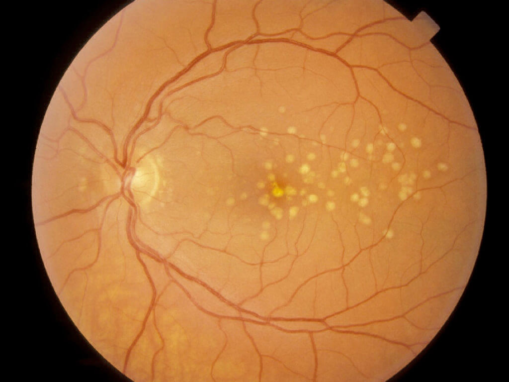
What is the finding in this fundus?
What is the finding in this fundus?
Drusen
What is the finding in this fundus?
Drusen
168
Dx?
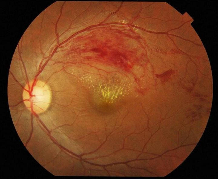
Dx?
Branched retinal vein occlusion
Branched retinal vein occlusion
169
Diagnosis?

Diagnosis?
Central retinal vein occlusion
Central retinal vein occlusion
170
Diagnosis in this patient with sudden painless loss of vision?
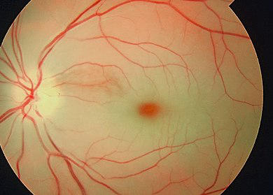
Diagnosis in this patient with sudden painless loss of vision?
Central Retinal Artery Occlusion
Central Retinal Artery Occlusion
171
Management of central retinal artery occlusion should be done in hrs
Management of central retinal artery occlusion should be done in 4-6hrs
172
Management steps of central retinal artery occlusion
1)
2) Anterior chamber paracentesis: sudden↓ in IOP, may dislodge the embolus
3) IV acetazolamide / IV mannitol(to ↓IOP)
4) Carbogen therapy (5%CO2, 95% O2)
1)
2) Anterior chamber paracentesis: sudden↓ in IOP, may dislodge the embolus
3) IV acetazolamide / IV mannitol(to ↓IOP)
4) Carbogen therapy (5%CO2, 95% O2)
Management steps of central retinal artery occlusion
1) Ocular massage
2) Anterior chamber paracentesis: sudden↓ in IOP, may dislodge the embolus
3) IV acetazolamide / IV mannitol(to ↓IOP)
4) Carbogen therapy (5%CO2, 95% O2)
1) Ocular massage
2) Anterior chamber paracentesis: sudden↓ in IOP, may dislodge the embolus
3) IV acetazolamide / IV mannitol(to ↓IOP)
4) Carbogen therapy (5%CO2, 95% O2)
173
Management steps of central retinal artery occlusion
1) Ocular massage
2)
3) IV acetazolamide / IV mannitol(to ↓IOP)
4) Carbogen therapy (5%CO2, 95% O2)
1) Ocular massage
2)
3) IV acetazolamide / IV mannitol(to ↓IOP)
4) Carbogen therapy (5%CO2, 95% O2)
Management steps of central retinal artery occlusion
1) Ocular massage
2) Anterior chamber paracentesis: sudden↓ in IOP, may dislodge the embolus
3) IV acetazolamide / IV mannitol(to ↓IOP)
4) Carbogen therapy (5%CO2, 95% O2)
1) Ocular massage
2) Anterior chamber paracentesis: sudden↓ in IOP, may dislodge the embolus
3) IV acetazolamide / IV mannitol(to ↓IOP)
4) Carbogen therapy (5%CO2, 95% O2)
174
Management steps of central retinal artery occlusion
1) Ocular massage
2) Anterior chamber paracentesis: sudden↓ in IOP, may dislodge the embolus
3)
4) Carbogen therapy (5%CO2, 95% O2)
1) Ocular massage
2) Anterior chamber paracentesis: sudden↓ in IOP, may dislodge the embolus
3)
4) Carbogen therapy (5%CO2, 95% O2)
Management steps of central retinal artery occlusion
1) Ocular massage
2) Anterior chamber paracentesis: sudden↓ in IOP, may dislodge the embolus
3) IV acetazolamide / IV mannitol(to ↓IOP)
4) Carbogen therapy (5%CO2, 95% O2)
1) Ocular massage
2) Anterior chamber paracentesis: sudden↓ in IOP, may dislodge the embolus
3) IV acetazolamide / IV mannitol(to ↓IOP)
4) Carbogen therapy (5%CO2, 95% O2)
175
Management steps of central retinal artery occlusion
1) Ocular massage
2) Anterior chamber paracentesis: sudden↓ in IOP, may dislodge the embolus
3) IV acetazolamide / IV mannitol(to ↓IOP)
4)
1) Ocular massage
2) Anterior chamber paracentesis: sudden↓ in IOP, may dislodge the embolus
3) IV acetazolamide / IV mannitol(to ↓IOP)
4)
Management steps of central retinal artery occlusion
1) Ocular massage
2) Anterior chamber paracentesis: sudden↓ in IOP, may dislodge the embolus
3) IV acetazolamide / IV mannitol(to ↓IOP)
4) Carbogen therapy (5%CO2, 95% O2)
1) Ocular massage
2) Anterior chamber paracentesis: sudden↓ in IOP, may dislodge the embolus
3) IV acetazolamide / IV mannitol(to ↓IOP)
4) Carbogen therapy (5%CO2, 95% O2)
176
Cattle trucking on fundoscopy is seen in
Cattle trucking on fundoscopy is seen in CRAO
177
Hollenhorst plaques are seen in
Hollenhorst plaques are seen in Cholesterol emboli
178
Fundus in a patient after head injury: Dx?
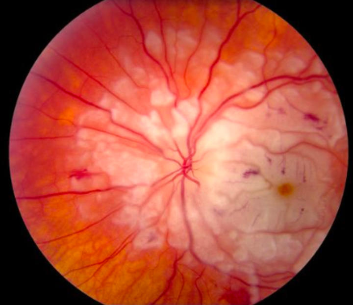
Fundus in a patient after head injury: Dx?
Purtschers retinopathy
Purtschers retinopathy
179
This finding on fundus fluorescein angiography is called and is diagnostic of cystoid macular edema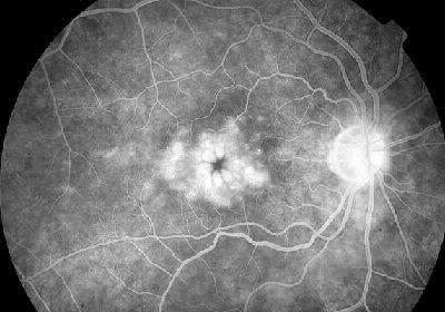
This finding on fundus fluorescein angiography is called Flower Petal pattern and is diagnostic of cystoid macular edema
180
This finding on fundus fluorescein angiography is called Flower Petal pattern and is diagnostic of
This finding on fundus fluorescein angiography is called Flower Petal pattern and is diagnostic of cystoid macular edema
181
syndrome is cystoid macular edema after cataract surgery as a post-operative complication
Irvine-Gass syndrome is cystoid macular edema after cataract surgery as a post-operative complication
182
Irvine-Gass syndrome is after cataract surgery as a post-operative complication
Irvine-Gass syndrome is cystoid macular edema after cataract surgery as a post-operative complication
183
Irvine-Gass syndrome is cystoid macular edema after surgery as a post-operative complication
Irvine-Gass syndrome is cystoid macular edema after cataract surgery as a post-operative complication
184
Type 1 DM diagnosed. When does he need to get fundus exam?
Type 1 DM diagnosed. When does he need to get fundus exam?
After 5yrs
After 5yrs
185
Type 2 DM diagnosed. When does the patient get a fundus exam?
Type 2 DM diagnosed. When does the patient get a fundus exam?
Right at diagnosis
Right at diagnosis
186
Patient with h/o diabetes and hypertension underwent FFA.
Dx?
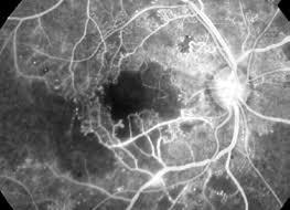
Dx?
Patient with h/o diabetes and hypertension underwent FFA.
Dx?
Macular ischemia (along with PDR)
Dx?
Macular ischemia (along with PDR)
187
Gas used for pneumatic retinopexy to treat retinal detachments?
Gas used for pneumatic retinopexy to treat retinal detachments?
Sulphur hexafluoride
Sulphur hexafluoride
188
Following waves on electroretinogram signify what:
a-wave:
b-wave: Muller cells and bipolar cells
c-wave: Retinal Pigment epithelium
a-wave:
b-wave: Muller cells and bipolar cells
c-wave: Retinal Pigment epithelium
Following waves on electroretinogram signify what:
a-wave: Rods & Cones
b-wave: Muller cells and bipolar cells
c-wave: Retinal Pigment epithelium
a-wave: Rods & Cones
b-wave: Muller cells and bipolar cells
c-wave: Retinal Pigment epithelium
189
Following waves on electroretinogram signify what:
a-wave: Rods & Cones
b-wave:
c-wave: Retinal Pigment epithelium
a-wave: Rods & Cones
b-wave:
c-wave: Retinal Pigment epithelium
Following waves on electroretinogram signify what:
a-wave: Rods & Cones
b-wave: Muller cells and bipolar cells
c-wave: Retinal Pigment epithelium
a-wave: Rods & Cones
b-wave: Muller cells and bipolar cells
c-wave: Retinal Pigment epithelium
190
Following waves on electroretinogram signify what:
a-wave: Rods & Cones
b-wave: Muller cells and bipolar cells
c-wave:
a-wave: Rods & Cones
b-wave: Muller cells and bipolar cells
c-wave:
Following waves on electroretinogram signify what:
a-wave: Rods & Cones
b-wave: Muller cells and bipolar cells
c-wave: Retinal Pigment epithelium
a-wave: Rods & Cones
b-wave: Muller cells and bipolar cells
c-wave: Retinal Pigment epithelium
191
syndrome is most commonly associated with retinitis pigmentosa
Usher syndrome is most commonly associated with retinitis pigmentosa
192
is characterized by a deficiency of apolipoprotein B-48 and B-100
Abetalipoproteinemia is characterized by a deficiency of apolipoprotein B-48 and B-100
193
Abetalipoproteinemia is characterized by a deficiency of apolipoprotein and
Abetalipoproteinemia is characterized by a deficiency of apolipoprotein B-48 and B-100
194
What pathological RBC is seen in abetalipoproteinemia?
What pathological RBC is seen in abetalipoproteinemia?
Acanthocytes
Acanthocytes
195
What vitamins are deficient in patients with abetalipoproteinemia?
What vitamins are deficient in patients with abetalipoproteinemia?
Fat soluble(especially Vitamin A and Vitamin E)
Fat soluble(especially Vitamin A and Vitamin E)
196
What apolipoproteins will be absent in abetalipoproteinemia?
(1)
(2)
(3)
(1)
(2)
(3)
What apolipoproteins will be absent in abetalipoproteinemia?
(1) Chylomicrons
(2) VLDL
(3) LDL
(1) Chylomicrons
(2) VLDL
(3) LDL
197
In Abetalipoproteinemia there will be due to Vitamin E deficiency resulting in Ataxia
In Abetalipoproteinemia there will be spinocerebellar degeneration due to Vitamin E deficiency resulting in Ataxia
198
In Abetalipoproteinemia there will be spinocerebellar degeneration due to deficiency resulting in Ataxia
In Abetalipoproteinemia there will be spinocerebellar degeneration due to Vitamin E deficiency resulting in Ataxia
199
In Abetalipoproteinemia there will be spinocerebellar degeneration due to Vitamin E deficiency resulting in
In Abetalipoproteinemia there will be spinocerebellar degeneration due to Vitamin E deficiency resulting in Ataxia
200
What occular condition is associated with abetalipoproteinemia?
What occular condition is associated with abetalipoproteinemia?
Retinitis Pigmentosa
Retinitis Pigmentosa
201
What are the GI symptoms of abetalipoproteinemia?
What are the GI symptoms of abetalipoproteinemia?
Diarrhea due to fat malabsorption
Diarrhea due to fat malabsorption
202
What is the cause of ataxia in abetalipoproteinemia?
What is the cause of ataxia in abetalipoproteinemia?
Vitamin E deficiency leading to spinocerebellar degeneration
Vitamin E deficiency leading to spinocerebellar degeneration
203
Which mitochondrial inheritance disease is associated with retinitis pigmentosa?
Which mitochondrial inheritance disease is associated with retinitis pigmentosa?
Kearn-Sayre syndrome
Kearn-Sayre syndrome
204
In the early disease, retinitis pigmentosa affects
In the early disease, retinitis pigmentosa affects rods
205
Gold standard for the diagnosis of retinitis pigmentosa?
Gold standard for the diagnosis of retinitis pigmentosa?
Electroretinography(ERG)
Electroretinography(ERG)
206
In retinitis pigmentosa, triad of fundoscopy findings are seen:
Waxy disc pallor
Bone-spicule retinal pigmentation
Waxy disc pallor
Bone-spicule retinal pigmentation
In retinitis pigmentosa, triad of fundoscopy findings are seen:
Arteriolar attenuation
Waxy disc pallor
Bone-spicule retinal pigmentation
Arteriolar attenuation
Waxy disc pallor
Bone-spicule retinal pigmentation
207
In retinitis pigmentosa, triad of fundoscopy findings are seen:
Arteriolar attenuation
Arteriolar attenuation
In retinitis pigmentosa, triad of fundoscopy findings are seen:
Arteriolar attenuation
Waxy disc pallor
Bone-spicule retinal pigmentation
Arteriolar attenuation
Waxy disc pallor
Bone-spicule retinal pigmentation
208
Perimetry findings of retinits pigmentosa?
Perimetry findings of retinits pigmentosa?
Ring scotoma, which develops into tunnel vision
Ring scotoma, which develops into tunnel vision
209
MC form of juvenile macular dystrophy is
MC form of juvenile macular dystrophy is Stargadt's Disease
210
Stargadt disease has loss of vision.
Stargadt disease has central loss of vision.
211
appearance of fundus is seen in Best's disease.
Scrambled egg appearance of fundus is seen in Best's disease.
212
Scrambled egg appearance of fundus is seen in disease.
Scrambled egg appearance of fundus is seen in Best's disease.
213
Appearance of fundus is seen in Stargadt's disease
Beaten Bronze Appearance of fundus is seen in Stargadt's disease
214
Beaten Bronze Appearance of fundus is seen in disease
Beaten Bronze Appearance of fundus is seen in Stargadt's disease
215
ERG in Stargardt disease is
ERG in Stargardt disease is Normal
216
Silent fluorocein angiography is seen in
Silent fluorocein angiography is seen in Stargadt disease
217
Electrooculogram is abnormal in
Electrooculogram is abnormal in Best disease
218
Flat electrooculogram is an arden ratio of less than
Flat electrooculogram is an arden ratio of less than 1.25
219
Normal Arden ratio is
Normal Arden ratio is >1.85
220
The only disease with normal Electroretinogram(ERG) and abnormal Electrooculogram is
The only disease with normal Electroretinogram(ERG) and abnormal Electrooculogram is Best disease
221
disease is an idiopathic vascular malformation in retina leading to exudative lesions
Coat's disease is an idiopathic vascular malformation in retina leading to exudative lesions
222
Red reflex in Coat's disease is
Red reflex in Coat's disease is Abnormal
223
What demographic is most affected by Coat's disease?
What demographic is most affected by Coat's disease?
Boys aged 6-8yrs
Boys aged 6-8yrs
224
In a 7yr old boy, what disease of the retina shows 1) Vascular telangiectasias, microaneurysms
2) Subretinal yellow exudates
3) Retinal Detachment
2) Subretinal yellow exudates
3) Retinal Detachment
In a 7yr old boy, what disease of the retina shows 1) Vascular telangiectasias, microaneurysms
2) Subretinal yellow exudates
3) Retinal Detachment
Coat's Disease
2) Subretinal yellow exudates
3) Retinal Detachment
Coat's Disease
225
Recurrent vitreous hemorrhage is seen in
Recurrent vitreous hemorrhage is seen in Eales disease
226
IOC of vitreous hemorrhage?
IOC of vitreous hemorrhage?
USG B-scan
USG B-scan
227
Sub-hyaloid hemorrhage is
Sub-hyaloid hemorrhage is boat-shaped
228
is the collection of emulsified silicone oil in the anterior chamber
Reverse hypopyon is the collection of emulsified silicone oil in the anterior chamber
229
Reverse hypopyon is the collection of in the anterior chamber
Reverse hypopyon is the collection of emulsified silicone oil in the anterior chamber
230
Nuclei of muller cells of retina lie in the
Nuclei of muller cells of retina lie in the inner nuclear layer
231
During intrauterine life, the retinal vessels reach the nasal ora at wks gestation and temporal ora by wks
During intrauterine life, the retinal vessels reach the nasal ora at 36wks gestation and temporal ora by 40wks
232
This macula is classically associated with what drug intake?
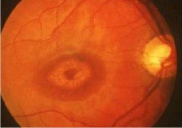
This macula is classically associated with what drug intake?
Chloroquine
Chloroquine
233
Normal IOP is mmHg
Normal IOP is 10-21mmHg
234
Eyeball hypotony is defined at an IOP of less than mmHg
Eyeball hypotony is defined at an IOP of less than 5mmHg
235
Shifting fluid sign is seen in
Shifting fluid sign is seen in exudative retinal detachment
236
Is family history a risk factor for primary open angle glaucoma?
Is family history a risk factor for primary open angle glaucoma?
Yes
Yes
237
corneas will have a falsely low intra-ocular pressure.
Thin corneas will have a falsely low intra-ocular pressure.
238
Thin corneas will have a falsely intra-ocular pressure.
Thin corneas will have a falsely low intra-ocular pressure.
239
What does corneal thickness have to do with glaucoma (as far as risk for developing glaucoma)?
What does corneal thickness have to do with glaucoma (as far as risk for developing glaucoma)?
Thin corneas are an independent risk factor for development of glaucoma
Thin corneas are an independent risk factor for development of glaucoma
240
Which race is predisposed to developing open angle glaucoma?
Which race is predisposed to developing open angle glaucoma?
African American and Hispanics
African American and Hispanics
241
Which race is predisposed to developing angle closure glaucoma?
Which race is predisposed to developing angle closure glaucoma?
Asians
Asians
242
are at an increased risk of developing angle closure glaucoma
Hypermyopes are at an increased risk of developing angle closure glaucoma
243
Most common mechanism for angle closure glaucoma is
Most common mechanism for angle closure glaucoma is pupillary block
244
Pathogenesis of Pigment Dispersion Glaucoma?
.
.
Pathogenesis of Pigment Dispersion Glaucoma?
Pigmented back-surface of the iris rubs against the radial zonules supporting the lens. Little flecks of pigment are shed into the aqueous and end up clogging the trabecular meshwork drain.
Pigmented back-surface of the iris rubs against the radial zonules supporting the lens. Little flecks of pigment are shed into the aqueous and end up clogging the trabecular meshwork drain.
245
Pathogenesis of pseudoexfoliation syndrome?
Pathogenesis of pseudoexfoliation syndrome?
Basement membrane-like material is deposited throughout the body. This material adheres to the anterior lens capsule, creating a rough surface. As the overlying iris dilates and contracts with daily activity, pigment is rubbed off and clogs the trabecular drain.
Basement membrane-like material is deposited throughout the body. This material adheres to the anterior lens capsule, creating a rough surface. As the overlying iris dilates and contracts with daily activity, pigment is rubbed off and clogs the trabecular drain.
246
If residual lens epithelial cells are left behind after cataract surgery, it can lead to formation of
If residual lens epithelial cells are left behind after cataract surgery, it can lead to formation of posterior capsular opacification(PCO)
247
Causes of cystoid macular edema can be remembered by the mnemonic DEPRIVENS
Epinephrine drops
Pars planitis\/uveitis
Retinitis pigmentosa
Irvine-Gass Syndrome
Vein Occlusion(CRVO/BRVO)
E2 Prostaglandins
Niacin/Nicotinic acid(excess)
Surgery(eg glaucoma procedures/laser photocoagulation)
Epinephrine drops
Pars planitis\/uveitis
Retinitis pigmentosa
Irvine-Gass Syndrome
Vein Occlusion(CRVO/BRVO)
E2 Prostaglandins
Niacin/Nicotinic acid(excess)
Surgery(eg glaucoma procedures/laser photocoagulation)
Causes of cystoid macular edema can be remembered by the mnemonic DEPRIVENS
Diabetes
Epinephrine drops
Pars planitis\/uveitis
Retinitis pigmentosa
Irvine-Gass Syndrome
Vein Occlusion(CRVO/BRVO)
E2 Prostaglandins
Niacin/Nicotinic acid(excess)
Surgery(eg glaucoma procedures/laser photocoagulation)
Diabetes
Epinephrine drops
Pars planitis\/uveitis
Retinitis pigmentosa
Irvine-Gass Syndrome
Vein Occlusion(CRVO/BRVO)
E2 Prostaglandins
Niacin/Nicotinic acid(excess)
Surgery(eg glaucoma procedures/laser photocoagulation)
248
Causes of cystoid macular edema can be remembered by the mnemonic DEPRIVENS
Diabetes
Pars planitis\/uveitis
Retinitis pigmentosa
Irvine-Gass Syndrome
Vein Occlusion(CRVO/BRVO)
E2 Prostaglandins
Niacin/Nicotinic acid(excess)
Surgery(eg glaucoma procedures/laser photocoagulation)
Diabetes
Pars planitis\/uveitis
Retinitis pigmentosa
Irvine-Gass Syndrome
Vein Occlusion(CRVO/BRVO)
E2 Prostaglandins
Niacin/Nicotinic acid(excess)
Surgery(eg glaucoma procedures/laser photocoagulation)
Causes of cystoid macular edema can be remembered by the mnemonic DEPRIVENS
Diabetes
Epinephrine drops
Pars planitis\/uveitis
Retinitis pigmentosa
Irvine-Gass Syndrome
Vein Occlusion(CRVO/BRVO)
E2 Prostaglandins
Niacin/Nicotinic acid(excess)
Surgery(eg glaucoma procedures/laser photocoagulation)
Diabetes
Epinephrine drops
Pars planitis\/uveitis
Retinitis pigmentosa
Irvine-Gass Syndrome
Vein Occlusion(CRVO/BRVO)
E2 Prostaglandins
Niacin/Nicotinic acid(excess)
Surgery(eg glaucoma procedures/laser photocoagulation)
249
Causes of cystoid macular edema can be remembered by the mnemonic DEPRIVENS
Diabetes
Epinephrine drops
Retinitis pigmentosa
Irvine-Gass Syndrome
Vein Occlusion(CRVO/BRVO)
E2 Prostaglandins
Niacin/Nicotinic acid(excess)
Surgery(eg glaucoma procedures/laser photocoagulation)
Diabetes
Epinephrine drops
Retinitis pigmentosa
Irvine-Gass Syndrome
Vein Occlusion(CRVO/BRVO)
E2 Prostaglandins
Niacin/Nicotinic acid(excess)
Surgery(eg glaucoma procedures/laser photocoagulation)
Causes of cystoid macular edema can be remembered by the mnemonic DEPRIVENS
Diabetes
Epinephrine drops
Pars planitis\/uveitis
Retinitis pigmentosa
Irvine-Gass Syndrome
Vein Occlusion(CRVO/BRVO)
E2 Prostaglandins
Niacin/Nicotinic acid(excess)
Surgery(eg glaucoma procedures/laser photocoagulation)
Diabetes
Epinephrine drops
Pars planitis\/uveitis
Retinitis pigmentosa
Irvine-Gass Syndrome
Vein Occlusion(CRVO/BRVO)
E2 Prostaglandins
Niacin/Nicotinic acid(excess)
Surgery(eg glaucoma procedures/laser photocoagulation)
250
Causes of cystoid macular edema can be remembered by the mnemonic DEPRIVENS
Diabetes
Epinephrine drops
Pars planitis\/uveitis
Irvine-Gass Syndrome
Vein Occlusion(CRVO/BRVO)
E2 Prostaglandins
Niacin/Nicotinic acid(excess)
Surgery(eg glaucoma procedures/laser photocoagulation)
Diabetes
Epinephrine drops
Pars planitis\/uveitis
Irvine-Gass Syndrome
Vein Occlusion(CRVO/BRVO)
E2 Prostaglandins
Niacin/Nicotinic acid(excess)
Surgery(eg glaucoma procedures/laser photocoagulation)
Causes of cystoid macular edema can be remembered by the mnemonic DEPRIVENS
Diabetes
Epinephrine drops
Pars planitis\/uveitis
Retinitis pigmentosa
Irvine-Gass Syndrome
Vein Occlusion(CRVO/BRVO)
E2 Prostaglandins
Niacin/Nicotinic acid(excess)
Surgery(eg glaucoma procedures/laser photocoagulation)
Diabetes
Epinephrine drops
Pars planitis\/uveitis
Retinitis pigmentosa
Irvine-Gass Syndrome
Vein Occlusion(CRVO/BRVO)
E2 Prostaglandins
Niacin/Nicotinic acid(excess)
Surgery(eg glaucoma procedures/laser photocoagulation)
251
Causes of cystoid macular edema can be remembered by the mnemonic DEPRIVENS
Diabetes
Epinephrine drops
Pars planitis\/uveitis
Retinitis pigmentosa
Vein Occlusion(CRVO/BRVO)
E2 Prostaglandins
Niacin/Nicotinic acid(excess)
Surgery(eg glaucoma procedures/laser photocoagulation)
Diabetes
Epinephrine drops
Pars planitis\/uveitis
Retinitis pigmentosa
Vein Occlusion(CRVO/BRVO)
E2 Prostaglandins
Niacin/Nicotinic acid(excess)
Surgery(eg glaucoma procedures/laser photocoagulation)
Causes of cystoid macular edema can be remembered by the mnemonic DEPRIVENS
Diabetes
Epinephrine drops
Pars planitis\/uveitis
Retinitis pigmentosa
Irvine-Gass Syndrome
Vein Occlusion(CRVO/BRVO)
E2 Prostaglandins
Niacin/Nicotinic acid(excess)
Surgery(eg glaucoma procedures/laser photocoagulation)
Diabetes
Epinephrine drops
Pars planitis\/uveitis
Retinitis pigmentosa
Irvine-Gass Syndrome
Vein Occlusion(CRVO/BRVO)
E2 Prostaglandins
Niacin/Nicotinic acid(excess)
Surgery(eg glaucoma procedures/laser photocoagulation)
252
Causes of cystoid macular edema can be remembered by the mnemonic DEPRIVENS
Diabetes
Epinephrine drops
Pars planitis\/uveitis
Retinitis pigmentosa
Irvine-Gass Syndrome
E2 Prostaglandins
Niacin/Nicotinic acid(excess)
Surgery(eg glaucoma procedures/laser photocoagulation)
Diabetes
Epinephrine drops
Pars planitis\/uveitis
Retinitis pigmentosa
Irvine-Gass Syndrome
E2 Prostaglandins
Niacin/Nicotinic acid(excess)
Surgery(eg glaucoma procedures/laser photocoagulation)
Causes of cystoid macular edema can be remembered by the mnemonic DEPRIVENS
Diabetes
Epinephrine drops
Pars planitis\/uveitis
Retinitis pigmentosa
Irvine-Gass Syndrome
Vein Occlusion(CRVO/BRVO)
E2 Prostaglandins
Niacin/Nicotinic acid(excess)
Surgery(eg glaucoma procedures/laser photocoagulation)
Diabetes
Epinephrine drops
Pars planitis\/uveitis
Retinitis pigmentosa
Irvine-Gass Syndrome
Vein Occlusion(CRVO/BRVO)
E2 Prostaglandins
Niacin/Nicotinic acid(excess)
Surgery(eg glaucoma procedures/laser photocoagulation)
253
Causes of cystoid macular edema can be remembered by the mnemonic DEPRIVENS
Diabetes
Epinephrine drops
Pars planitis\/uveitis
Retinitis pigmentosa
Irvine-Gass Syndrome
Vein Occlusion(CRVO/BRVO)
Niacin/Nicotinic acid(excess)
Surgery(eg glaucoma procedures/laser photocoagulation)
Diabetes
Epinephrine drops
Pars planitis\/uveitis
Retinitis pigmentosa
Irvine-Gass Syndrome
Vein Occlusion(CRVO/BRVO)
Niacin/Nicotinic acid(excess)
Surgery(eg glaucoma procedures/laser photocoagulation)
Causes of cystoid macular edema can be remembered by the mnemonic DEPRIVENS
Diabetes
Epinephrine drops
Pars planitis\/uveitis
Retinitis pigmentosa
Irvine-Gass Syndrome
Vein Occlusion(CRVO/BRVO)
E2 Prostaglandins
Niacin/Nicotinic acid(excess)
Surgery(eg glaucoma procedures/laser photocoagulation)
Diabetes
Epinephrine drops
Pars planitis\/uveitis
Retinitis pigmentosa
Irvine-Gass Syndrome
Vein Occlusion(CRVO/BRVO)
E2 Prostaglandins
Niacin/Nicotinic acid(excess)
Surgery(eg glaucoma procedures/laser photocoagulation)
254
Causes of cystoid macular edema can be remembered by the mnemonic DEPRIVENS
Diabetes
Epinephrine drops
Pars planitis\/uveitis
Retinitis pigmentosa
Irvine-Gass Syndrome
Vein Occlusion(CRVO/BRVO)
E2 Prostaglandins
Surgery(eg glaucoma procedures/laser photocoagulation)
Diabetes
Epinephrine drops
Pars planitis\/uveitis
Retinitis pigmentosa
Irvine-Gass Syndrome
Vein Occlusion(CRVO/BRVO)
E2 Prostaglandins
Surgery(eg glaucoma procedures/laser photocoagulation)
Causes of cystoid macular edema can be remembered by the mnemonic DEPRIVENS
Diabetes
Epinephrine drops
Pars planitis\/uveitis
Retinitis pigmentosa
Irvine-Gass Syndrome
Vein Occlusion(CRVO/BRVO)
E2 Prostaglandins
Niacin/Nicotinic acid(excess)
Surgery(eg glaucoma procedures/laser photocoagulation)
Diabetes
Epinephrine drops
Pars planitis\/uveitis
Retinitis pigmentosa
Irvine-Gass Syndrome
Vein Occlusion(CRVO/BRVO)
E2 Prostaglandins
Niacin/Nicotinic acid(excess)
Surgery(eg glaucoma procedures/laser photocoagulation)
255
Causes of cystoid macular edema can be remembered by the mnemonic DEPRIVENS
Diabetes
Epinephrine drops
Pars planitis\/uveitis
Retinitis pigmentosa
Irvine-Gass Syndrome
Vein Occlusion(CRVO/BRVO)
E2 Prostaglandins
Niacin/Nicotinic acid(excess)
Diabetes
Epinephrine drops
Pars planitis\/uveitis
Retinitis pigmentosa
Irvine-Gass Syndrome
Vein Occlusion(CRVO/BRVO)
E2 Prostaglandins
Niacin/Nicotinic acid(excess)
Causes of cystoid macular edema can be remembered by the mnemonic DEPRIVENS
Diabetes
Epinephrine drops
Pars planitis\/uveitis
Retinitis pigmentosa
Irvine-Gass Syndrome
Vein Occlusion(CRVO/BRVO)
E2 Prostaglandins
Niacin/Nicotinic acid(excess)
Surgery(eg glaucoma procedures/laser photocoagulation)
Diabetes
Epinephrine drops
Pars planitis\/uveitis
Retinitis pigmentosa
Irvine-Gass Syndrome
Vein Occlusion(CRVO/BRVO)
E2 Prostaglandins
Niacin/Nicotinic acid(excess)
Surgery(eg glaucoma procedures/laser photocoagulation)
256
On fundus exam, what optic disc finding is seen in pathological myopia?
On fundus exam, what optic disc finding is seen in pathological myopia?
Pale disc with Temporal myopic crest
Pale disc with Temporal myopic crest
257
are blackish spots due to retinal pigment epithelium(RPE) hyperplasia that occur as a reparative response to choroidal neovascularization and sub-retinal hemorrhage. It is seen in Pathological Myopia
Foster Fuchs spots are blackish spots due to retinal pigment epithelium(RPE) hyperplasia that occur as a reparative response to choroidal neovascularization and sub-retinal hemorrhage. It is seen in Pathological Myopia
258
Foster Fuchs spots are blackish spots due to hyperplasia that occur as a reparative response to choroidal neovascularization and sub-retinal hemorrhage. It is seen in Pathological Myopia
Foster Fuchs spots are blackish spots due to retinal pigment epithelium(RPE) hyperplasia that occur as a reparative response to choroidal neovascularization and sub-retinal hemorrhage. It is seen in Pathological Myopia
259
Foster Fuchs spots are blackish spots due to retinal pigment epithelium(RPE) hyperplasia that occur as a reparative response to choroidal neovascularization and sub-retinal hemorrhage. It is seen in
Foster Fuchs spots are blackish spots due to retinal pigment epithelium(RPE) hyperplasia that occur as a reparative response to choroidal neovascularization and sub-retinal hemorrhage. It is seen in Pathological Myopia
260
Tesselated fundus(aka Tigroid fundus) is seen in
Tesselated fundus(aka Tigroid fundus) is seen in pathological myopia
261
appearance of fundus in pathological myopia is due to retinal pigment epithelium thinning and ↑visibility of choroidal vasculature.
Tigroid appearance of fundus in pathological myopia is due to retinal pigment epithelium thinning and ↑visibility of choroidal vasculature.
262
Tigroid appearance of fundus in pathological myopia is due to retinal pigment epithelium thinning and ↑visibility of .
Tigroid appearance of fundus in pathological myopia is due to retinal pigment epithelium thinning and ↑visibility of choroidal vasculature.
263
The arrow marked finding in pathological myopia is called as 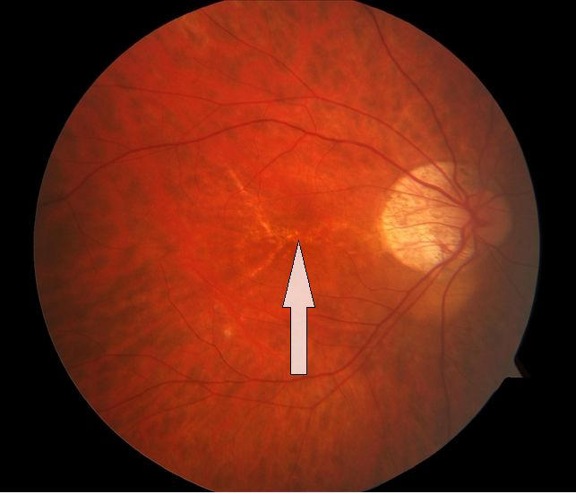
The arrow marked finding in pathological myopia is called as lacquer cracks
264
Diagnosis?
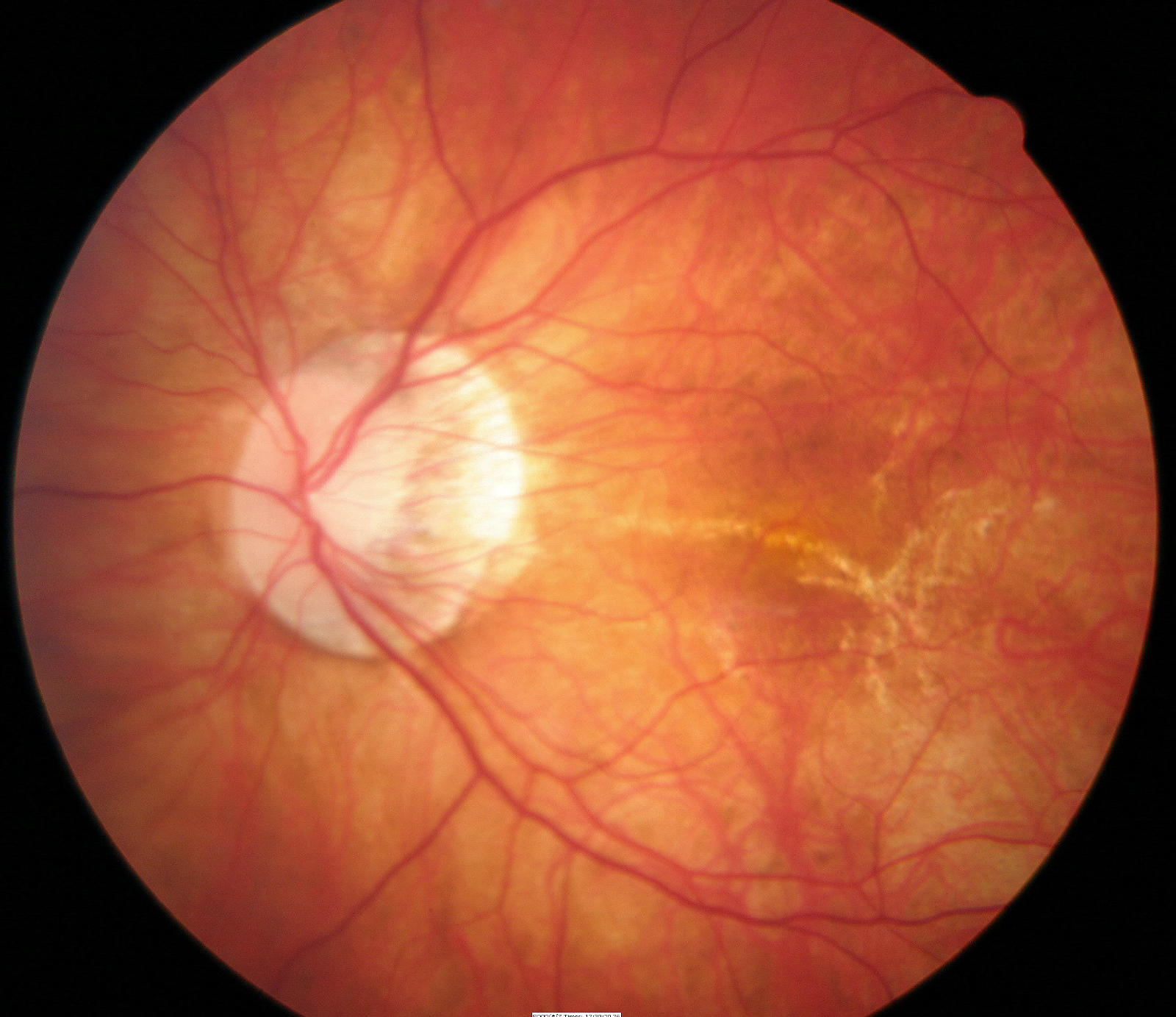
Diagnosis?
Diagnosis?
Pathological myopia
Diagnosis?
Pathological myopia
265
Diagnosis?
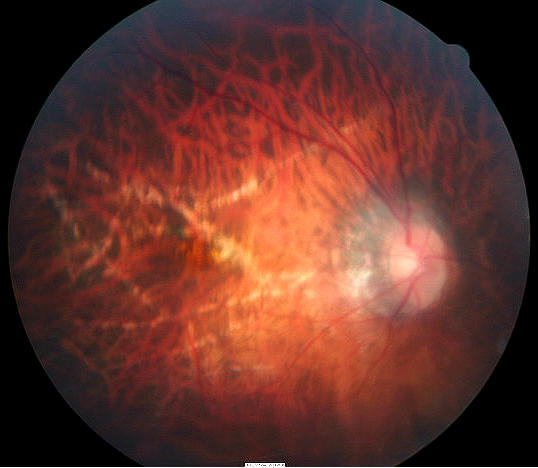
Diagnosis?
Pathological myopia
Pathological myopia
266
Diagnosis?
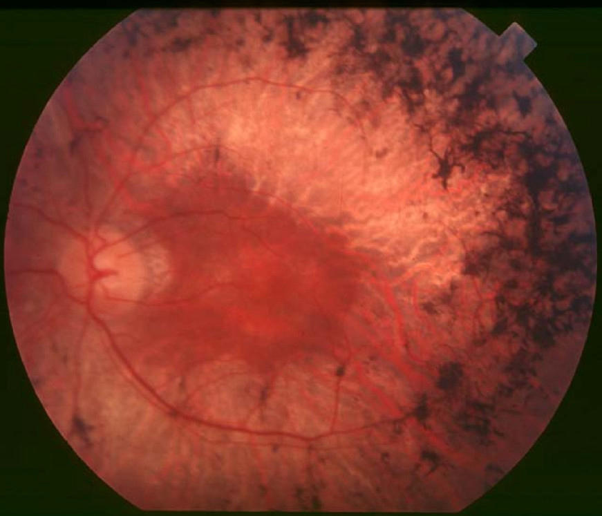
Diagnosis?
Retinitis Pigmentosa
Retinitis Pigmentosa
267
retinal detachment is one of the complications of pathological myopia
Rhegmatogenous retinal detachment is one of the complications of pathological myopia
268
MC complication of pathological myopia is
MC complication of pathological myopia is macular degeneration
269
In high myopia, it is better to slightly -correct for distance.
In high myopia, it is better to slightly under-correct for distance.
270
laser is used in LASIK procedure.
Excimer laser is used in LASIK procedure.
271
The nearest point at which small objects can be clearly seen is called the
The nearest point at which small objects can be clearly seen is called the near point
272
Accomodative squint is a complication of uncorrected long standing hypermetropia in children.
Accomodative convergent squint is a complication of uncorrected long standing hypermetropia in children.
273
Pseudopapillitis is seen in
Pseudopapillitis is seen in hypermetropia
274
Shot silk appearance of retina is seen in .
Shot silk appearance of retina is seen in hypermetropia.
275
Conductive keratoplasty is used for the treatment of
Conductive keratoplasty is used for the treatment of hypermetropia
276
Normal astigmatism seen in eyes due to eyelid pressure is the rule
Normal astigmatism seen in eyes due to eyelid pressure is with the rule
277
With the rule astigmatism is more commonly seen in
With the rule astigmatism is more commonly seen in young
278
Against the rule astigmatism is more commonly seen in
Against the rule astigmatism is more commonly seen in elderly
279
If the vision improves on pinhole test, the pathology is
If the vision improves on pinhole test, the pathology is optical error
280
If the vision doesn't improve on pinhole test, the pathology is
If the vision doesn't improve on pinhole test, the pathology is organic pathology
281
With the rule astigmatism means that vertical meridian is myotic
With the rule astigmatism means that vertical meridian is more myotic
282
Identify the type of the refractive error?
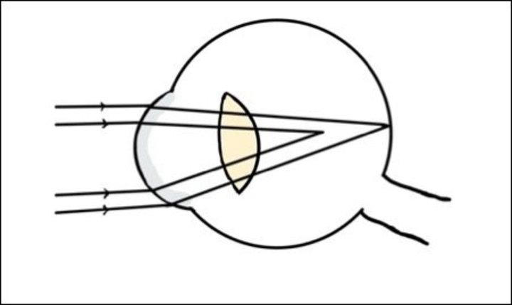
Identify the type of the refractive error?
Simple myopic astigmatism
Simple myopic astigmatism
283
Identify the type of refractive error:
Identify the type of refractive error:
Simple hypermetropic astigmatism
Simple hypermetropic astigmatism
284
Identify the type of refractive error?
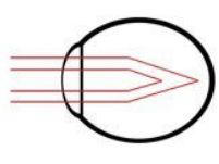
Identify the type of refractive error?
Compound myopic astigmatism
Compound myopic astigmatism
285
Identify the type of refractive error?
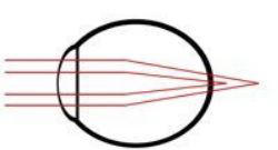
Identify the type of refractive error?
Compound hypermetropic astigmatism
Compound hypermetropic astigmatism
286
Identify the type of refractive error?
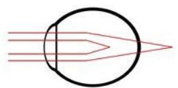
Identify the type of refractive error?
Mixed(myopic & hypermetropic) astigmatism
Mixed(myopic & hypermetropic) astigmatism
287
Patient was prescribed glasses of power "-1 DC X 180 degrees".What is the type of his refractive error?
Patient was prescribed glasses of power "-1 DC X 180 degrees".What is the type of his refractive error?
Simple myopic asigmatism
Simple myopic asigmatism
288
Patient was prescribed glasses of power "+2 DC X 180 degrees".What is the type of his refractive error?
Patient was prescribed glasses of power "+2 DC X 180 degrees".What is the type of his refractive error?
Simple hypermetropic asigmatism
Simple hypermetropic asigmatism
289
Patient was prescribed glasses of power "-1DS +2 DC X 180 degrees".What is the type of his refractive error?
Patient was prescribed glasses of power "-1DS +2 DC X 180 degrees".What is the type of his refractive error?
Mixed astigmatism
Mixed astigmatism
290
Patient was prescribed glasses of power "-1DS +1 DC X 180 degrees".What is the type of his refractive error?
Patient was prescribed glasses of power "-1DS +1 DC X 180 degrees".What is the type of his refractive error?
Simple myopic
Simple myopic
291
Patient was prescribed glasses of power "-1DS +1 DC X 180 degrees".
The type os astigmatism in this patient is the rule.
The type os astigmatism in this patient is the rule.
Patient was prescribed glasses of power "-1DS +1 DC X 180 degrees".
The type os astigmatism in this patient is against the rule.
The type os astigmatism in this patient is against the rule.
292
Patient was prescribed glasses of power "-1DS -1 DC X 90 degrees".
What is the type of his refractive error?
What is the type of his refractive error?
Patient was prescribed glasses of power "-1DS -1 DC X 90 degrees".
What is the type of his refractive error?
Compound myopic
What is the type of his refractive error?
Compound myopic
293
During near accomodation, ciliary muscles
During near accomodation, ciliary muscles contract
294
The reading glasses of a 47yr old man should be about D
The reading glasses of a 47yr old man should be about +1.5D
295
is a condition in which there is a difference in the size of the image formed by the two eyes
Aniseikonia is a condition in which there is a difference in the size of the image formed by the two eyes
296
is a condition in which iris vibrates during eye movement.
Iridodonesis is a condition in which iris vibrates during eye movement.
297
Iridodonesis is primarily seen in and
Iridodonesis is primarily seen in lens subluxation and aphakia
298
In a normal eye, how many purkinje images are visible?
In a normal eye, how many purkinje images are visible?
4
4
299
In a pseudophakic eye, how many purkinje images are visible?
In a pseudophakic eye, how many purkinje images are visible?
4
4
300
In a aphakic eye, how many purkinje images are visible?
In a aphakic eye, how many purkinje images are visible?
2
2
494 weitere Karten in OutLearn. Ganzes Deck öffnen
Mehr Karteikarten: Medizin und Gesundheit
Skin and Bones: Integumentary and Skeletal SystemMedizin und Gesundheit · EnglischTaylorMedical Terminology ReviewMedizin und Gesundheit · EnglischKatlynKEPars anatomicaMedizin und Gesundheit · Englischeinem OutLearn-Studierenden5- Glandes annexesMedizin und Gesundheit · FranzösischOcéane1- synapse + GABAMedizin und Gesundheit · FranzösischOcéanePenicilina benzatínica, hidroclorotiazida, glibenclamidaMedizin und Gesundheit · SpanischLAURAingredients Medizin und Gesundheit · EnglischashAccommodation et amétropiesMedizin und Gesundheit · FranzösischOcéaneFeedback LoopsMedizin und Gesundheit · EnglischviolaEnterotoxicosisMedizin und Gesundheit · EnglischcaeMedical Cardiac Arrest DirectiveMedizin und Gesundheit · EnglischKaliCirculatory and Cardiovascular System AnatomyMedizin und Gesundheit · Englischgnourt7
Alle Decks: Medizin und Gesundheit
Lerne diese Decks in der App
Kostenlos auf iPhone, Android, Mac und im Web. Füge jedes Deck mit einem Tipp zu deinem Konto hinzu.