Hypersensitivity and Immunology
Diese Karteikarten lernenKostenlos · Spaced Repetition · iPhone, Android, Mac & Web
Alle Karten
1
What is hypersensitivity?
An exaggerated immune response to a normally harmless antigen that causes tissue damage.
2
What is the Gell-Coombs classification, and when was it introduced? EXAM
A classification of four hypersensitivity types based on immune injury mechanisms, introduced in 1963.
3
What are the four types of hypersensitivity? EXAM
Type I: immediate, anaphylaxis;
Type II: cytotoxic, immune mediated hemolytic anemia;
Type III: immune complex-mediated, glomerulonephritis
ype IV: delayed, T-cell-mediated, allergic contact dermatitis
Type II: cytotoxic, immune mediated hemolytic anemia;
Type III: immune complex-mediated, glomerulonephritis
ype IV: delayed, T-cell-mediated, allergic contact dermatitis
4
Which hypersensitivity types involve antibodies, and which does not? EXAM
Types I, II, and III involve antibodies; Type IV is T-cell-mediated.
5
What occurs during the sensitization phase of hypersensitivity?
Initial antigen exposure primes the immune system without causing an allergic reaction.
6
What occurs during the effector phase of hypersensitivity?
A subsequent exposure or persistent antigen may trigger an immune reaction.
second exposure
second exposure
7
Can hypersensitivity develop without a second antigen exposure?
Yes, if the antigen persists after initial sensitization.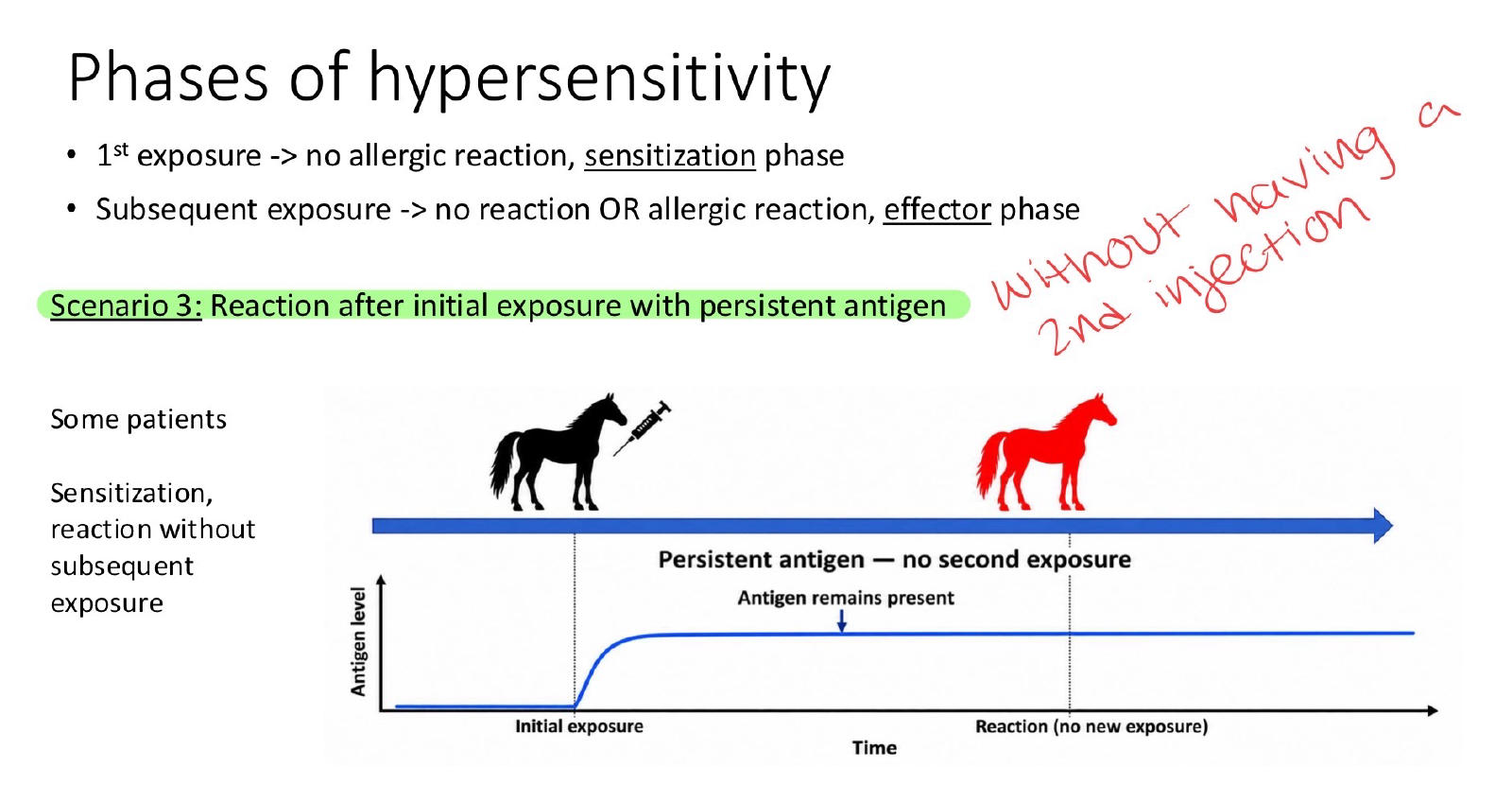
8
What is allergy? Which is the most common?
abnormal immune response to typically harmless substances
- most allergies are type 1
- most allergies are type 1
9
What is an allergen?
An antigen that causes an allergic reaction.
10
What is atopy?
A genetic predisposition to develop IgE-mediated allergies against normally harmless substances (dust, pollen)
11
What are the four routes of allergen exposure?
Ingestion, inhalation, percutaneous exposure, and skin contact.
12
Which immune components mediate Type I hypersensitivity?
IgE antibodies and mast cells.
13
What happens during the first exposure in Type I hypersensitivity?
Dendritic cells present allergens to naïve T cells, promoting Th2 differentiation and IgE production.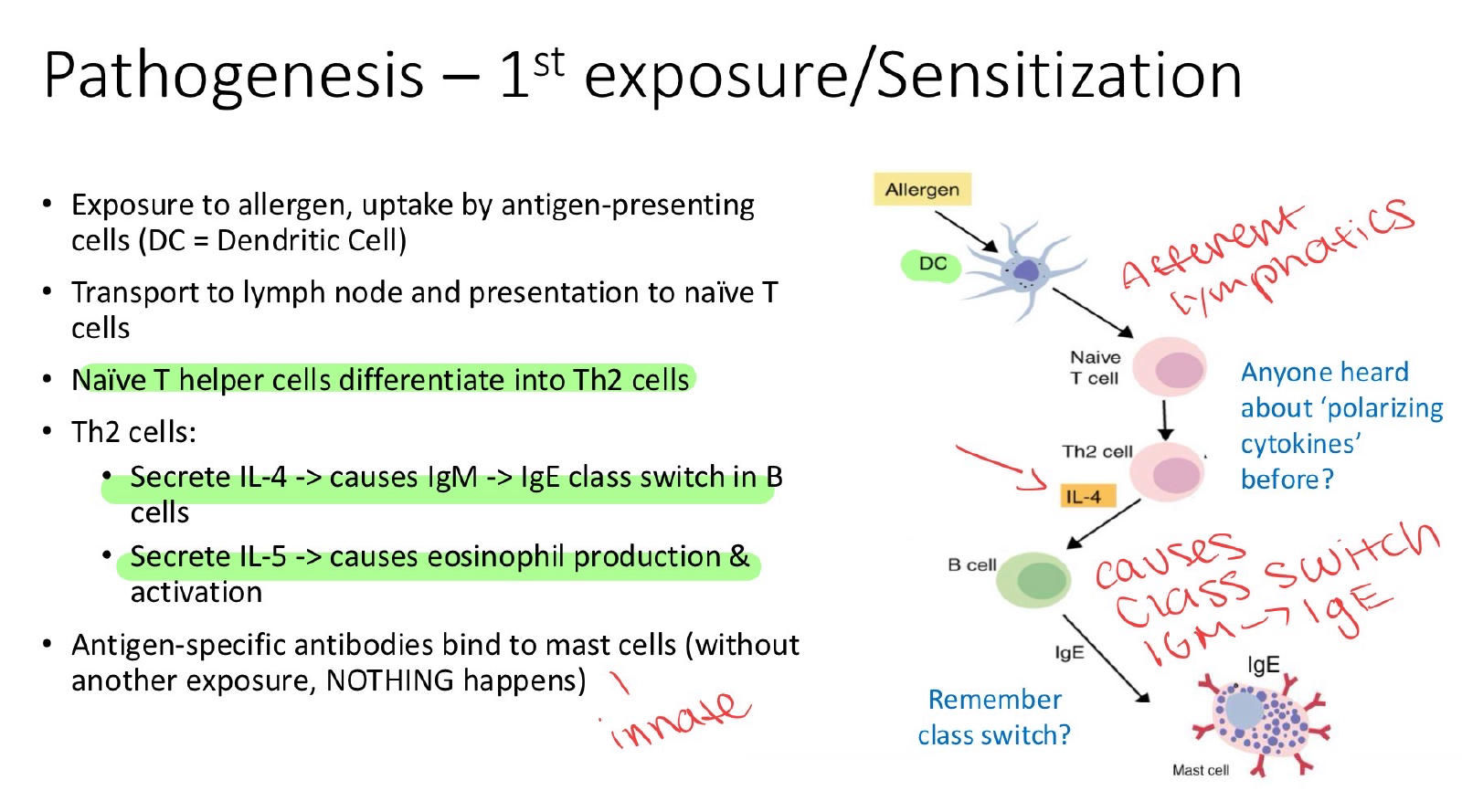
14
Which Th2 cytokine causes B cells to class switch from IgM to IgE?
IL-4.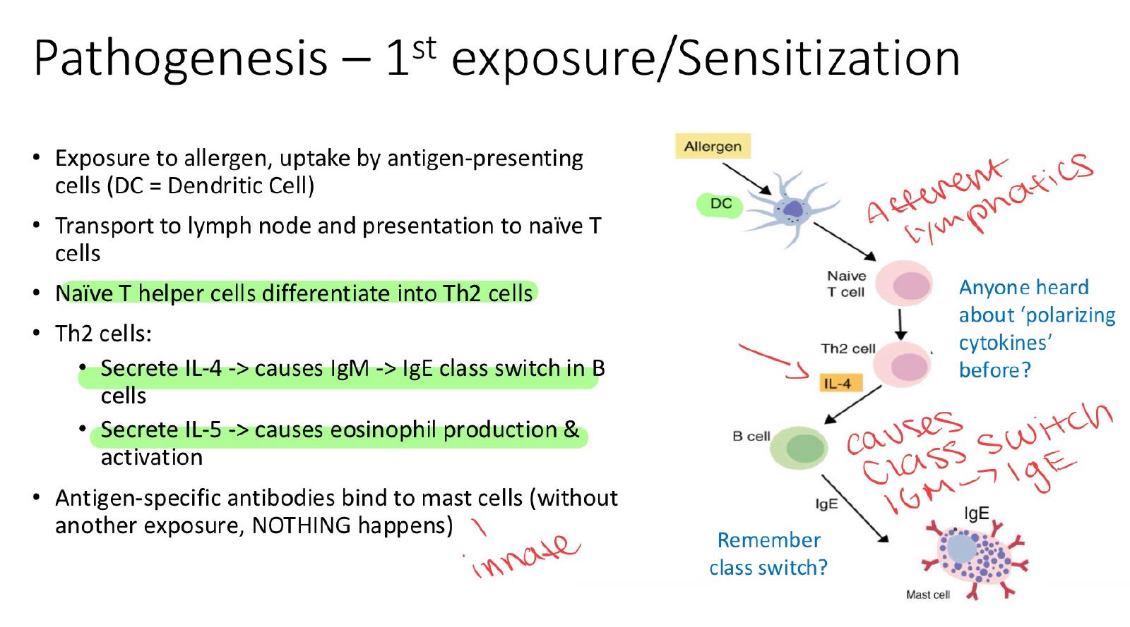
15
Which Th2 cytokine promotes eosinophil production and activation?
IL-5.
16
What happens to IgE after the first allergen exposure?
IgE binds mast cells, sensitizing them without causing degranulation.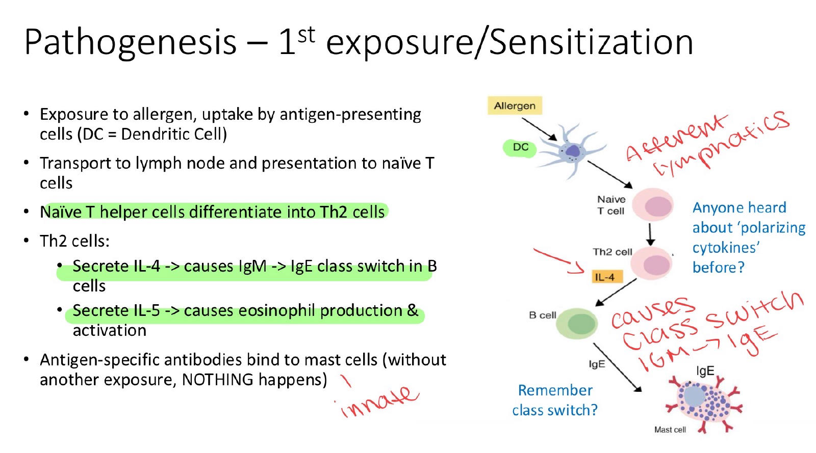
17
What triggers mast cell degranulation during Type I hypersensitivity?
Allergen cross-linking of mast cell-bound IgE during subsequent exposure.
18
How do eosinophils contribute to Type I hypersensitivity?
They release major basic protein and eosinophil cationic protein, causing tissue injury.
19
Which immune components mediate Type II hypersensitivity?
IgG or IgM antibodies and complement.
20
What is the main mechanism of Type II hypersensitivity?
Antibodies bind cell-surface antigens, causing cell destruction; cytotoxic effect
21
What are common examples of Type II hypersensitivity?
Neonatal isoerythrolysis, immune-mediated hemolytic anemia, and incompatible blood transfusion reactions.
22
What causes neonatal isoerythrolysis in foals?
An Aa-positive foal ingests anti-Aa antibodies from its sensitized Aa-negative mare's colostrum.
23
How are RBCs destroyed during neonatal isoerythrolysis? EXAM
Maternal antibodies bind foal RBCs and activate complement-mediated hemolysis.
24
What is immune-mediated hemolytic anemia (IMHA)?
IgG or IgM antibodies target RBC surface antigens, causing RBC destruction.
25
What are the two mechanisms of RBC destruction in IMHA?
Extravascular hemolysis by macrophages and intravascular hemolysis by complement.
26
What is a hapten, and how can it cause drug-induced Type II hypersensitivity?
A small molecule that becomes immunogenic when bound to a carrier; drugs can bind RBCs and trigger antibody-mediated destruction.
A hapten is a molecule too small to trigger the immune system on its own, but it becomes a target once it sticks to a bigger protein.
A drug is a sticker that glues onto the RBC surface and changes how it looks. The guard sees a "new" cell and attacks. The drug is acting as a hapten (small Lego piece clipped onto a big base).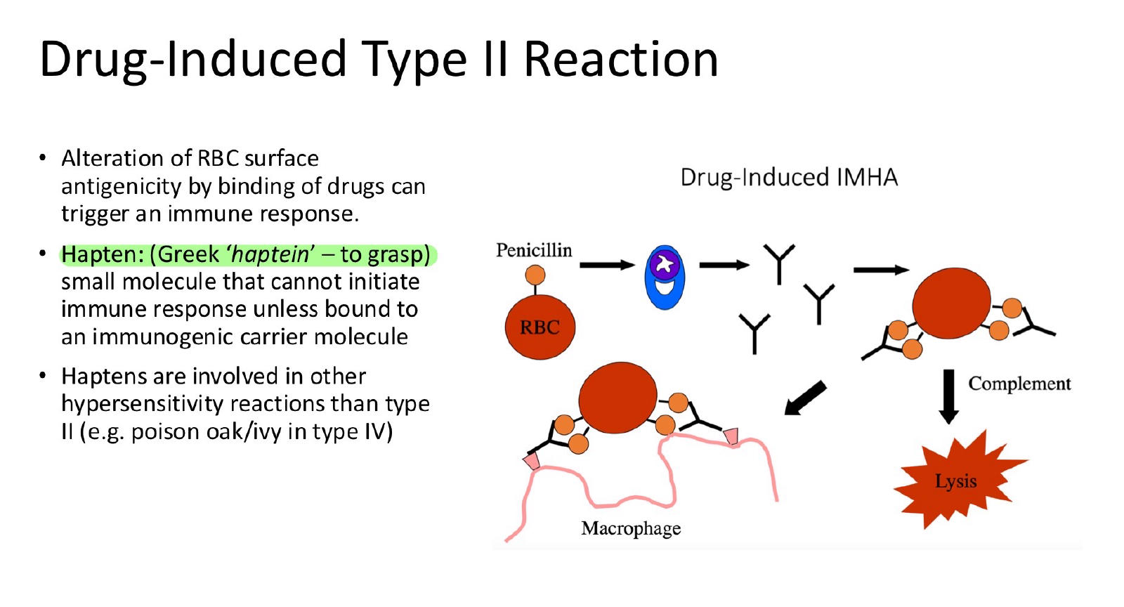
A hapten is a molecule too small to trigger the immune system on its own, but it becomes a target once it sticks to a bigger protein.
A drug is a sticker that glues onto the RBC surface and changes how it looks. The guard sees a "new" cell and attacks. The drug is acting as a hapten (small Lego piece clipped onto a big base).
27
What is the main mechanism of Type III hypersensitivity?
Antigen-antibody immune complexes deposit in tissues, activate complement, and cause inflammation.
28
What is serum sickness, and when did it occur after horse anti-tetanus serum administration?
A systemic Type III reaction that developed approximately 10 days after administration.
29
How does the antigen-antibody ratio affect immune complex clearance? type 3
Antibody excess promotes clearance; antigen excess favors complex deposition and tissue injury.
• Antibody excess: complement activates, and complexes are cleared by mononuclear phagocytes (the body handles it).
• Antigen excess: complexes are NOT cleared, so they deposit in blood vessel walls, activate leukocytes, and cause tissue injury.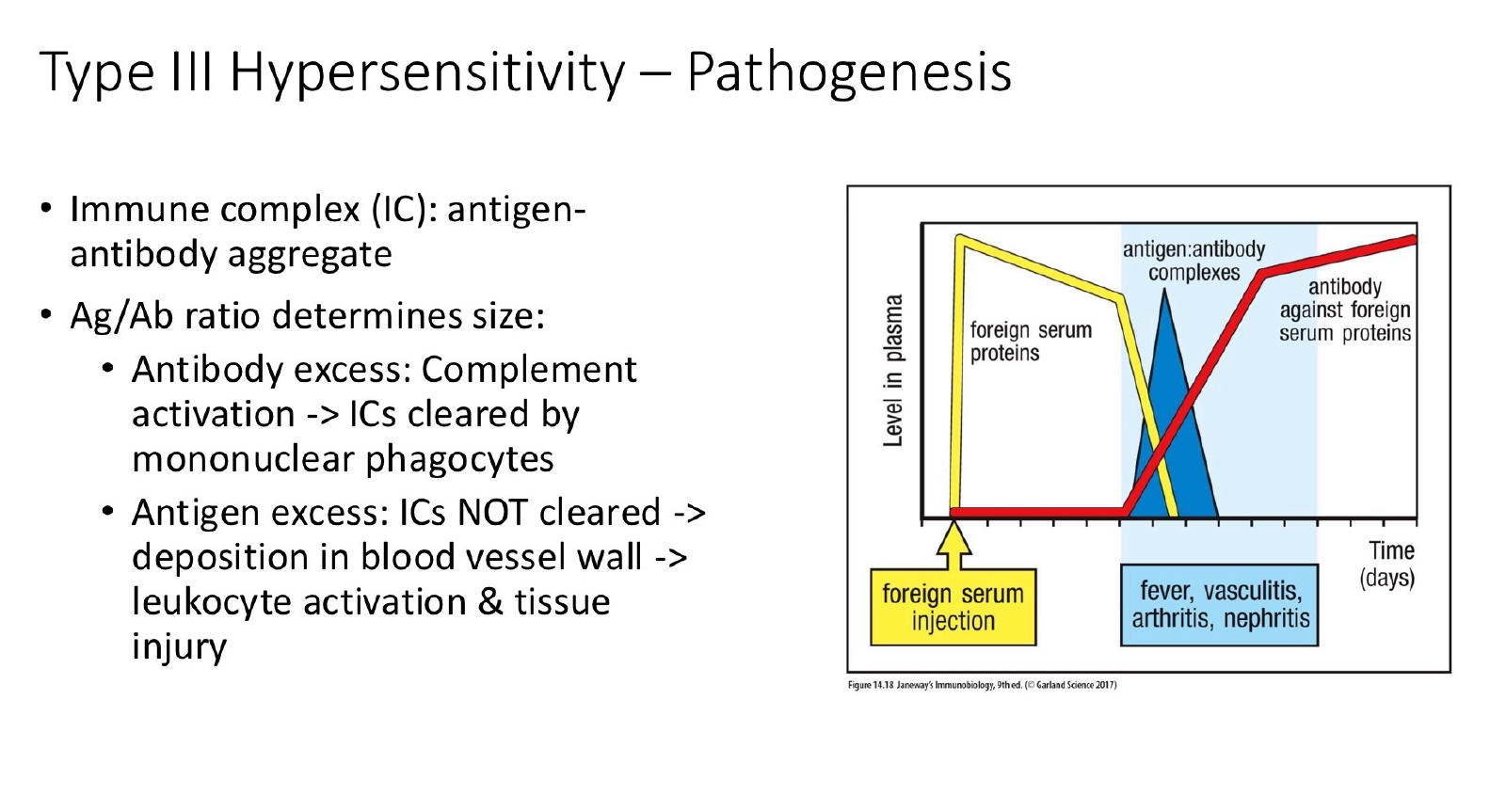
• Antibody excess: complement activates, and complexes are cleared by mononuclear phagocytes (the body handles it).
• Antigen excess: complexes are NOT cleared, so they deposit in blood vessel walls, activate leukocytes, and cause tissue injury.
30
What are the three major steps of Type III hypersensitivity?
Immune complex deposition, classical complement activation, and neutrophil recruitment causing tissue damage.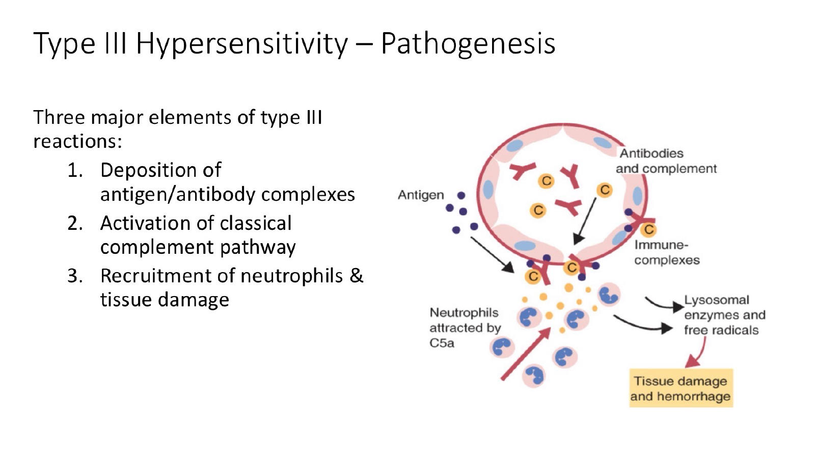
31
How does immune complex glomerulonephritis develop in dogs?
Immune complexes deposit in glomeruli, causing inflammation, structural damage, and impaired kidney function.
32
What is purpura hemorrhagica in horses?
A Type III hypersensitivity reaction occurring 2–4 weeks after infection or vaccination, often involving Streptococcus equi, causing edema, urticaria, and hemorrhages.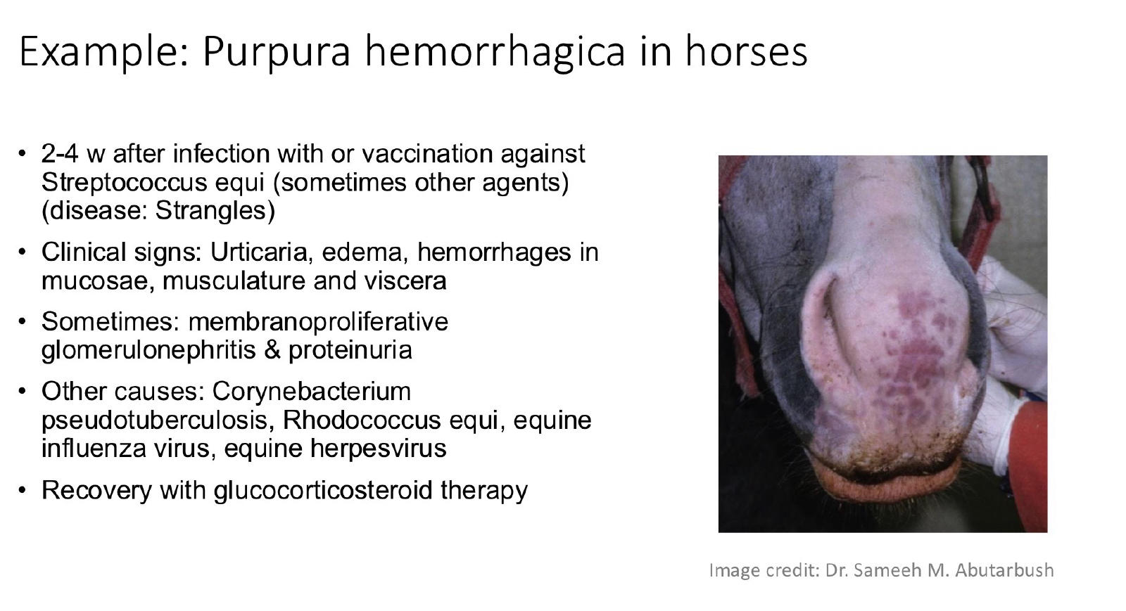
33
Which immune cells mediate Type IV hypersensitivity?
T cells and myeloid cells, especially macrophages.
34
How quickly does Type IV hypersensitivity typically develop? Poison oak
Delayed, generally 24–72 hours after exposure.
35
What is the tuberculin skin test used for in cattle?
Detecting immune sensitization to mycobacterial antigens as part of tuberculosis control.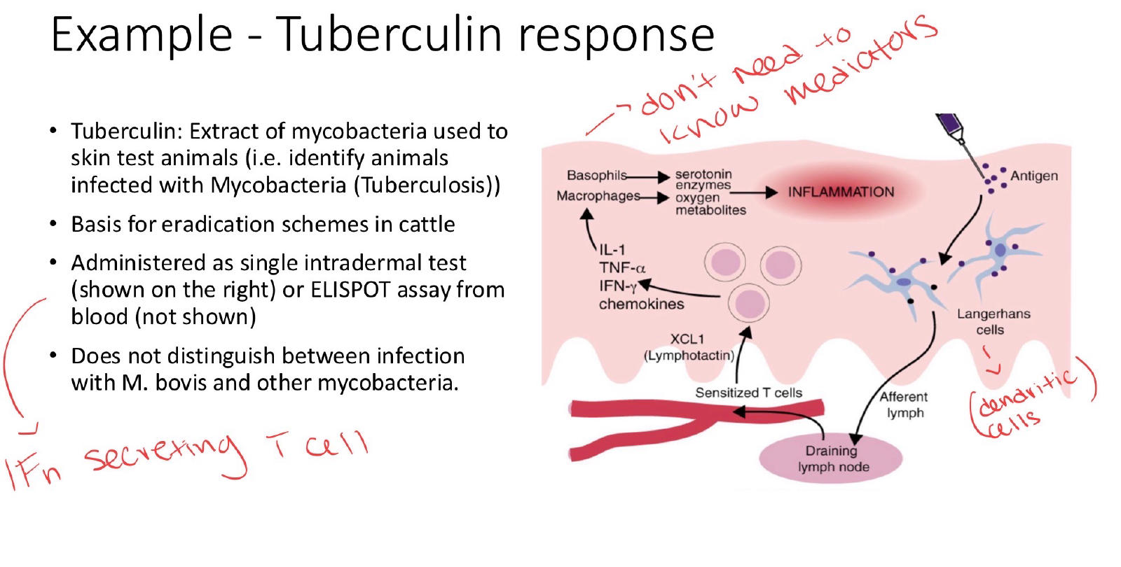
36
How does allergic contact dermatitis develop?
Haptens bind skin proteins, Langerhans cells present antigens to T cells, and activated T cells cause inflammation and keratinocyte damage.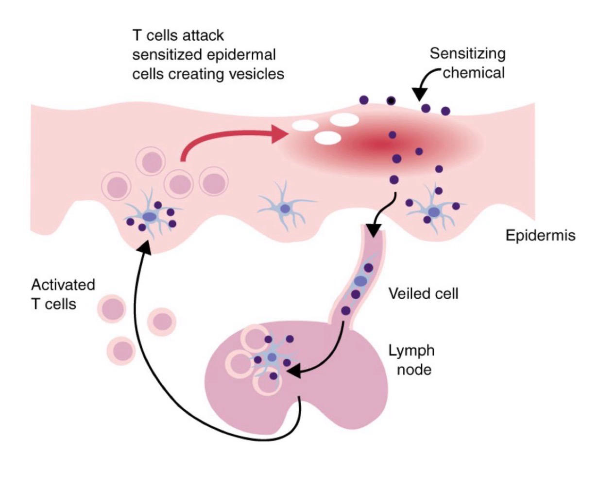
37
What causes poison oak contact dermatitis?
Urushiol binds skin proteins to form neoantigens, triggering a delayed Type IV reaction after sensitization.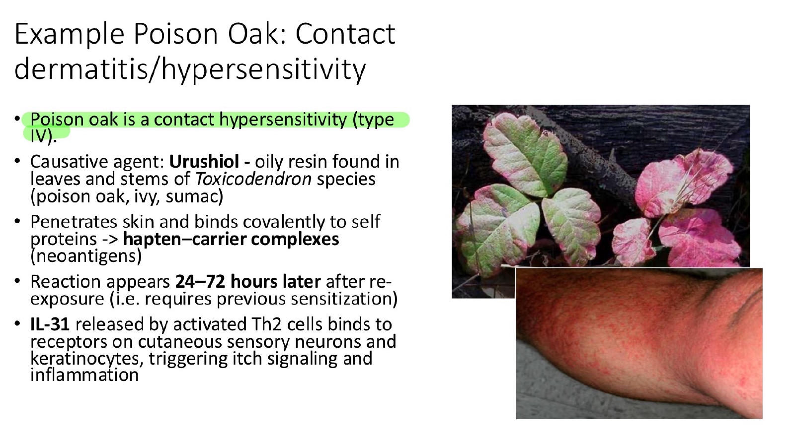
38
What is the role of IL-31 in poison oak hypersensitivity?
IL-31 from activated Th2 cells stimulates sensory neurons and keratinocytes, causing itching and inflammation.
39
How does nickel cause allergic contact dermatitis?
Nickel acts as a hapten, binds skin proteins, activates T cells and macrophages, and causes keratinocyte destruction.
40
What is the difference between hypersensitivity and autoimmunity?
Hypersensitivity describes the immune injury mechanism; autoimmunity describes an immune response against self-antigens.
41
Why can IMHA and lupus nephritis be classified as both autoimmune and hypersensitivity diseases?
IMHA targets self-RBCs through Type II cytotoxicity; lupus nephritis involves self-directed immune complexes causing Type III injury.
Mehr Karteikarten: Medizin und Gesundheit
Respiratory System FlashcardsMedizin und Gesundheit · EnglischVickyBone PathologyMedizin und Gesundheit · EnglischEshaHypertensionMedizin und Gesundheit · EnglischSaraExam 2: Part 2 ReviewMedizin und Gesundheit · EnglischAMaduva spinariiMedizin und Gesundheit · RumänischRyan2- SNC: MSMedizin und Gesundheit · FranzösischOcéaneOtic DisordersMedizin und Gesundheit · Englischkay24Exam 1 StatPearlsMedizin und Gesundheit · EnglischMadiLecture 8 - HypersensitivityMedizin und Gesundheit · EnglischKimMuscle Relaxation PhysiologyMedizin und Gesundheit · EnglischnewSkull AnatomyMedizin und Gesundheit · EnglischAlyssaBio 115: Lymphatic and Immune SystemsMedizin und Gesundheit · EnglischDillon
Alle Decks: Medizin und Gesundheit
Lerne diese Decks in der App
Kostenlos auf iPhone, Android, Mac und im Web. Füge jedes Deck mit einem Tipp zu deinem Konto hinzu.

