KUB Radiography and Anatomy
Diese Karteikarten lernenKostenlos · Spaced Repetition · iPhone, Android, Mac & Web
Alle Karten
1
WHAT MUST THE EXAMINATION ANSWER?
morphology, lumen, function,vessels, procedure
2
can prevent the wrong protocol even when the modality is correct
complete request
3
INFORMATION THAT CHANGES THE EXAMINATION
Clinical details, safety and protcol details
4
translates the clinical question into phases, sequences, views, timing, and dose.
Protocoling
5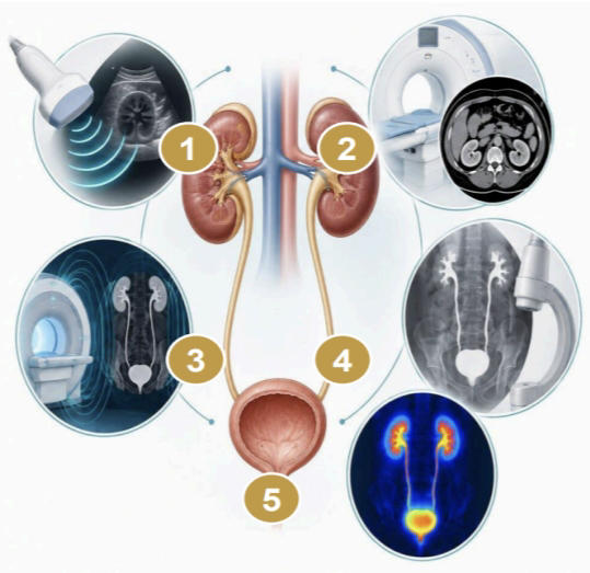
1. Right kidney, 2. Left kidney; 3. Right ureter; 4. Left ureter
5. Urinary bladder
5. Urinary bladder
6
does not have contrast
Plain KUB
7
what is shown in the middle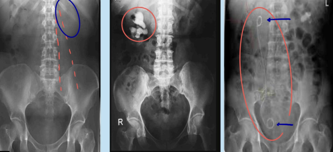
Staghorn calculus
8
what is shown on the right image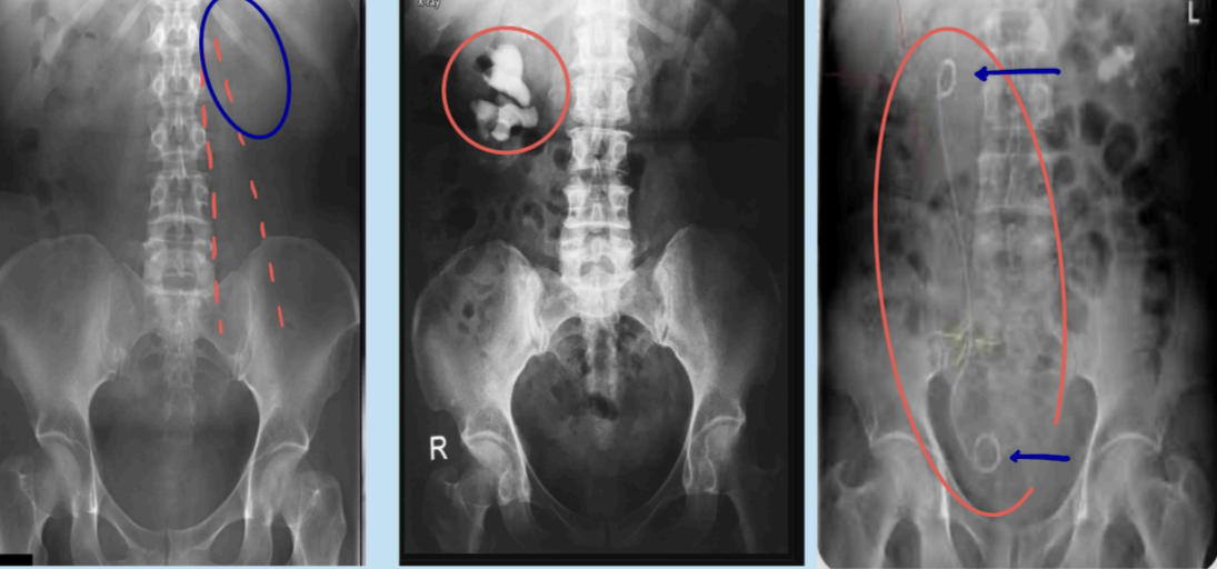
Ureteral sign or Double-J stent
9
include both kidneys and the bladder region
diagnostic KUB
10
Coverage and exposure
Both renal fossae = included superiorly.
Pubic symphysis and bladder region = included
inferiorly.; Spinous processes = centered between pedicles.
Pubic symphysis and bladder region = included
inferiorly.; Spinous processes = centered between pedicles.
11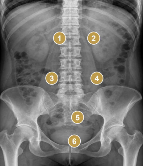
1. Right renal outline, 2. Left renal outline, 3. Right psoas margin, 4. Left psoas margin, 5. Pelvic brim/expected ureter course, 6. Pubic symphysis and bladder region
12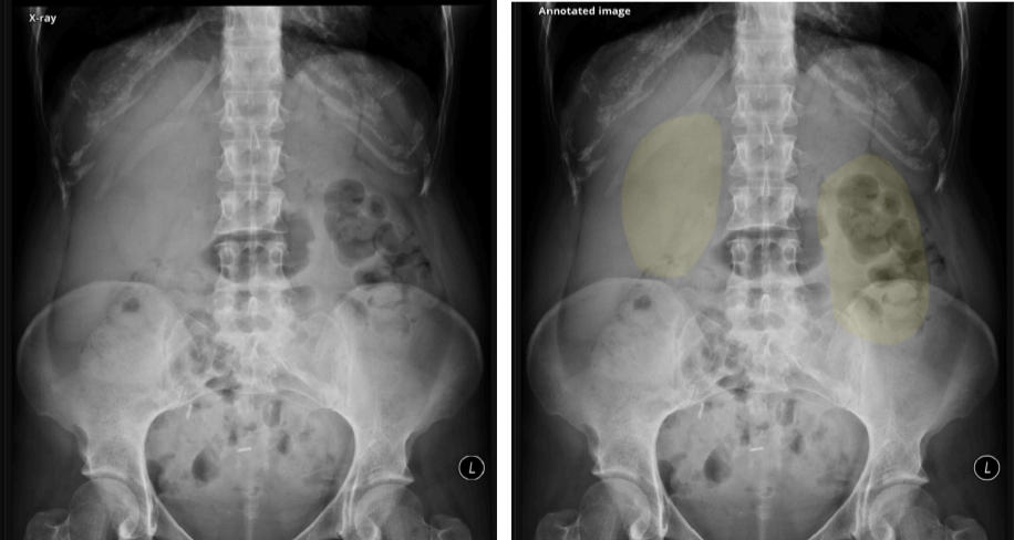
Plain KUB (X-RAY)
13
KUB LIMITATIONS–AND THE ESCALATION RULE
Bowel gas, stool, overlying bones obscure small calculi.; Radiolucent stones and most soft-tissue diseases are not seen. (eg. abscesses); Projection imaging cannot reliably localize a calcication in depth.
14
Shows iodinated contrast moving through the urinary tract during lling, drainage, or voiding.
Enables real-time positioning and targeted spot images.
Enables real-time positioning and targeted spot images.
FLUOROSCOPY
15
FLUOROSCOPIC EXAMINATION IN FOUR MOVES
Prepare, scout, observe, finish
16
What IVU demonstrates
Renal uptake and parenchymal symmetry; Opacication and morphology; Drainage into the bladder over time
17
timed examination: uptake, concentration, excretion,,and drainage.
INTRAVENOUS UROGRAPHY
18
more superior that IVU to show urinary tract and surrounding tissues cross sectionally
CT Urography
19
reference image for every subsequent phase
scout
20
patient-specic; preparation should never be reduced to a single laboratory value.
Contrast safety
21
FIRST PHASE: NEPHROGENIC PHASE
about 1-3 minutes after injection
22
● Opacication of the bilateral kidneys
● It reects renal perfusion, ltration, and tubular concentration
● But it is not a direct measurement of the eGFR
● It reects renal perfusion, ltration, and tubular concentration
● But it is not a direct measurement of the eGFR
FIRST PHASE: NEPHROGENIC PHASE
23
STEP 4: EARLY EXCRETORY/PYELOGRAPHIC PHASE
around 5-10 minutes; adapt to the patient
24
Calyces and renal pelvis begin to opacify, proximal ureters may appear intermitently with peristalsis
STEP 4: EARLY EXCRETORY/PYELOGRAPHIC PHASE
25
Use only when the added upper-tract details are needed.
Avoid if obstruction or trauma is suspected
Temporary distal ureter compression (using sand bag)
26
Done in 30 degrees, problem-solving projection
OBLIQUE VIEWS
27
STEP 5: FULL-LENGTH EXCRETORY IMAGE
10-15 minutes
28
Collecting systems, ureters, bladder = assessed on a full-length image; ureteric lling may remain segmental
STEP 5: FULL-LENGTH EXCRETORY IMAGE
29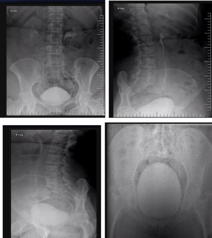
Excretory Phase
30
○ Poor or asymmetric renal function.
○ Suspected obstruction with incomplete ureteric opacication.
○ Possible urinary leak, stula, or ectopic insertion.
○ Suspected obstruction with incomplete ureteric opacication.
○ Possible urinary leak, stula, or ectopic insertion.
DELAYED IMAGES, 20-30 minutes or later
31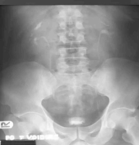
Post-void Image
32
used to check for the residual volume (quantify)
UTZ
33
urinary retention
50cc and above
34
Retrograde contrast dye injected through a urinary catheter to outline the bladder. It is used to check for bladder leak, trauma, fistula, structural or functional abnormalities
CONVENTIONAL CYSTOGRAPHY
35
Assessing bladder leak after trauma
fill to detrusorcontraction or to at least 300 mL
36
postoperative injury,
fill to a smaller volume
(100-150 mL or until resistance is felt)
(100-150 mL or until resistance is felt)
37
evaluate presence or absence of Vesicoureteral Reux (VUR)
VOIDING CYSTOURETHROGRAPHY (VCUG)
38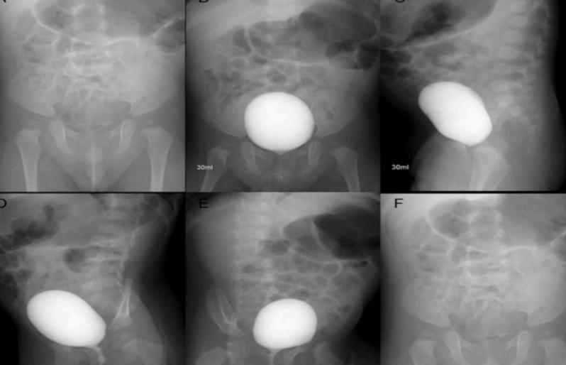
VCUG
39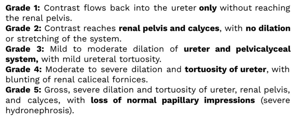
VESICOURETERAL REFLUX GRADING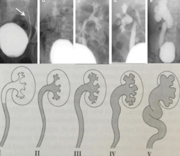
40
if tissue remains obstructive
after birth or initial treatment—is a congenital birth defect
affecting only males where extra aps of tissue block the
urethra.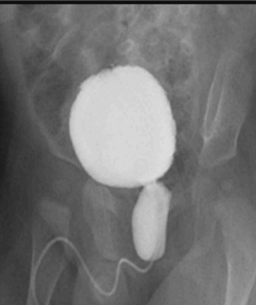
after birth or initial treatment—is a congenital birth defect
affecting only males where extra aps of tissue block the
urethra.
POSTERIOR URETHRAL VALVES (PUV)
PERSISTENT URETHRAL VALVES
PERSISTENT URETHRAL VALVES
41
➢ Elongated urinary bladder with conical shape towards
dome of the bladder like a tree. Bladder wall hypertrophy
gives rise to an irregular outline.
➢ Typically occurs in those with sacral abnormalities at birth.
➢ Diagnosed through voiding cystogram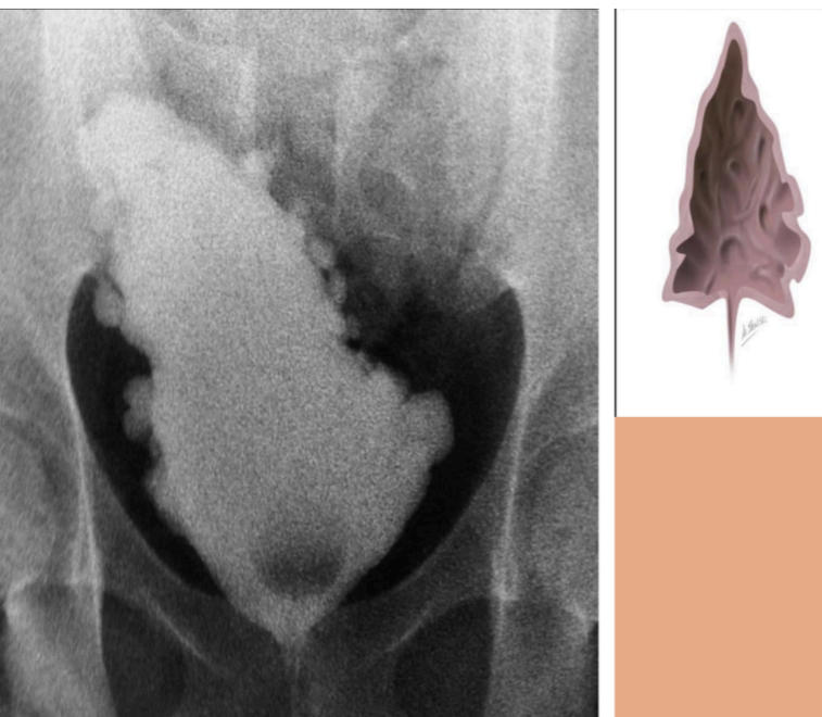
dome of the bladder like a tree. Bladder wall hypertrophy
gives rise to an irregular outline.
➢ Typically occurs in those with sacral abnormalities at birth.
➢ Diagnosed through voiding cystogram
NEUROGENIC BLADDER
42
Christmas tree or pine cone bladder.
NEUROGENIC BLADDER
43
Fluoroscopic X-Ray Examination that inserts catheter to
the penis to inject contrast to check for damage,
narrowing and blockage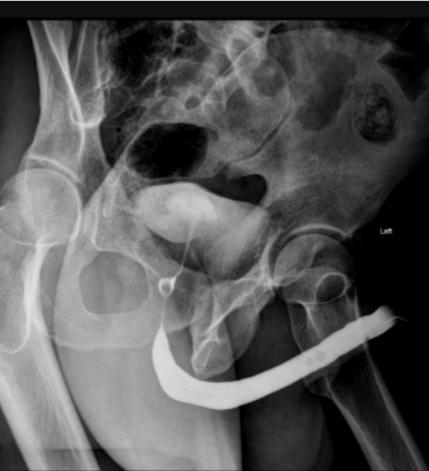
the penis to inject contrast to check for damage,
narrowing and blockage
RETROGRADE URETHOGRAPHY (RUG)
44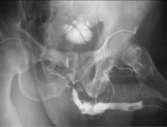
Urethral transection
45
abnormal narrowing of urethra caused by scar tissue; More common in males than in females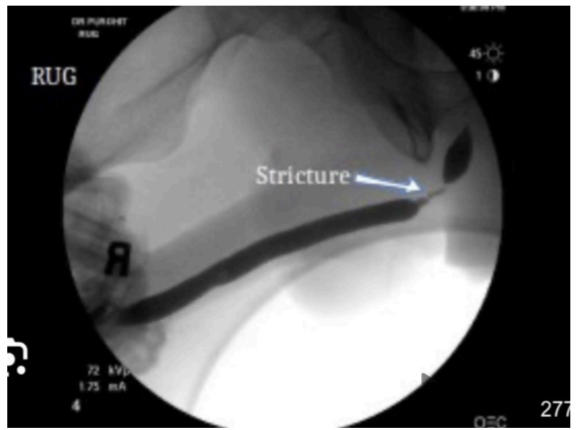
URETHRAL STRICTURE
46
Can guide stent placement or sampling—but requires
instrumentation; Evaluates upper urinary tract
instrumentation; Evaluates upper urinary tract
RETROGRADE
PYELOGRAPHY
47
Assesses drainage, obstruction, leak, and catheter position.
ANTEROGRADE PYELOGRAPHY
48
Known radiopaque stone follow-up, Device position:
KUB
49
New acute ank pain
CT or ultrasound
50
Bladder leak
cystography or CT cystography
51
Reux or voiding urethra
VCUG
52
Anterior urethral trauma/stricture
retrograde urethrography
53
BLADDER ULTRASOUND: METHOD OF EXAMINATION
fill; two planes; measure; recheck
54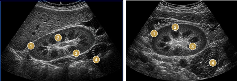
1. Renal cortex; 2. Medullary pyramids; 3. Echogenic renal sinus; 4. Renal pelvis region
55
Cortex thickness
6 mm
56
parenchymal thickness
15-20 mm
57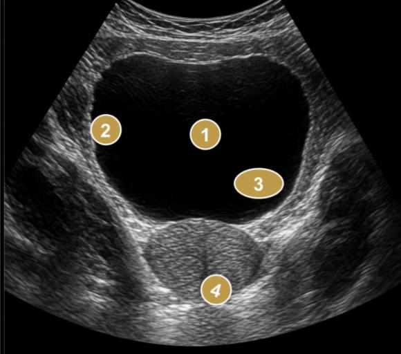
1. Anechoic urine-lled lumen; 2. Bladder wall; 3. Trigone/ ureteric
insertion region; 4. Prostate or adjacent pelvic tissues
insertion region; 4. Prostate or adjacent pelvic tissues
58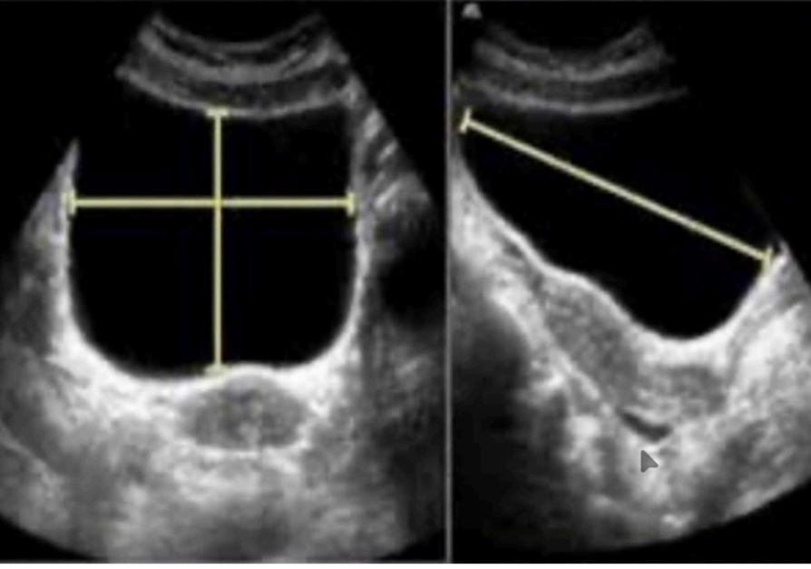
Urinary Bladder
59
well-distended adult urinary bladder typically holds
300-400 mL (up to 500 mL comfortably).
60
Wall thickness of bladder
≤ 3mm when the bladder is well-distended.
○ Up to 5 mm when not well distended
○ Up to 5 mm when not well distended
61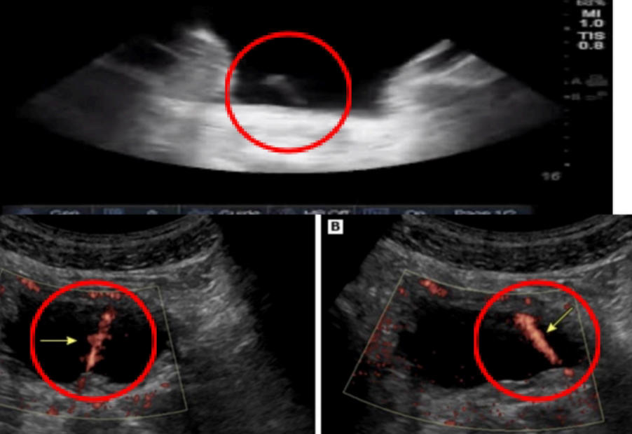
Ultrasound images of the urinary bladder
62
Ureteric jets can be assessed using
color Doppler ultrasound
63
➢ Dilation of renal pelvis and calyces.
➢ Renal cortical thinning in chronic severe disease.
➢ Bladder distension, ureteric jets, or a visible,proximal/distal cause in selected patients.
➢ Renal cortical thinning in chronic severe disease.
➢ Bladder distension, ureteric jets, or a visible,proximal/distal cause in selected patients.
HYDRONEPHROSIS
64
an anatomical sign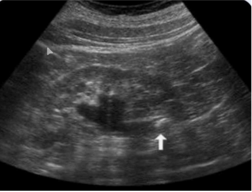
Hydronephrosis
65
➢ Useful for selected focal renal lesions, trauma, perfusion,
transplant questions.
transplant questions.
CONTRAST-ENHANCED ULTRASOUND (CEUS)
66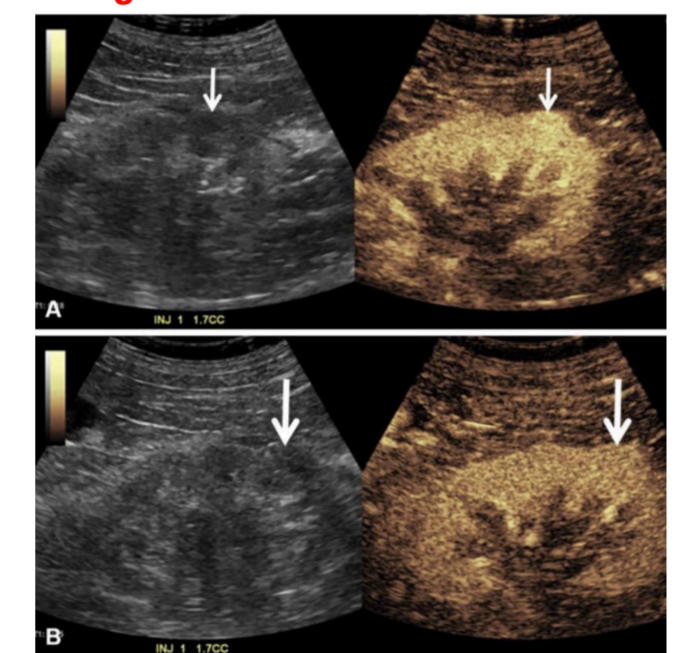
oncocytoma
67
Excellent detection of calculi, gas, hemorrhage, fat, and calcication.
COMPUTED TOMOGRAPHY
68
Patient with nephrolithiasis
CT Stonogram
69
Hounseld unit (HU) of 1Uric acid stones
200-400 HU
70
Hounseld unit (HU) of Calcium oxalate stones
600-1200 HU
71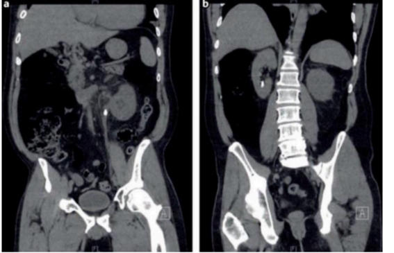
Patient with nephrolithiasis
72
can be used to predict responsiveness to shockwave lithotripsy
CT atenuation
73
Morphologic detail of renal parenchyma and collecting system
CT UROGRAPHY
74
Important nephrographic parameters:
Time of appearance of contrast agent in kidneys
Time of disappearance
Density of nephrogram and pyelogram
Time of disappearance
Density of nephrogram and pyelogram
75
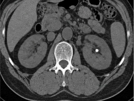
RENAL CT PHASE 0 : UNENHANCED BASELINE
Before Contrast: 0 second
76
Unenhanced renal parenchyma. We see stones, fat, hemorrhage, gas, and calcication; retain native attenuation.
RENAL CT PHASE 0 : UNENHANCED BASELINE
77
ARTERIAL PHASE
15–25 seconds after injection or bolus-triggered; Contrast in aorta and some in renal arteries
78
BEST FOR:
○ Renal arteries, active bleeding, aneurysm, stenosis,
and planning.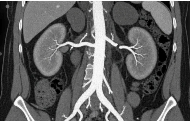
○ Renal arteries, active bleeding, aneurysm, stenosis,
and planning.
ARTERIAL PHASE
79
RENAL CT PHASE 1 : CORTICOMEDULLARY PHASE
25–50 seconds after injection
80
EXPECTED: Cortex enhances strongly while medulla remains lower in atenuation. Vessels are conspicuous.
➢ ➢ BEST FOR: Hypervascular masses, vascular anatomy, cortical perfusion, and trauma.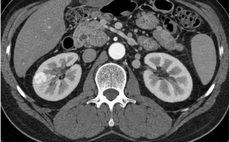
➢ ➢ BEST FOR: Hypervascular masses, vascular anatomy, cortical perfusion, and trauma.
RENAL CT PHASE 1 : CORTICOMEDULLARY PHASE
81
PORTAL VENOUS PHASE
60–75 seconds after injection
82
➢ EXPECTED:
○ Abdominal viscera and veins enhance well. Renal parenchyma approaches uniformity.
➢ BEST FOR:
○ Infection, complications, venous thrombosis, trauma, and metastases.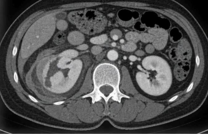
○ Abdominal viscera and veins enhance well. Renal parenchyma approaches uniformity.
➢ BEST FOR:
○ Infection, complications, venous thrombosis, trauma, and metastases.
PORTAL VENOUS PHASE
83
RENAL CT PHASE 3 : NEPHROGRAPHIC PHASE
About 80–120 seconds after injection
84
➢ EXPECTED:
○ Cortex and medulla enhance more uniformly, increasing contrast between parenchyma and many lesions.
➢ BEST FOR:
○ Renal masses, infection, infarct, and laceration.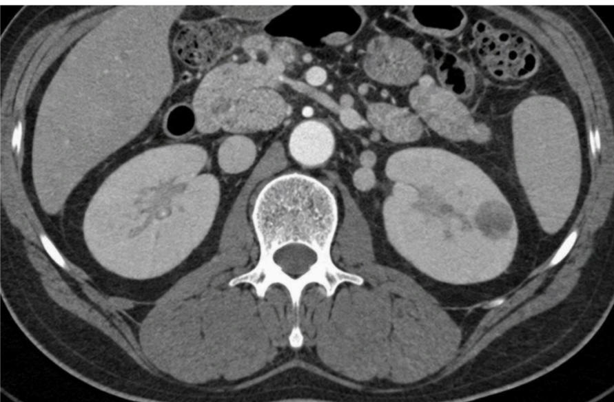
○ Cortex and medulla enhance more uniformly, increasing contrast between parenchyma and many lesions.
➢ BEST FOR:
○ Renal masses, infection, infarct, and laceration.
RENAL CT PHASE 3 : NEPHROGRAPHIC PHASE
85
DELAYED OR EQUILIBRIUM PHASE
About 2 minutes after injection
86
➢ EXPECTED:
○ Vascular contrast equilibrates. Some lesions wash out or enhance progressively. Early urine may opacify.
➢ BEST FOR:
○ Washout, delayed enhancement, renal or adrenal questions, and brosis.
○ Vascular contrast equilibrates. Some lesions wash out or enhance progressively. Early urine may opacify.
➢ BEST FOR:
○ Washout, delayed enhancement, renal or adrenal questions, and brosis.
DELAYED OR EQUILIBRIUM PHASE
87
RENAL CT PHASE 3 : NEPHROGRAPHIC PHASE
Usually 3-15 minutes after injection
88
➢ EXPECTED
○ Contrast reaches calyces, renal pelves, ureters, and bladder. Filling defects happens due to peristalsis
➢ BEST FOR
○ Urothelial lesions, collecting system, ureteric narrowing, and leak.
○ Contrast reaches calyces, renal pelves, ureters, and bladder. Filling defects happens due to peristalsis
➢ BEST FOR
○ Urothelial lesions, collecting system, ureteric narrowing, and leak.
RENAL CT PHASE 3 : NEPHROGRAPHIC PHASE
89
TARTGETED DELAY PHASE
15-30 minutes—or longer when physiology requires
90
➢ EXPECTED
○ Additional opacication may reveal slow excretion, a distal ureter, or a leak absent earlier.
➢ BEST FOR
○ Obstruction, ureteric injury, postoperative leak, or delayed excretion
○ Additional opacication may reveal slow excretion, a distal ureter, or a leak absent earlier.
➢ BEST FOR
○ Obstruction, ureteric injury, postoperative leak, or delayed excretion
TARTGETED DELAY PHASE
91
Renal gas or your Emphysematous pyelonephritis
NON CONTRAST CT FOR INFECTION AND
COMPLICATIONS
COMPLICATIONS
92
fat stranding
presence of infection of inflammation
93
Purpose
○ Hematuria or suspected upper-tract urothelial disease.
○ Assess kidneys, collecting systems, ureters, and bladder.
○ Detects lling defects, wall thickening, obstruction, or another cause.
○ Hematuria or suspected upper-tract urothelial disease.
○ Assess kidneys, collecting systems, ureters, and bladder.
○ Detects lling defects, wall thickening, obstruction, or another cause.
CT UROGRAPHY
94
CT anatomy: unenhanced axial image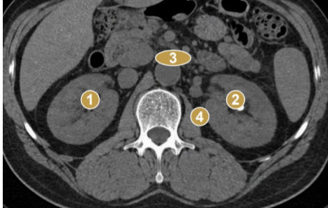
1. Right kidney 2. Left kidney 3. Aorta 4.Psoas muscle
95
nephrographic phase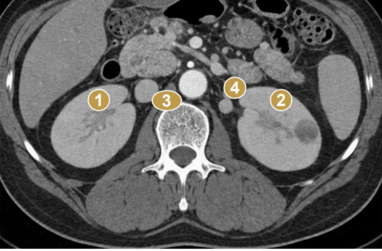
1. Right enhancing renal parenchyma 2. Left enhancing renal parenchyma 3. Right
renal sinus/pelvis region 4. Left renal sinus/pelvis region
renal sinus/pelvis region 4. Left renal sinus/pelvis region
96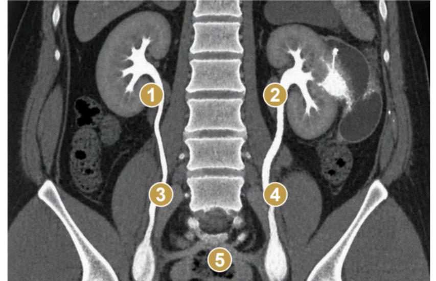
excretory phase. 1. Right collecting system 2. Left collecting system 3. Right ureter 4. Left
ureter 5. Urinary bladder
ureter 5. Urinary bladder
Mehr Karteikarten: Medizin und Gesundheit
les bases de la surveillance des traitementMedizin und Gesundheit · FranzösischClarafracturesMedizin und Gesundheit · EnglischelijahCh14 (from the book)Medizin und Gesundheit · EnglischJESSQTRANPathology: Inflammation and HealingMedizin und Gesundheit · EnglischJESSQTRANGiardia lambliaMedizin und Gesundheit · EnglischJESSQTRANMCB 134 Cancer Biology (Lectures 19-23)Medizin und Gesundheit · EnglischAshIntroduction to MusclesMedizin und Gesundheit · Englischeinem OutLearn-StudierendenMed Term Prefix & Suffix ReviewMedizin und Gesundheit · EnglischKatlynKEpredlaktie + rukaMedizin und Gesundheit · LateinBronislavaRespiratory systemMedizin und Gesundheit · EnglischKayleighBlood supply to the brain and spinal cordMedizin und Gesundheit · EnglischSammieDrugs for Quiz 2Medizin und Gesundheit · Englischcathsims
Alle Decks: Medizin und Gesundheit
Lerne diese Decks in der App
Kostenlos auf iPhone, Android, Mac und im Web. Füge jedes Deck mit einem Tipp zu deinem Konto hinzu.

