Bontrager Chapter 7: Pelvis and Proximal Femur
Diese Karteikarten lernenKostenlos · Spaced Repetition · iPhone, Android, Mac & Web
Alle Karten
1
The largest and strongest bone of the body is the
femur
2
A small depression located in the center of the femoral head is the
fovea capitis
3
The lesser trochanter is located on the _________________ (medial or lateral) aspect of the proximal femur. It projects _______________ (anteriorly or posteriorly) from the junction between the neck & shaft.
Medial; posteriorly
4
Because of the alignment between the femoral head & pelvis, the lower limb must be rotated ____________ degrees internally to place the femoral neck parallel to the plane of the IR to achieve a true AP projection.
15 to 20
5
True/False: The terms pelvis & pelvic girdle are not synonyms.
True
6
List the 4 bones that make up the pelvis
Right & left hip bones, sacrum, & coccyx
7
List the 2 bones that make up the pelvic girdle
Right & left hip bones
8
List 2 additional terms used for the bones identified in the pelvic girdle.
Ossa coxae &/or innominate bones
9
List the 3 divisions of the hip bone.
A. Ilium
B. Ischium
C. Pubis
B. Ischium
C. Pubis
10
All 3 divisions of the hip bone eventually fuse at the _____________ at the age of ________________.
acetabulum; midteens
11
What are the 2 important landmarks found on the ilium?
A. Crest of ilium (iliac crest)
B. Anterior superior iliac spine (ASIS)
B. Anterior superior iliac spine (ASIS)
12
Which bony landmark is found on the most inferior aspect of the posterior pelvis?
Ischial tuberosity
13
What is the name of the joint found between the superior rami of the pubic bones?
Symphysis pubis
14
The ____________ of the pelvis is the largest foramen in the skeletal system.
Obturator foramen
15
The upper margin of the greater trochanter is approximately (A) ________________ inches above the level of the superior border of the symphysis pubis, & the ischial tuberosity is about (B) _________________ inches below.
A. 1 inch (2.5 cm)
B. 1.5 to 2 inches (4 to 5 cm)
B. 1.5 to 2 inches (4 to 5 cm)
16
An imaginary plane that divides the pelvic region into the greater & lesser pelvis is called the _______________.
Pelvic brim
17
List the alternate terms for:
A. Greater pelvis
B. Lesser pelvis
A. Greater pelvis
B. Lesser pelvis
A. False pelvis
B. True pelvis
B. True pelvis
18
List the major function of the:
A. Greater pelvis
B. Lesser pelvis
A. Greater pelvis
B. Lesser pelvis
A. Supports the lower abdominal organs & fetus
B. Forms the actual birth canal
B. Forms the actual birth canal
19
List the 3 aspects of the lesser pelvis, which also describe the birth route during the deliver process.
A. Inlet (superior aperture)
B. Cavity
C. Outlet (inferior aperture)
B. Cavity
C. Outlet (inferior aperture)
20
Which hip bone? Possesses a large tuberosity found at the most inferior aspect of the pelvis.
Ischium
21
Which hip bone? Lesser sciatic notch
Ischium
22
Which hip bone? Ala
Ilium
23
Which hip bone? Posterior superior iliac spine (PSIS)
Ilium
24
Which hip bone? Possesses a slightly movable joint
Pubis
25
Which hip bone? Anterior superior iliac spine (ASIS)
Ilium
26
Which hip bone? Forms the anterior, inferior aspect of the lower pelvic girdle
Pubis
27
Which hip bone? Articulates with the sacrum to form the sacroiliac (SI) joints
Ilium
28
In the past, which radiographic examination was performed to measure the fetal head in comparison with the maternal pelvis to predict possible birthing problems? What imaging modality has replaced it?
Cephalopelvimetry; Sonography (ultrasound)
29
Male or Female? Wide, more flared ilia
Female
30
Male or Female? Pubic arch angle of 110 degrees
Female
31
Male or Female? A heart-shaped pelvic inlet
Male
32
Male or Female? Narrow ilia that are less flared
Male
33
Male or Female? Pubic arch angle of 75 degrees
Male
34
Male or Female? Ischial spines protruding less into pelvic inlet
Female
35
Classification, Mobility type, & Movement type: Hip Joint
Synovial, Diarthrodial, Ball & socket (spheroidal)
36
Classification, Mobility type, & Movement type: Sacroiliac
Synovial, Amphiarthrodial, limited
37
Classification, Mobility type, & Movement type: Symphysis pubis
Cartilaginous, Amphiarthrodial, limited
38
Classification, Mobility type, & Movement type: Acetabulum (union)
Cartilaginous, Synarthrodial, non-movable
39
Which 2 bony landmarks need to be palpated for hip localization? From the midpoint of the imaginary line created by these 2 landmarks, where would the femoral neck be located?
A. ASIS
B. Symphysis pubis (or greater trochanter if palpation of this landmark is not permitted by institution) Approximately 2.5 inches (6 to 7 cm) below the midpoint of the line
B. Symphysis pubis (or greater trochanter if palpation of this landmark is not permitted by institution) Approximately 2.5 inches (6 to 7 cm) below the midpoint of the line
40
A second method for locating the femoral head is to palpate the ____________ and go ____________ inches (________ cm) medial at the level of the ___________, which is ___________ inches (__________ cm) distal to the original palpation point.
ASIS; 1 to 2 inches (3 to 5 cm); symphysis pubis &/or greater trochanter; 3 to 4 inches (8 to 10 cm)
41
To achieve a true AP position of the proximal femur, the lower limb must be rotated ____________ degrees internally.
15 to 20
42
Which structures on an AP pelvis or hip radiograph indicate whether the proximal head & neck are in position for a true AP projection?
Lesser trochanter should not be visible, or should only be slightly visible, on the radiograph
43
Which physical sign may indicate that a patient has a hip fracture?
The patient's foot is rotated externally
44
Which projection should be taken first & reviewed by a radiologist before attempting to rotate the hip into a lateral position (if trauma is suspected)?
AP pelvis
45
Gonadal shielding should be used for all patients of reproductive age, unless
It covers anatomic structures of primary interest
46
Should a gonadal shield be used for a hip study on a young female? If yes, describe how it should be placed on the patient.
Yes. Use a shaped ovarian shield with top of shield at level of ASIS & bottom at symphysis pubis
47
Should a gonadal shield be used for a hip study on a young male? If yes, describe how it should be placed on the patient.
Yes, the top of the shield should be placed at the inferior margin of the symphysis pubis
48
Which of the following conditions is a common clinical indication for performing pelvic & hip examinations on a pediatric (newborn) patient?
A. Osteoporosis
B. Developmental dysplasia of hip (DDH)
C. Ankylosing spondylitis
D. Osteoarthritis
A. Osteoporosis
B. Developmental dysplasia of hip (DDH)
C. Ankylosing spondylitis
D. Osteoarthritis
B. DDH
49
True/False: Geriatric patients are often more prone to hip fractures because of their increased incidence of osteoporosis.
True
50
True/False: The soft-tissue of an obese patient is indicative of the actual size of the bony pelvis.
False
51
Which of the following imaging modalities can be used on a newborn to assess hip joint stability during movement of the lower limbs?
A. Sonography
B. Computed tomography
C. MRI
D. Nuclear medicine
A. Sonography
B. Computed tomography
C. MRI
D. Nuclear medicine
A. Sonography
52
Which of the following imaging modalities is most sensitive in diagnosing early signs of metastatic carcinoma of the pelvis?
A. Sonography
B. Computed tomography
C. MRI
D. Nuclear medicine
A. Sonography
B. Computed tomography
C. MRI
D. Nuclear medicine
D. Nuclear medicine
53
Clinical indication defined? A degenerative joint disease
Osteoarthritis
54
Clinical indication defined? Most common fracture in older patients because of high incidence of osteoporosis or avascular necrosis
Proximal hip fracture
55
Clinical indication defined? A malignant tumor of the cartilage of the hip
Chondrosarcoma
56
Clinical indication defined? A disease producing extensive calcification of the longitudinal ligament of the spinal column
Ankylosing spondylitis
57
Clinical indication defined? A fracture resulting from a severe blow to one side of the pelvis
Pelvic ring fracture
58
Clinical indication defined? Malignancy spread to bone via the circulatory & lymphatic systems or direct invasion
Metastatic carcinoma
59
Clinical indication defined? Now referred to as developmental dysplasia of the hip
Congenital dislocation
60
Which of the following modalities will best demonstrate a possible pelvic ring fracture?
A. CT
B. Nuclear medicine
C. MRI
D. Sonography
A. CT
B. Nuclear medicine
C. MRI
D. Sonography
A. CT
61
True/False: Both joints must be included on an AP & lateral projection of the femur even if a fracture of the proximal femur is evident.
True. If an AP & lateral femur study is ordered, both joints must be demonstrated on each projection.
62
Where is the CR placed for an AP pelvis projection?
Midway between ASIS & symphysis pubis
63
The CR for the AP pelvis projection is approximately __________ inch(es) (cm) inferior to the level of the ASIS.
2 inches (5 cm)
64
Which specific positioning error is present when the left iliac wing is elongated on an AP pelvis radiograph?
Rotation toward left side
65
Which specific positioning error is present when the left obturator foramen is more open that the right side on an AP pelvis radiograph?
Rotation toward right side
66
Traumatic (T) or not traumatic (NT): Axiolateral, inferosuperior (Danelius-Miller) projection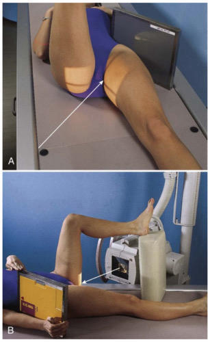
T
67
Traumatic (T) or not traumatic (NT): Unilateral "frog-leg" (modified Cleaves method)
NT
68
Traumatic (T) or not traumatic (NT): AP bilateral "frog-leg" (modified Cleaves method)
NT
69
Traumatic (T) or not traumatic (NT): Modified axiolateral (Clements-Nakayama method)
T
70
Traumatic (T) or not traumatic (NT): AP axial for pelvic outlet (Taylor method)
T
71
Which of the following projections is recommended to demonstrate the superoposterior wall of the acetabulum?
A. AP axial inlet
B. PA axial oblique
C. Axiolateral inferosuperior
D. Modified axiolateral
A. AP axial inlet
B. PA axial oblique
C. Axiolateral inferosuperior
D. Modified axiolateral
B. PA axial oblique
72
When gonadal shielding is not used, ______(males/females) receive a greater gonadal dose with an AP pelvis projection.
Females (because of location of CR & reproductive organs)
73
How many degrees are the femurs abducted (from the vertical plane) for the bilateral frog-leg projection?
40 to 45 degrees
74
Where is the CR placed for a bilateral frog-leg (modified Cleaves method) projection?
3 inches (7.5 cm) below level of ASIS (1 inch [2.5 cm] above symphysis pubis)
75
Where is the CR placed for an AP unilateral frog-leg projection?
Midfemoral neck
76
Which CR angle is required for the AP axial-outlet (Taylor method) projection for a female patient?
A. 15 to 25 degrees caudad
B. 30 to 45 cephalad
C. 20 to 35 cephalad
D. None (CR is perpendicular)
A. 15 to 25 degrees caudad
B. 30 to 45 cephalad
C. 20 to 35 cephalad
D. None (CR is perpendicular)
B. 30 to 45 degrees cephalad
77
Which type of pathology is best demonstrated with the posterior oblique (Judet method)?
A. Acetabular fractures
B. Anterior pelvic bone fractures
C. Proximal femur fractures
D. Femoral neck fractures
A. Acetabular fractures
B. Anterior pelvic bone fractures
C. Proximal femur fractures
D. Femoral neck fractures
A. Acetabular fractures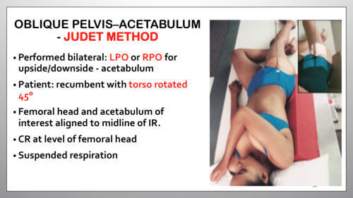
78
How much obliquity of the body is required for the posterior oblique projection (Judet method)?
A. None (CR is perpendicular)
B. 20 degrees
C. 30 degrees
D. 45 degrees
A. None (CR is perpendicular)
B. 20 degrees
C. 30 degrees
D. 45 degrees
D. 45 degrees
79
What type of CR angle is used for a PA axial oblique (Teufel) projection?
A. 15 degrees cephalad
B. 15 to 20 degrees cephalad
C. 5 degrees caudad
D. 12 degrees cephalad
A. 15 degrees cephalad
B. 15 to 20 degrees cephalad
C. 5 degrees caudad
D. 12 degrees cephalad
D. 12 degrees cephalad
80
How is the pelvis (body) positioned for a PA axial oblique (Teufel) projection?
A. PA with 45 degrees rotated away from affected side
B. Prone or erect PA - no rotation
C. PA 35 to 40 degrees toward affected side
D. AP with 40 degrees away from affected side
A. PA with 45 degrees rotated away from affected side
B. Prone or erect PA - no rotation
C. PA 35 to 40 degrees toward affected side
D. AP with 40 degrees away from affected side
C. PA 35 to 40 degrees toward the affected side
81
True/False: Any orthopedic device or appliance of the hip should be seen in its entirety on an AP hip radiograph.
True
82
The axiolateral (inferosuperior) projection is designed for ______________ (traumatic or non-traumatic) situations.
Traumatic
83
How is the unaffected leg positioned for the axiolateral hip projection?
If is flexed & elevated to prevent it from being superimposed over the affected hip
84
Which of the following factors does not apply to an axiolateral (inferosuperior) projection of the hip on a male patient?
A. IR parallel to femoral neck
B. 80 to 90 kV
C. Use of gonadal shielding
D. Use of a stationary grid
A. IR parallel to femoral neck
B. 80 to 90 kV
C. Use of gonadal shielding
D. Use of a stationary grid
C. Use of gonadal shielding
85
True/False: An AP pelvis projection using 90 kV & 8 mAs results in less patient dose than a projection using 80 kV & 12 mAs (for both males & females).
True
86
True/ False: The unaffected foot during an axiolateral (inferosuperior) projection can be burned if allowed to rest on the collimator.
True
87
The modified axiolateral (Clements-Nakayama method) projection requires the CR to be angled ____________ degrees posterior from horizontal.
15 to 20
88
A. Which special projection of the hip demonstrates the anterior & posterior rims of the acetabulum & the ilioischial & iliopubic columns? (Include projection name & method name.)
B. Which CR angle (if any) is used for this projection?
B. Which CR angle (if any) is used for this projection?
A. Posterior oblique projections of acetabulum (Judet method)
B. 0 degrees (perpendicular)
B. 0 degrees (perpendicular)
89
What is the name of a special projection of the pelvis used to assess trauma to pubic & ischial structures? (Include projection name & method name.)
AP axial outlet projection (Taylor method)
90
Proper name? Axiolateral (inferosuperior)
Danelius-Miller
91
Proper name? Modified axiolateral
Clements-Nakayama
92
Proper name? AP bilateral or unilateral frog-leg
Modified Cleaves
93
Proper name? Posterior axial oblique for acetabulum
Teufel
94
Proper name? AP axial for pelvic "outlet" bones
Taylor
95
Proper name? Posterior oblique for acetabulum
Judet
96
What is the optimal amount of hip abduction applied for the unilateral "frog-leg" projection to demonstrate the femoral neck without distortion?
A. 45 degrees from vertical
B. 90 degrees from vertical
C. 10 degrees from vertical
D. 20 to 30 degrees from vertical
A. 45 degrees from vertical
B. 90 degrees from vertical
C. 10 degrees from vertical
D. 20 to 30 degrees from vertical
D. 20 to 30 degrees from vertical
97
True/False: The Lauenstein/Hickey method for the unilateral "frog-leg" projection will produce distortion of the femoral neck.
True
98
How much is the IR tilted for the modified axiolateral projection of the hip?
15 degrees from the vertical
99
True/False: Gonadal shielding can be used for males for the axiolateral (inferosuperior) projection of the hip.
False
100
Situation: A radiograph of an AP pelvis projection shows that the lesser trochanters are readily demonstrated on the medial side of the proximal femurs. The patient is ambulatory but has a history of early osteoarthritis in both hips. Which positioning modification needs to be made to prevent this positioning error?
Rotate the lower limbs 15 to 20 degrees internally to place the proximal femurs in a true AP position. (With general chronic pain, the lower limbs usually can be rotated safely.)
101
Situation: A radiograph of an AP pelvis shows that the right iliac wing is foreshortened as compared with the left side. Which specific positioning error was made?
The patient is rotated toward the left - left posterior oblique (LPO)
102
Situation: A radiograph of a unilateral frog-leg (modified Cleaves) projection produces distortion of the femoral neck. Based on the AP hip projection, the radiologist suspects a nondisplaced fracture of the femoral neck. What can the technologist do to define this region better?
Repeat the exposure & only abduct the femur 20 to 30 degrees from vertical. (It will produce less distortion of the femoral neck.)
103
Situation: A radiograph of an axiolateral (inferosuperior) projection shows that the posterior aspect of the acetabulum & femoral head were cut off of the bottom of the image. The emergency room physician requests that the position be repeated. What can be done to avoid this problem on the repeat exposure?
If possible, elevate the patient at least 2 inches (5 cm) by placing sheets or blankets beneath the patient
104
Situation: A patient enters the ER with a pelvis injury resulting from a motor vehicle accident. The initial AP pelvis projection demonstrates a possible defect or fracture of the left acetabulum. No other fractures are detected & the patient is able to move comfortably. What additional projections can be taken to demonstrate a possible acetabular fracture?
The PA axial oblique (Teufel method) or posterior oblique (Judet method) can be taken to demonstrate aspects of the acetabulum more completely.
105
Situation: A radiograph from a modified axiolateral projection of the hip shows excessive grid lines on the image, which also appears underexposed. What can be done to avoid this problem during the repeat exposure?
Ensure that the CR is centered to near the midline of the grid cassette & the face of the IR is perpendicular to the CR.
106
Situation: A portable AP and lateral hip study is ordered for a patient who is in recovery following hip replacement surgery. The radiograph of the AP hip reveals that the upper portion of the acetabular prosthesis is slightly cut off but is included on the lateral projection. Should the technologist repeat the AP projection? Why or why not?
Yes. Any orthopedic appliance or prosthesis must be seen in its entirety in both projections.
107
Situation: A patient with hip pain from a fall enters the emergency room. The physician orders a left hip study. When moved to the radiographic table, the patient complains loudly about the pain in the left hip. Which positioning routine should be used for this patient?
AP pelvis & axiolateral (inferosuperior) left hip. The AP pelvis radiograph should be taken initially without leg rotation; the radiograph must be reviewed by the physician & checked for fractures or dislocations before attempting an internal rotation of the left leg for the axiolateral (inferosuperior) projection.
108
Situation: A patient has just been moved to his hospital room after a bilateral hip replacement surgery. The surgeon has ordered a postoperative hip routine for both hips. Which specific positioning routine should be used? (The patient can be brought to the radiology department.)
AP pelvis & modified axiolateral - Clements-Nakayama method
109
Situation: A patient with a possible pelvic ring fracture from a trauma enters the emergency room. The AP pelvis projection, which was taken to determine whether the right acetabulum was fractured, is inconclusive. Which other radiographic projection can be taken to better visualize the acetabulum? What other imaging modality can be used to determine the presence of a pelvic ring fracture?
Posterior oblique - Judet method. CT is often judged superior in detecting pelvic ring fractures
110
Situation: A physician orders a study for inlet & outlet projections of the pelvis. Which projections could be performed to meet this request?
AP axial for pelvic "outlet" (Taylor method) & AP axial for pelvic "inlet" projections & possibly the posterior oblique (Judet method) projections to provide another perspective of the inlet & outlet regions of the pelvis. (If unsure of the request routine, contact the physician for clarification.)
111
Situation: A technologist notices that his AP pelvis projections often demonstrate a moderate degree of rotation. What positioning technique can the technologist perform to eliminate (or at least minimize) rotation on his AP pelvis projections?
Palpate both ASIS & ensure they are equal distance from the tabletop. To verify no rotation is still present, ensure that the iliac wings are symmetric, as seen on the radiograph.
112
Situation: A very young child comes to the radiology department with a clinical history of DDH. What is the most common positioning routine for this condition?
AP pelvis & bilateral "frog-leg" (modified Cleaves) projections
Mehr Karteikarten: Medizin und Gesundheit
DOSAGE FORMSMedizin und GesundheitLlanceVideo 2Medizin und Gesundheitjocyt9180 MidtermMedizin und Gesundheit · EnglischTaylorGonorrheaMedizin und Gesundheit · EnglischcaeEPS⚕️Medizin und Gesundheit · SpanischGonzálezEmbryology: Pharyngeal ApparatusMedizin und Gesundheit · EnglischSaraPulmonary Function Tests (PFT) ReviewMedizin und Gesundheit · EnglischcathsimsEndocrinology and BloodMedizin und Gesundheit · EnglischJESSQTRANNeurology: Fainting, Seizures and EpilepsyMedizin und Gesundheit · EnglischTejuBlood Anatomy (Chapter 18)Medizin und Gesundheit · Englischcaeanatomy places + functionsMedizin und Gesundheit · EnglischhanaPrimary Hemostasis Medizin und Gesundheit · EnglischCHRISTY
Alle Decks: Medizin und Gesundheit
Lerne diese Decks in der App
Kostenlos auf iPhone, Android, Mac und im Web. Füge jedes Deck mit einem Tipp zu deinem Konto hinzu.